
The Vitrotest ELISA range covers a wide spectrum of immunodiagnostic applications, including HIV infection and viral hepatitis, TORCH infections, parasitic diseases, allergies, tumor markers, hormones, and metabolic disorders. Vitrotest diagnostic kits combine ease of use, reproducibility of results, and simplicity of the testing procedure, supported by standardized incubation times, microplates with detachable wells, clear color development, as well as ready-to-use TMB and сonjugate solutions. TMB, Wash Tween and Stop Solution are interchangeable across Vitrotest kits.
Original synthetic control materials help enhance biosafety. Evaluation of the kits using commercial serum panels and alignment of quantitative methods with international standards help ensure consistent product performance.
-
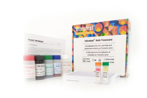
The test kit Vitrotest® Anti-Toxocara is an enzyme linked immunosorbent assay (ELISA) for the detection of IgG class antibodies to Toxocara canis in human serum or plasma.
Determination of IgG antibodies to Toxocara canis in the test kit Vitrotest® Anti-Toxocara is based on a solid phase, indirect ELISA in a two-step incubation procedure.
○ ТК058 - 96 tests
○ ТК112 - 192 tests- Solid phase: breakable microplate ELISA is coated Toxocara canis larvae antigens.
- Conjugate: a monoclonal antibodies to human IgG conjugated to horseradish peroxidase.
- Chromogen: ready to use TMB solution.
- Volume of sample for analysis: 10 μl.
- Assay time: 1h 15 min.
Toxocariasis is an widespread parasitic disease of humans caused by infection with the second stage larvae of two main species of parasitic nematodes, Toxocara canis and Toxocara cati. The role of toxocaras in human disease was disclosed in the 1950th by Wilder and Beaver et al. who first identified toxocara larvae in ocular and visceral tissues respectively.
Adult worms of T. canis and T.cati live within the lumen of the small intestine of dogs and cats respectively which serve as definitive hosts for these helminths. Unembryonated eggs produced by adult worms are shed in the feces of the definitive hosts. Eggs embryonate in the environment, and could be ingested by definitive hosts as well as accidental, paratenic hosts. After ingestion by the definitive hosts, microscopic (300 µm long and 20 µm in diameter) larvae hatch in the intestine, penetrate the intestinal wall, reach lungs via bloodstream, penetrate alveolar walls, ascend the bronchial tree to the throat where they are swallowed, and mature into adult worms. In humans and other paratenic hosts, the larvae are unable to undergo the full development cycle described above; instead, they are carried by the circulation to a wide variety of organs and tissues (liver, heart, lungs, brain, muscle, eyes), and cause severe local reactions that are the basis of toxocariasis.
The degree of host damage, and the concomitant signs and symptoms, varies with regard to which tissue has been invaded; the liver, lungs, and CNS (including the eyes) appear to be most sensitive. In the eye, migrating larvae can damage the retina, inducing granulomatous reactions leading to impaired sight or even loss of sight. The number of migrating larvae and the age of the host are two additional factors defining the presence and severity of symptoms.
Lifespan of the larvae could be as long as several years, and clinical disease could present in any time during this period, or even later, representing pathological immune response to dying or dead larvae.
For most people, an infection with these helminths causes no symptoms. Possible symptoms symptoms are nonspecific and include fever, fatigue, anorexia, or lymphadenopathy. Pulmonary symptoms and abdominal symptoms are similar to those under many other diseases; they are present when larvae migrate to the lungs or abdominal organs, respectively. Neurologic findings are diverse and also nonspecific. -
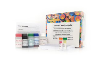
The test kit Vitrotest® Anti-Trichinella is an enzyme linked immunosorbent assay (ELISA) for the detection of IgG class antibodies to Trichinella spiralis in human serum or plasma.
Determination of IgG antibodies to Trichinella spiralis in the test kit Vitrotest® Anti-Trichinella is based on a solid phase, indirect ELISA in a two-step incubation procedure.
○ ТК067 - 96 tests- Solid phase: breakable microplate ELISA is coated Trichinella spiralis larvae antigens.
- Conjugate: a monoclonal antibodies to human IgG conjugated to horseradish peroxidase.
- Chromogen: ready to use TMB solution.
- Volume of sample for analysis: 10 μl.
- Assay time: 1h 15 min.
Trichinosis is a helminthiasis caused by a nematode Trichinella spiralis. Trichinellas are small, thread-like worms covered with a striped cuticle.
Transmission occurs by ingestion of meat containing encapsulated trichinella larvae. During digestion under the action of the gastric juice larvae release from capsules penetrate the submucosal layer of the small intestine, adhere to the mucosa and begin to proliferate. Soon after fertilization of females, the males die and the females start producing larvae, which enter the blood and lymphatic vessels through tissue mucosa, spread throughout the body and settle in striated muscles. Thereafter this capsule is impregnated with calcium salts leading to calcification. The larvae remain viable for many years.
The incubation period of human trichinosis lasts 10-25 days. Trichinosis is characterized by fever, myalgia, facial swelling, skin rash, blood eosinophilia, and in severe cases – by damage to internal organs and central nervous system.
The diagnosis of trichinosis is based on clinical signs, epidemiological history, serological tests (complement fixation tests, the reaction of indirect hemagglutination) and ELISA. The latter method is recommended by OIE for serological diagnosis of trichinosis.
The most specific and successful method to confirm infestation is the detection of IgG antibodies to trichinella antigens in the blood. These antibodies could be determined from 2-3 to 4-6 weeks after the eating of contaminated meat. Specific IgE class antibodies are also present in the blood during the acute stage of the disease, however, they are rarely detected due to the short period of their circulation in the bloodstream. During the early stage of invasion, specific antibodies might be still undetectable. Therefore, another sample should be taken in 1-2 weeks to confirm or reject suspected trichinosis. Seroconversion usually occurs 2-5 weeks after infection depending on the infectious dose. Assessment of antibody dynamics is a very informative marker of therapy effectiveness. In cases of ineffective or untimely therapy, specific antibodies are detected for up to 20 years if trichinosis is effectively treated in the first two weeks after infection, antibodies disappear within a year. -
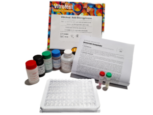
The test kit Vitrotest® Anti-Strongyloides is an enzyme linked immunosorbent assay (ELISA) for the detection of IgG class antibodies to Strongyloides stercoralis in human serum or plasma.
Determination of IgG antibodies to Strongyloides stercoralis in the test kit Vitrotest® Anti-Strongyloides is based on a solid phase, indirect ELISA in a two-step incubation procedure.
- ТК146 - 96 tests
- Solid phase: breakable microplate ELISA is coated Strongyloides stercoralis antigens.
- Conjugate: a monoclonal antibodies to human IgG conjugated to horseradish peroxidase.
- Chromogen: ready to use TMB solution.
- Volume of sample for analysis: 10 μl.
- Assay time: 40 min.
Strongyloidiasis is a chronic parasitic infection of humans caused by Strongyloides stercoralis. This helminth is predominantly found in tropical and subtropical regions but can also be present in temperate climates. It is estimated that approximately 300 to 600 million people worldwide are infected.
Primary infection occurs when S. stercoralis larvae penetrate human skin through direct contact with contaminated soil. The larvae migrate via the bloodstream and lymphatic system to the respiratory tract, where they are then coughed up and swallowed, reaching the intestines. In the intestines, the parasites develop into adult worms, lay eggs, and produce larvae that can be excreted in the feces. A unique feature of S. stercoralis is its ability to cause autoinfection, as larvae may re-enter the intestines or perianal skin without leaving the host, leading to persistent infection.
In immunocompetent individuals, uncomplicated strongyloidiasis may be asymptomatic or present with mild cutaneous and gastrointestinal symptoms. Often, the only sign of infection is unexplained peripheral eosinophilia. However, in cases of heavy infestation or in immunosuppressed patients, the disease can cause severe manifestations such as abdominal pain, watery diarrhea, constipation, weight loss, vomiting, or small bowel obstruction. Hyperinfection syndrome is the most severe manifestation of the disease, with high mortality rates.
Parasitological methods (microscopy, culture) are traditionally used for diagnosis. The sensitivity and specifi city of these tests, however, are not high. The standard stool examination has a sensitivity of only 21% (5). Serological methods, especially enzyme-linked immunosorbent assay (ELISA), are highly sensitive and convenient for detecting antibodies to S. stercoralis larvae. ELISA is widely used for screening, diagnosing strongyloidiasis, and monitoring treatment effi cacy, as antibody titers signifi cantly decline within the fi rst 6 months following successful eradication therapy.
-
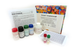
The test kit Vitrotest Ureaplasma-IgG is an enzyme linked immunosorbent assay (ELISA) for the qualitative and semiquantitative determination of IgG class antibodies to Ureaplasma urealyticum in human serum or plasma.
Determination of IgG antibodies to U.urealyticum in the test kit Vitrotest Ureaplasma-IgG is based on a solid phase indirect ELISA in a two-step incubation procedure.
○ ТК028 - 96 tests
- Solid phase: breakable microplate ELISA is coated with recombinant antigens of U. urealyticum.
- Conjugate: a monoclonal antibodies to human IgG conjugated to horseradish peroxidase.
- Chromogen: ready to use TMB solution.
- Volume of sample for analysis: 20 μl.
- Assay time: 30/30/15 min.
-
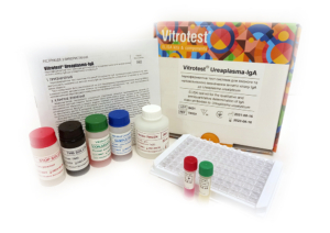
The test kit Vitrotest Ureaplasma-IgA is an enzyme linked immunosorbent assay (ELISA) for the qualitative and semiquantitative determination of IgA class antibodies to Ureaplasma urealyticum in human serum or plasma.
Determination of IgA antibodies to U.urealyticum in the test kit Vitrotest Ureaplasma-IgA is based on a solid phase indirect ELISA in a two-step incubation procedure.
○ ТК029 - 96 tests
- Solid phase: breakable microplate ELISA is coated with recombinant antigens of U. urealyticum.
- Conjugate: a monoclonal antibodies to human IgA conjugated to horseradish peroxidase.
- Chromogen: ready to use TMB solution.
- Volume of sample for analysis: 20 μl.
- Assay time: 30/30/15 min.
-
The test kit Vitrotest Ureaplasma-IgМ is an enzyme linked immunosorbent assay (ELISA) for the qualitative and semiquantitative determination of IgM class antibodies to Ureaplasma urealiticum in human serum or plasma.
Determination of IgM antibodies to Ureaplasma urealiticum in the test kit Vitrotest Ureaplasma-IgМ is based on a solid phase, «IgM-capture» ELISA in a two-step incubation procedure.
○ ТК096 - 96 tests
- Solid phase: breakable microplate ELISA is coated with monoclonal anti-IgM antibodies.
- Conjugate: recombinant U. urealiticum antigen, conjugated to horseradish peroxidase.
- Chromogen: ready to use TMB solution.
- Volume of sample for analysis: 10 μl.
- Assay time: 30/30/30 min.
-
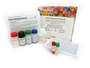
The test kit Vitrotest Mycoplasma hominis-IgG is an enzyme linked immunosorbent assay (ELISA) for the qualitative and semiquantitative determination of IgG class antibodies to Mycoplasma hominis in human serum or plasma.
Determination of IgG antibodies to M. hominis in the test kit Vitrotest Mycoplasma hominis-IgG is based on a solid phase indirect ELISA in a two-step incubation procedure.
○ ТК024 - 96 tests
- Solid phase: breakable microplate ELISA is coated with native Mycoplasma hominis antigens.
- Conjugate: a monoclonal antibodies to human IgG conjugated to horseradish peroxidase.
- Chromogen: ready to use TMB solution.
- Volume of sample for analysis: 10 μl.
- Assay time: 30/30/15 min.
-
The test kit Vitrotest Mycoplasma hominis-IgA is an enzyme linked immunosorbent assay (ELISA) for the qualitative and semiquantitative determination of IgA class antibodies to Mycoplasma hominis in human serum or plasma.
Determination of IgA antibodies to M. hominis in the test kit Vitrotest Mycoplasma hominis-IgA is based on a solid phase indirect ELISA in a two-step incubation procedure.
○ ТК025 - 96 tests
- Solid phase: breakable microplate ELISA is coated with native Mycoplasma hominis antigens.
- Conjugate: a monoclonal antibodies to human IgA conjugated to horseradish peroxidase.
- Chromogen: ready to use TMB solution.
- Volume of sample for analysis: 10 μl.
- Assay time: 30/30/15 min.
-
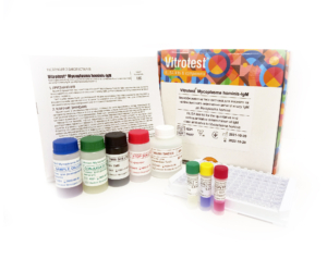
The test kit Vitrotest Mycoplasma hominis-IgM is an enzyme linked immunosorbent assay (ELISA) for the qualitative and semiquantitative determination of IgM class antibodies to Mycoplasma hominis in human serum or plasma.
Determination of IgM antibodies to Mycoplasma hominis in the test kit Vitrotest Mycoplasma hominis-IgM is based on a solid phase, «IgM-capture» ELISA in a two-step incubation procedure.
○ ТК097 - 96 tests
- Solid phase: breakable microplate ELISA is coated with monoclonal anti-IgM antibodies.
- Conjugate: recombinant M. hominis antigen, conjugated to horseradish peroxidase.
- Chromogen: ready to use TMB solution.
- Volume of sample for analysis: 10 μl.
- Assay time: 30/30/30 min.
-
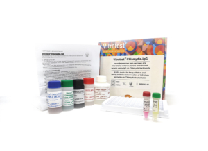
The test kit Vitrotest Chlamydia-IgG is an enzyme linked immunosorbent assay (ELISA) for the qualitative and semiquantitative determination of IgG class antibodies to Chlamydia trachomatis in human serum or plasma.
Determination of IgG antibodies to Chlamydia trachomatis in the test kit Vitrotest Chlamydia-IgG is based on a solid phase indirect ELISA in a two-step incubation procedure.
○ ТК014 - 96 tests
- Solid phase: breakable microplate ELISA is coated with recombinant Chlamydia trachomatis antigens.
- Conjugate: a monoclonal antibodies to human IgG conjugated to horseradish peroxidase.
- Chromogen: ready to use TMB solution.
- Volume of sample for analysis: 20 μl.
- Assay time: 30/30/15 min.
-
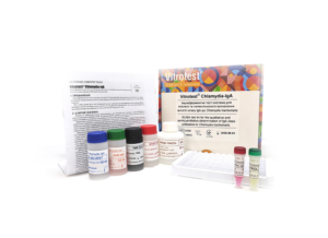
The test kit Vitrotest Chlamydia-IgА is an enzyme linked immunosorbent assay (ELISA) for the qualitative and semiquantitative determination of IgА class antibodies to Chlamydia trachomatis in human serum or plasma.
Determination of IgА antibodies to Chlamydia trachomatis in the test kit Vitrotest Chlamydia-IgА is based on a solid phase indirect ELISA in a two-step incubation procedure.
○ ТК015 - 96 tests
- Solid phase: breakable microplate ELISA is coated with recombinant Chlamydia trachomatis antigens.
- Conjugate: a monoclonal antibodies to human IgА conjugated to horseradish peroxidase.
- Chromogen: ready to use TMB solution.
- Volume of sample for analysis: 20 μl.
- Assay time: 30/30/15 min.
-
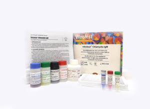
The test kit Vitrotest Chlamydia-IgМ is an enzyme linked immunosorbent assay (ELISA) for the qualitative determination of IgM class antibodies to Chlamydia trachomatis in human serum or plasma.
Determination of IgM antibodies to Chlamydia trachomatis in the test kit Vitrotest Chlamydia-IgМ is based on a solid phase «IgM-capture» ELISA in a two-step incubation procedure.
○ ТК036 - 96 tests
- Solid phase: breakable microplate ELISA is coated with monoclonal anti-IgM antibodies.
- Conjugate: recombinant antigen MOMP of Chlamydia trachomatis, conjugated to horseradish peroxidase.
- Chromogen: ready to use TMB solution.
- Volume of sample for analysis: 10 μl.
- Assay time: 30/30/30 min.
-
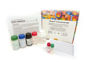
The test kit Vitrotest Trichomonas-IgG is an enzyme linked immunosorbent assay (ELISA) for the qualitative and semiquantitative determination of IgG class antibodies to Trichomonas vaginalis in human serum or plasma.
Determination of IgG antibodies to Trichomonas vaginalis in the test kit Vitrotest Trichomonas-IgG is based on a solid phase, indirect ELISA in a two-step incubation procedure.
○ ТК061 - 96 tests
- Solid phase: breakable microplate ELISA is coated with Trichomonas vaginalis antigens.
- Conjugate: a monoclonal antibodies to human IgG conjugated to horseradish peroxidase.
- Chromogen: ready to use TMB solution.
- Volume of sample for analysis: 20 μl.
- Assay time: 30/30/15 min.
-
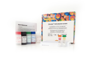
The test-kit Vitrotest Helicobacter screen is an enzyme linked immunosorbent assay (ELISA) for the detection of IgG, IgA and IgM specific antibodies to Helicobacter pylori in human serum or plasma.
Detection of antibodies to H. pylori in the test-kit Vitrotest Helicobacter screen is based on a solid phase, indirect ELISA in a two-step incubation procedure.
○ ТК045 - 96 tests
- Solid phase: breakable microplate ELISA is coated with recombinant antigens of H. pylori.
- Conjugate: a monoclonal antibodies to human IgA, IgG and IgM conjugated to horseradish peroxidase.
- Chromogen: ready to use TMB solution.
- Volume of sample for analysis: 10 μl.
- Assay time: 30/30/15 min.
-
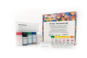
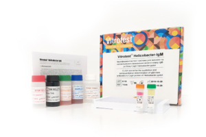
The test kit Vitrotest Helicobacter-IgG is an enzyme linked immunosorbent assay (ELISA) for the quantitative and semiquantitative determination of IgG class antibodies to CagA protein of Helicobacter pylori in human serum or plasma.
Determination of IgG antibodies to CagA protein of H. pylori in the test kit Vitrotest Helicobacter-IgG is based on a solid phase, indirect ELISA in a two-step incubation procedure.
○ ТК046 - 96 tests
- Solid phase: breakable microplate ELISA is coated with recombinant CagA protein of H. pylori.
- Conjugate: a monoclonal antibodies to human IgG conjugated to horseradish peroxidase.
- Chromogen: ready to use TMB solution.
- Volume of sample for analysis: 10 μl.
- Assay time: 30/30/15 min.
- Linearity range: 1,25-200 U/ml.
-
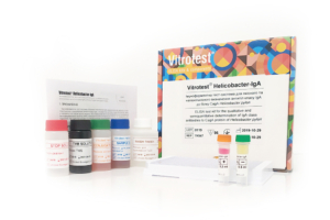
The test kit Vitrotest Helicobacter-IgА is an enzyme linked immunosorbent assay (ELISA) for the qualitative and semiquantitative determination of IgА class antibodies to CagA protein of Helicobacter pylori in human serum or plasma.
Determination of IgА antibodies to CagA protein of H. pylori in the test kit Vitrotest Helicobacter-IgА is based on a solid phase, indirect ELISA in a two-step incubation procedure.
○ ТК047 - 96 tests
- Solid phase: breakable microplate ELISA is coated with recombinant CagA protein of H. pylori.
- Conjugate: a monoclonal antibodies to human IgА conjugated to horseradish peroxidase.
- Chromogen: ready to use TMB solution.
- Volume of sample for analysis: 10 μl.
- Assay time: 30/30/15 min.
-
The test kit Vitrotest Helicobacter-IgМ is an enzyme linked immunosorbent assay (ELISA) for the qualitative and semiquantitative determination of IgМ class antibodies to CagA protein of Helicobacter pylori in human serum or plasma.
Determination of IgМ antibodies to CagA protein of H. pylori in the test kit Vitrotest Helicobacter-IgМ is based on a solid phase, indirect ELISA in a two-step incubation procedure.
○ ТК048 - 96 tests
- Solid phase: breakable microplate ELISA is coated with recombinant CagA protein of H. pylori.
- Conjugate: a monoclonal antibodies to human IgМ conjugated to horseradish peroxidase.
- Chromogen: ready to use TMB solution.
- Volume of sample for analysis: 10 μl.
- Assay time: 30/30/15 min.
-
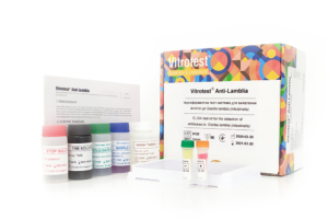
The test kit Vitrotest Anti-Lamblia is an enzyme linked immunosorbent assay (ELISA) for the detection of total antibodies to Giardia lamblia (intestinalis) in human serum or plasma.
Determination of total antibodies to Giardia lamblia in the test kit Vitrotest Anti-Lamblia is based on a solid phase, indirect ELISA in a two-step incubation procedure.
○ ТК030 - 96 tests
○ ТК108 - 192 tests
- Solid phase: breakable microplate ELISA is coated Giardia lamblia purified antigens.
- Conjugate: a monoclonal antibodies to human IgG, IgA and IgM conjugated to horseradish peroxidase.
- Chromogen: ready to use TMB solution.
- Volume of sample for analysis 10 μl.
- Assay time: 30/30/15 min.
Giardia lamblia (intestinalis) causes giardiasis, a parasitic infestation that can manifest as latent parasitic carriage or in more pronounced forms, such as intestinal dysfunction. Giardiasis has been recorded on all five continents and in most countries worldwide. Infection rates vary from less than 1% to 50%. In many developing countries, where basic sanitary conditions are lacking, infection with Giardia in children by the age of 2 is almost universal. In contrast, in developed countries, the infection rate of G.lamblia is only 3–7%. The disease affects all age groups, but preschool-aged children are the most affected demographic.
The primary route of transmission for G.lamblia is fecal-oral. The parasite has a simple two-stage life cycle. After the host ingests cysts, they release trophozoites in the duodenum, which then attach to the mucosal lining of the small intestine.
Trophozoites exist only on the surface of the mucosa in the upper part of the small intestine. Therefore, Giardia mechanically blocks the mucosa and disrupts membrane digestion and the motor activity of the small intestine. Giardia impairs the absorption of fats, carbohydrates, vitamins C and B12, and leads to secondary bacterial infections. Symptoms of giardiasis can include diarrhea, fatigue, bloating, apathy, weight loss, decreased appetite, pallor, and muscle twitching. From the gastrointestinal tract perspective, giardiasis mainly manifests as enterocolitis with catarrhal symptoms.
Numerous findings indicate the role of the humoral immune response in the elimination of G.lamblia. As shown in an experimental human infection model, IgM antibody levels significantly increased on days 14-21 post-infection and gradually declined following therapy. In contrast, IgG antibody levels remained elevated after successful treatment. The dynamics of IgA levels were similar to those of IgM.
The diagnosis of giardiasis is based on clinical history, symptoms, and the presence of cysts in fecal samples or trophozoites in material obtained from the small intestine during duodenal aspiration or duodenal biopsy. Alternative methods include the detection of G.lamblia antigen in feces and the determination of specific antibodies against Giardia in the patient’s serum. Serological testing is considered a valuable adjunct in the diagnosis of giardiasis. In addition to aiding in diagnosis, it can be useful for assessing the patient’s immune response and for epidemiological studies. -
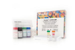
The test kit Vitrotest Lamblia-IgM is an enzyme linked immunosorbent assay (ELISA) for the detection of IgM class antibodies to Giardia lamblia (intestinalis) in human serum or plasma.
Determination of IgM antibodies to Giardia lamblia in the test kit Vitrotest Lamblia-IgM is based on a solid phase, indirect ELISA in a two-step incubation procedure.
○ ТК031 - 96 tests
- Solid phase: breakable microplate ELISA is coated Giardia lamblia purified antigens.
- Conjugate: a monoclonal antibodies to human IgM conjugated to horseradish peroxidase.
- Chromogen: ready to use TMB solution.
- Volume of sample for analysis 10 μl.
- Assay time: 30/30/15 min.
Giardia lamblia (intestinalis) causes giardiasis, a parasitic infestation that can manifest as latent parasitic carriage or in more pronounced forms, such as intestinal dysfunction. Giardiasis has been recorded on all five continents and in most countries worldwide. Infection rates vary from less than 1% to 50%. In many developing countries, where basic sanitary conditions are lacking, infection with Giardia in children by the age of 2 is almost universal. In contrast, in developed countries, the infection rate of G.lamblia is only 3–7%. The disease affects all age groups, but preschool-aged children are the most affected demographic.
The primary route of transmission for G.lamblia is fecal-oral. The parasite has a simple two-stage life cycle. After the host ingests cysts, they release trophozoites in the duodenum, which then attach to the mucosal lining of the small intestine.
Trophozoites exist only on the surface of the mucosa in the upper part of the small intestine. Therefore, Giardia mechanically blocks the mucosa and disrupts membrane digestion and the motor activity of the small intestine. Giardia impairs the absorption of fats, carbohydrates, vitamins C and B12, and leads to secondary bacterial infections. Symptoms of giardiasis can include diarrhea, fatigue, bloating, apathy, weight loss, decreased appetite, pallor, and muscle twitching. From the gastrointestinal tract perspective, giardiasis mainly manifests as enterocolitis with catarrhal symptoms.
Numerous findings indicate the role of the humoral immune response in the elimination of G.lamblia. As shown in an experimental human infection model, IgM antibody levels significantly increased on days 14-21 post-infection and gradually declined following therapy. In contrast, IgG antibody levels remained elevated after successful treatment. The dynamics of IgA levels were similar to those of IgM.
The diagnosis of giardiasis is based on clinical history, symptoms, and the presence of cysts in fecal samples or trophozoites in material obtained from the small intestine during duodenal aspiration or duodenal biopsy. Alternative methods include the detection of G.lamblia antigen in feces and the determination of specific antibodies against Giardia in the patient’s serum. Serological testing is considered a valuable adjunct in the diagnosis of giardiasis. In addition to aiding in diagnosis, it can be useful for assessing the patient’s immune response and for epidemiological studies. -
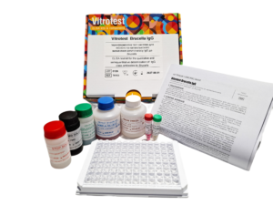
The test kit Vitrotest® Brucella-IgG is an enzyme linked immunosorbent assay (ELISA) for the qualitative and semiquantitative determination of igG class antibodies to Brucella in human serum or plasma.
Determination of IgG antibodies to Brucella in the test kit Vitrotest® Brucella-IgG is based on a solid phase, indirect ELISA in a two-step incubation procedure.
- TK153 - 96 tests
- Solid phase: strip ELISA plate pre-coated with antigens of Brucella.
- Conjugate: monoclonal antibodies to human IgG conjugated with horseradish peroxidase.
- Chromogen: ready to use TMB solution.
- Sample volume: 10 μl.
- Assay time: 1 h 15 min.
Brucellosis is a zoonotic infectious disease caused by bacteria of the genus Brucella. Globally, 1.6–2.1 million human cases of brucellosis are reported annually. Among the countries with the highest reported incidence of brucellosis are Iran, Kyrgyzstan, Tajikistan, Kazakhstan, Azerbaijan, Turkmenistan, Armenia, and Uzbekistan.
The species most pathogenic to humans include Brucella melitensis, Brucella abortus, Brucella suis, and Brucella canis. Different species of Brucella vary in their reservoir hosts and degree of virulence: B. melitensis is considered the most pathogenic for humans, whereas B. abortus more commonly causes relatively milder forms of the disease.
Human infection occurs through contact with infected animals and their biological fluids, as well as through the consumption of animal-derived products (raw milk and insufficiently heat-treated meat), and by the airborne route during occupational exposure. The pathogen enters the human body through mucous membranes or damaged skin. Subsequently, the bacteria are transported by macrophages to lymphoid tissues, spread through the lymphatic system, and may potentially proliferate in multiple organs, causing localized and systemic infections. By persisting within host cells such as macrophages, Brucella spp. employ strategies to evade the host immune response, resulting in prolonged infection.
The incubation period of brucellosis usually ranges from 1 to 4 weeks but may extend to several months. Clinical manifestations are characterized by undulating fever, sweating (especially at night), weakness, and enlargement of the lymph nodes, liver, and spleen. In chronic cases, the musculoskeletal, nervous, and cardiovascular systems may also be affected.
Due to the nonspecific nature of its symptoms, the diagnosis of brucellosis is challenging and requires a comprehensive approach, including the evaluation of clinical findings, epidemiological history, and laboratory test results (bacterial culture, serological assays, and PCR). Serological methods used for the diagnosis of brucellosis include the agglutination test, complement fixation test, and enzyme-linked immunosorbent assay (ELISA).
Using the ELISA method, IgM antibodies are detected during the early stages of infection, whereas IgG antibodies are detected later and may persist for a long period after previous infection or successful treatment. A rapid decline in IgG antibody titers usually indicates a favorable response to antibiotic therapy, while persistently high or increasing titers may indicate treatment failure, residual disease, or an impending clinical relapse. Therefore, the detection of IgG antibodies specific to Brucella is an important marker of current or past infection, particularly in subacute and chronic forms of brucellosis. -
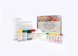
The test kit Vitrotest PSA Total is an enzyme linked immunosorbent assay (ELISA) for the quantitative determination of total prostate-specific antigen (PSA) in human serum or plasma.
Determination of concentration total PSA in the test kit Vitrotest PSA Total is based on on a solid-phase "sandwich" ELISA.
○ ТК104 - 96 tests
- Solid phase: breakable microplate ELISA is coated with monoclonal antibodies specific to human PSA.
- Conjugate: a monoclonal antibodies to human PSA conjugated to horseradish peroxidase.
- Chromogen: ready to use TMB solution.
- Volume of sample for analysis: 25 μl.
- Assay time: 60/15 min.
- Analytical sensitivity: 0,134 ng/ml.
- Linearity range: 0,25-30 ng/ml.
-
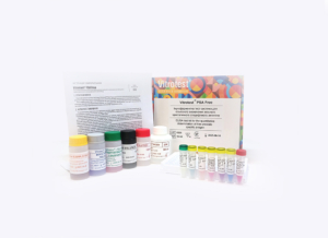
The test kit Vitrotest PSA Free is an enzyme linked immunosorbent assay (ELISA) for the quantitative determination of free prostate-specific antigen (PSA) in human serum or plasma.
Determination of concentration free PSA in the test kit Vitrotest PSA Free is based on on a solid-phase "sandwich" ELISA followed by the use of biotin-streptavidin interaction.
○ ТК105 - 96 tests
- Solid phase: breakable microplate ELISA is coated with monoclonal antibodies specific to human PSA.
- Biotinylated anti-PSA antibody solution: biotinylated monoclonal antibodies to human PSA.
- Streptavidin-HRP conjugate: streptavidin solution conjugated with horseradish peroxidase.
- Chromogen: ready to use TMB solution.
- Volume of sample for analysis: 25 μl.
- Assay time: 30/15/15 min.
- Analytical sensitivity: 0,096 ng/ml.
- Linearity range: 0,125-8 ng/ml.
-
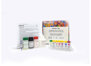
The test kit Vitrotest HE4 is an enzyme linked immunosorbent assay (ELISA) for the quantitative determination of antigen HE4 in human serum or plasma.
Determination of concentration antigen HE4 in the test kit Vitrotest HE4 is based on a solid-phase “sandwich” ELISA.
○ ТК107 - 96 tests
- Solid phase: breakable microplate ELISA is coated with monoclonal antibodies specific to antigen HE4.
- Conjugate: a monoclonal antibodies to antigen HE4 conjugated to horseradish peroxidase.
- Chromogen: ready to use TMB solution.
- Volume of sample for analysis: 10 μl.
- Assay time: 60/15 min.
- Analytical sensitivity: 4,85 pmol/l.
- Linearity range: 25-1000 pmol/l.
-
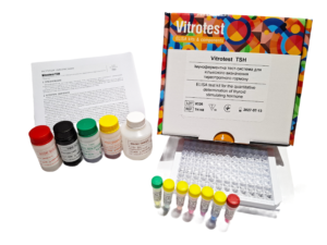
The test kit Vitrotest® TSH is an enzyme linked immunosorbent assay (ELISA) for the quantitative determination of thyroid-stimulating hormone (TSH) in human serum or plasma. Determination of thyroid-stimulating hormone concentration in the test kit Vitrotest® TSH is based on a solid-phase sandwich ELISA in a two-step incubation procedure.
- TK148 - 96 tests
- TK149 - 192 tests
- Solid phase: strip ELISA plate pre-coated with the first monoclonal antibodies specific to the β-subunit of human thyroid-stimulating hormone.
- Conjugate: monoclonal antibodies to human TSH conjugated with horseradish peroxidase.
- Chromogen: ready to use TMB solution.
- Sample volume: 50 μl.
- Assay time: 2 h.
- Analytical sensitivity: 0,106 μIU/ml.
- Linearity range: 0,125-16,4 µIU/ml.
According to the World Health Organization (WHO), thyroid diseases are the second most common endocrine disorders after diabetes mellitus. Over 200 million people worldwide suffer from various forms of thyroid dysfunction. In Ukraine, over the past 5 years, the number of people with thyroid diseases has increased fivefold. The main pathological conditions include hyperthyroidism, hypothyroidism, autoimmune thyroid diseases, benign and malignant neoplasms. A connection has also been established between thyroid dysfunction and other diseases such as diabetes, cardiovascular diseases, depression, oral diseases, and cancer.
A valuable biomarker of thyroid functional status widely used for screening, diagnosis, and monitoring of thyroid diseases is thyroid-stimulating hormone (TSH). TSH is a glycoprotein hormone synthesized by the anterior pituitary and is a key regulator of thyroid function. The TSH molecule consists of two different non-covalently bound subunits: the α-subunit, identical in amino acid sequence to the α-subunit of chorionic gonadotropin, luteinizing hormone, and follicle-stimulating hormone, and the hormone-specific β-subunit, which is unique. The main function of TSH is to stimulate the thyroid gland to synthesize and secrete thyroid hormones – thyroxine (T4) and triiodothyronine (T3). TSH binds to the TSH receptor on thyrocytes and activates intracellular signaling cascades that regulate iodine uptake, thyroid metabolism, thyroid growth, and hormone secretion. Through negative feedback, T3 and T4 inhibit TSH secretion.
In healthy adults, TSH serum levels are approximately 0.4 to 4.0 μIU/ml, although narrower ranges may be used to better detect subclinical hypothyroidism. Separate reference intervals for TSH are established for pregnant women, infants, and young children. TSH secretion has pulsatile and circadian patterns, and its concentration depends on factors such as age, sex, ethnicity, iodine intake, reproductive status, and body mass index.
Over the last three decades, laboratory methods used to determine TSH levels have significantly improved. Among immunochemical methods, ELISA has gained wide application due to its convenience, simplicity, high reproducibility, and sensitivity for determining thyroid-stimulating hormone in human serum and plasma.
The standardization of quantitative determination of TSH in human serum or plasma is ensured by the use of the WHO International Standard with assigned TSH concentration in μIU/ml for preparation of internal ELISA calibrators.
-
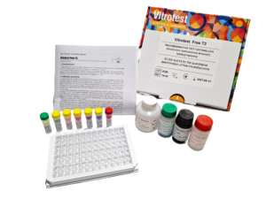
The test kit Vitrotest® Free T3 is an enzyme linked immunosorbent assay (ELISA) for the quantitative determination of free triiodothyronine (FT3) in human serum or plasma. Determination of free triiodothyronine concentration in the test kit Vitrotest® Free T3 is based on a competitive ELISA with a two-step incubation.
- TK150 - 96 tests
- Solid phase: strip ELISA plate pre-coated with triiodothyronine.
- Conjugate: buffer solution of monoclonal antibodies specific to triiodothyronine conjugated with horseradish peroxidase.
- Chromogen: ready to use TMB solution.
- Sample volume: 25 μl.
- Assay time: 1 h 15 min.
- Analytical sensitivity: 0,5 pg/ml.
According to data from the World Health Organization (WHO), thyroid diseases are the second most common endocrine disorders after diabetes mellitus. More than 200 million people worldwide suffer from various forms of thyroid dysfunction. In Ukraine, over the past five years, the number of people with thyroid diseases has increased fivefold. The main pathological conditions include hyperthyroidism, hypothyroidism, autoimmune thyroid diseases, benign and malignant neoplasms, etc.
The main hormones produced by the thyroid gland are thyroxine or tetraiodothyronine (T4, containing four iodine atoms) and triiodothyronine (T3, containing three iodine atoms). These hormones are lipophilic and circulate in the blood mainly bound to transport proteins: thyroxine-binding globulin (TBG), transthyretin (also known as thyroxine-binding prealbumin), and albumin. However, only free triiodothyronine and free thyroxine are functionally active, accounting for just 0.3% and 0.03% of total T3 and T4 in blood serum, respectively.
Free triiodothyronine is the biologically active form that directly enters target cells and binds to nuclear receptors to regulate gene expression and cellular function. Free thyroxine has minimal hormonal activity, but its long half-life (8 days) serves as a reservoir or prohormone for free triiodothyronine. All reactions required for the formation and release of T3 and T4 are controlled by thyroid-stimulating hormone (TSH) through a negative feedback mechanism.
Although determination of TSH and FT4 concentrations are considered the primary tests of choice for diagnosing thyroid dysfunction, measurement of FT3 levels is useful for detecting T3-thyrotoxicosis, complex or unusual manifestations of hyperthyroidism, monitoring thyroid disease treatment, and assessing metabolism.
Accumulated evidence indicates that changes in FT3 levels are also closely associated with a number of systemic disorders, such as cardiovascular diseases, dyslipidemia, type 2 diabetes mellitus, and liver dysfunction, highlighting the clinical importance of FT3 monitoring as a functional indicator of thyroid status.
-
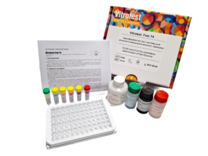
The test kit Vitrotest® Free T4 is an enzyme linked immunosorbent assay (ELISA) for the quantitative determination of free thyroxine (FT4) in human serum or plasma. Determination of free thyroxine concentration in the test kit Vitrotest® Free T4 is based on a competitive ELISA with a two-step incubation.
- TK151 - 96 tests
- Solid phase: strip ELISA plate pre-coated with monoclonal antibodies specific to thyroxine.
- Conjugate: buffer solution of thyroxine conjugated with horseradish peroxidase.
- Chromogen: ready to use TMB solution.
- Sample volume: 20 μl.
- Assay time: 1 h 15 min.
- Analytical sensitivity: 0,254 ng/dl.
According to WHO, thyroid diseases are the second most common endocrine disorders after diabetes mellitus. Over 200 million people worldwide suffer from various forms of thyroid dysfunction. In Ukraine, the number of people with thyroid diseases has increased fivefold over the past 5 years. The main pathological conditions include hyperthyroidism, hypothyroidism, autoimmune thyroid diseases, benign and malignant neoplasms.
The main hormones produced by the thyroid gland are thyroxine or tetraiodothyronine (T4, containing four iodine atoms) and triiodothyronine (T3, containing three iodine atoms). They are lipophilic and circulate in the blood mainly bound to transport proteins: thyroxine-binding globulin (TBG), transthyretin (thyroxine-binding prealbumin), and albumin. However, the biologically active forms are free triiodothyronine and free thyroxine, which represent only 0.3% and 0.03% of total T3 and T4 in serum, respectively.
Overall, FT4 has minimal hormonal activity, but its long half-life (8 days) serves as a reservoir or prohormone for active free triiodothyronine, which binds to nuclear receptors.
All processes required for the synthesis and release of T3 and T4 are controlled by thyroid-stimulating hormone (TSH), secreted by pituitary thyrotropic cells. TSH secretion is regulated through pituitary negative feedback: elevated free T4 and T3 levels suppress TSH synthesis and secretion, while decreased levels increase TSH secretion.
Determination of TSH and free thyroxine concentrations are the primary tests for diagnosing thyroid dysfunction. FT4 is an important marker for differentiating subclinical hyperthyroidism from overt hyperthyroidism or hypothyroidism, for investigating suspected abnormal TSH secretion, TSH-secreting pituitary adenomas, and for monitoring treatment of thyroid diseases.
-
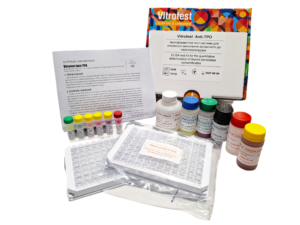
The test kit Vitrotest Anti-TPO is an enzyme linked immunosorbent assay (ELISA) for the quantitative determination of autoantibodies to thyroid peroxidase (TPO) in human serum or plasma. Determination of autoantibodies to thyroid peroxidase in the test kit Vitrotest Anti-TPO is based on a solid phase, indirect ELISA in a two-step incubation procedure.
- TK152 - 96 tests
- Solid phase: strip ELISA plate pre-coated with recombinant thyroid peroxidase.
- Conjugate: buffer solution of monoclonal antibodies to human IgG conjugated with horseradish peroxidase.
- Chromogen: ready to use TMB solution.
- Sample volume: 10 μl.
- Assay time: 1 h 15 min.
- Analytical sensitivity: 2,17 IU/ml.
- Linearity range: 3,125 - 410 IU/ml.
According to WHO, thyroid diseases are the second most widespread endocrine disorders after diabetes mellitus. Over 200 million people worldwide suffer from various types of thyroid dysfunction. In Ukraine, over the past 5 years, the number of people with thyroid diseases has increased fivefold.
Autoimmune thyroid diseases represent a diverse group of organ-specific autoimmune disorders, the most common of which are Hashimoto’s thyroiditis and Graves’ disease. The pathological process is associated with the formation of autoantibodies to thyroid peroxidase and thyroglobulin.
Thyroid peroxidase is a membrane-bound enzyme responsible for iodine oxidation and iodination of tyrosyl residues in the thyroglobulin molecule during the synthesis of thyroid hormones T3 and T4. Most anti-TPO antibodies belong to the IgG1 subclass, which activates complement. Additionally, anti-TPO antibodies can bind through their Fc fragment to natural killer cells, which in turn cause cytotoxic damage to thyrocytes.
Damage to thyroid cells, as well as direct enzyme inhibition, can lead to insufficient hormone production (hypothyroidism), sometimes preceded by transient hyperthyroidism. Besides thyroid disorders, elevated anti-TPO titers may also occur in a wide range of diseases: pernicious anemia, systemic lupus erythematosus, rheumatoid arthritis, insulin-dependent diabetes mellitus, breast cancer, and others. A correlation has also been established between anti-TPO antibodies and obstetric complications, such as postpartum thyroiditis or postpartum depression.
Anti-TPO antibodies may be present in individuals without clinical or laboratory signs of thyroid dysfunction. The presence of anti-TPO without overt disease is associated with a higher risk of developing autoimmune thyroiditis in the future and, together with TSH levels, is used to predict the development of hypo- and hyperthyroidism.
In modern laboratory diagnostics, ELISA is widely used for determining the concentration of autoantibodies to thyroid peroxidase due to its simplicity, convenience, high sensitivity, and specificity. Standardization of quantitative determination of anti-TPO in human serum or plasma is ensured by the use of the WHO International Standard with assigned concentration in IU/ml for preparation of internal ELISA calibrators.
-
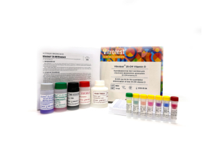
The test kit Vitrotest® 25-OH Vitamin Dis an enzyme linked immunosorbent assay (ELISA) for the quantitative determination of total 25-OH vitamin D (25-OH vitamin D2 and 25-OH vitamin D3) in human serum or plasma.
Determination of total 25-OH vitamin D concentration in the test kit Vitrotest® 25-OH Vitamin D is based on a competitive ELISA with a two-step incubation.
○ TK122 - 96 tests
○ TK124 - 192 tests- Solid phase: a stripped ELISA plate with wells pre-coated with monoclonal antibodies specific to 25-OH vitamin D.
- Biotinylated 25-OH vitamin D solution: biotinylated 25-OH vitamin D.
- Streptavidin-HRP conjugate: streptavidin conjugated with horseradish peroxidase.
- Chromogen: ready to use TMB solution.
- Volume of sample for analysis: 10 µl.
- Assay time: 1 h 30 min.
- Analytical sensitivity: 1,34 ng/ml.
Vitamin D is a fat-soluble steroid prohormone, whose active form is involved in the metabolism of phosphorus and calcium in the human body. Vitamin D, which is produced in the skin (D3, cholecalciferol) or obtained from food (D2, ergocalciferol), is biologically inert and requires two successive hydroxylations. First, 25-OH vitamin D (D2 and D3) is formed in the liver, which, in complex with a binding protein (vitamin D binding protein, VDBP), enters the bloodstream and is transported to the kidneys, where it is converted into the biologically active form 1,25-(OH)2 vitamin D and other metabolites whose roles are still being studied.
25-OH vitamin D is the main storage form of vitamin D in the human body and is present in the blood at much higher concentrations than its active form, 1,25-(OH)2 vitamin D. Its half-life is 2–3 weeks compared to 4-6 hours for 1,25-(OH)2 vitamin D. Additionally, the level of 1,25-(OH)2 vitamin D in serum does not provide information about the vitamin D status and is often normal or even elevated due to secondary hyperparathyroidism associated with vitamin D deficiency. For this reason, 25-OH vitamin D is the analyte of choice for determining the body’s vitamin D status.
According to the literature, the concentration of 25-OH vitamin D in human serum (plasma) within the range of 30-100 ng/ml is sufficient to support all necessary processes in the body that require vitamin D. Most experts consider a level of 25-OH vitamin D < 20 ng/ml to indicate vitamin D deficiency, while 20-30 ng/ml is considered an insufficient level. In Ukraine, only 3.3% of the population have a normal level of vitamin D, 14.9% have an insufficient level, and 81.8% live with its deficiency.
Hypovitaminosis D is widespread in the human population due to minimal exposure to sunlight and low dietary intake of vitamin D. As a result, vitamin D deficiency leads to secondary hyperparathyroidism, followed by loss of bone mass and osteoporosis. There is also evidence that vitamin D deficiency may have other adverse health effects, including an increased risk of malignant neoplasms, cardiovascular diseases, multiple sclerosis, diabetes, and more.
Currently, the reference method for determining the concentration of 25-OH vitamin D is tandem liquid chromatography with mass spectrometry (LC-MS/MS). However, in routine laboratory practice, this method is impractical due to its complexity and the need for specialized personnel. Therefore, immunoassay and immunochemiluminescent methods for determining 25-OH vitamin D have gained widespread use in diagnostic laboratories. -
The test kit Vitrotest Transglutaminase IgG is an enzyme linked immunosorbent assay (ELISA) for the quantitative determination of IgG class antibodies to tissue transglutaminase (human tTG) in human serum or plasma.
Determination of IgG class antibodies in the test kit Vitrotest Transglutaminase IgG is based on a solid phase, indirect ELISA in a two-step incubation procedure.
- TK155 - 96 tests
- Solid phase: strip ELISA plate pre-coated with recombinant human tissue transglutaminase 2.
- Conjugate: monoclonal antibodies to human IgG conjugated with horseradish peroxidase.
- Chromogen: ready to use TMB solution.
- Sample volume: 10 μl.
- Assay time: 30/30/15 min.
- Linearity range: 10 - 146 U/ml.
-
The test kit Vitrotest Transglutaminase IgA is an enzyme linked immunosorbent assay (ELISA) for the quantitative determination of IgA class antibodies to tissue transglutaminase (human tTG) in human serum or plasma.
Determination of IgA class antibodies in the test kit Vitrotest Transglutaminase IgA is based on a solid phase, indirect ELISA in a two-step incubation procedure.
- TK156 - 96 tests
- Solid phase: strip ELISA plate pre-coated with recombinant human tissue transglutaminase 2.
- Conjugate: monoclonal antibodies to human IgA conjugated with horseradish peroxidase.
- Chromogen: ready to use TMB solution.
- Sample volume: 10 μl.
- Assay time: 30/30/15 min.
- Linearity range: 7 - 164 U/ml.
-
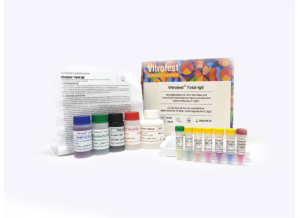
The test kit Vitrotest Total-IgE is an enzyme linked immunosorbent assay (ELISA) for the quantitative determination of total immunoglobulin E (IgE) in human serum or plasma.
Determination of total IgE content in the test kit Vitrotest Total-IgE is based on a solid-phase “sandwich” ELISA.
○ ТК070 - 96 tests
- Solid phase: breakable microplate ELISA is coated with monoclonal antibodies specific to human IgE.
- Conjugate: a monoclonal antibodies to human IgЕ conjugated to horseradish peroxidase.
- Chromogen: ready to use TMB solution.
- Volume of sample for analysis: 10 μl.
- Assay time: 30/30/15 min.
- Analytical sensitivity: 2,1 IU/ml.
- Linearity range: 2,1-1000 IU/ml.
-
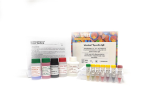
The test kit Vitrotest Specific-IgE is an enzyme linked immunosorbent assay (ELISA) for the quantitative determination of specific IgE class antibodies in human serum or plasma. The test kit is used together with biotinylated allergens Vitrotest XX-Biotin, where XX is a code of relevant allergen or mixture.
Quantitative determination of specific IgE antibodies in the Vitrotest Specific-IgE test kit is based on the principle of capturing IgE antibodies followed by the use of biotin-streptavidin interaction.
○ ТК071 - 192 tests
- Solid phase: breakable microplate ELISA is coated with monoclonal antibodies specific to human IgE.
- Biotinylated anti-IgE: biotinylated anti-IgE antibodies.
- Conjugate streptavidin-HRP: streptavidin сonjugated to horseradish peroxidase.
- Chromogen: ready to use TMB solution.
- Volume of sample for analysis: 50 μl.
- Assay time: 30/30/15/15 min.
- Analytical sensitivity: 0,080 IU/ml.
- Linearity range: 0,2-50 IU/ml.
-
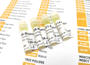
Biotinylated allergens Vitrotest XX-Biotin for the quantitative determination of specific IgE class antibodies in human serum or plasma. It is used exclusively with the Vitrotest Specific-IgE test kit.
○ 1 vial - 30 tests
Biotinylated allergens Vitrotest XX-Biotin, where XX is the code of the relevant allergen or mixture - each microtube contains 1.55 ml of a solution of biotinylated allergens, ready to use. The content of one bottle is enough to conduct 30 studies.
№ Allergen name BA-D1 Dermatophagoides pteronyssinus BA-D2 Dermatophagoides farina BA-Н1 House Dust Mixture № Allergen name BA-E1 Cat (epithelial) BA-E2 Dog (epithelial) BA-E3 Horse (epithelial) BA-E6 Guinea Pig (epithelial) BA-E70 Goose (feathers) BA-E78 Budgerigar (feathers) BA-E82 Rabbit (еpithelia) BA-E84 Hamster (epithelia) BA-E85 Chicken (feathers) BA-E86 Duck (feathers) № Allergen name BA-F1 Egg white BA-F2 Cow milk BA-F3 Сod BA-F4 Wheat flour BA-F5 Rye flour BA-F6 Barley flour BA-F7 Oats flour BA-F8 Corn flour BA-F9 Rice BA-F10 Sesamе BA-F11 Buckwheat flour BA-F13 Peanut BA-F14 Soybean BA-F17 Hazelnut BA-F20 Almond BA-F23 Crab meat BA-F24 Shrimp BA-F25 Tomato BA-F26 Pork BA-F27 Beef BA-F31 Carrot BA-F33 Orange BA-F34 Mandarin BA-F35 Potato BA-F37 Mussel BA-F41 Salmon BA-F44 Strawberry BA-F45 Baker’s yeast BA-F49 Apple BA-F75 Egg yolk BA-F76 Alpha-lactalbumin BA-F77 Beta-lactoglobulin BA-F78 Caseine BA-F79 Gluten BA-F83 Chicken meat BA-F84 Kiwi BA-F92 Banana BA-F93 Cocoa BA-F94 Pear BA-F205 Herring BA-F206 Mackerel BA-F208 Lemon BA-F213 Rabbit meat BA-F221 Coffee BA-F245 Whole Egg BA-F247 Honey BA-F256 Walnut BA-F259 Grape BA-F328 Fig BA-F343 Raspberry № Allergen name BA-T2 European Alder / Alnus glutinosa BA-T3 Silver Birch/Betula verrucosa BA-T4 Common Hazel / Corylus avellana BA-T7 Common Oak/ Quercus robur BA-Т12 Goat Willow/ Salix caprea BA-T14 Eastern Cottonwood /Populus deltoides № Allergen name BA-W1 Common Ragweed/ Ambrosia elatior BA-W6 Common Mugwort/ Artemisia vulgaris BA-W8 Common Dandelion/ Taraxacum vulgare BA-W9 Ribwort Plantain/ Plantago lanceolata BA-W10 White Goosefoot/ Chenopodium album BA-W12 European Goldenrod/ Solidago virgaurea № Allergen name BA-G2 Bermuda Grass/ Cynodon dactylon BA-G3 Cock’s-foot/ Dactylis glomerata BA-G4 Red Fescue/ Festuca elatior BA-G5 Perennial Ryegrass/ Lolium perenne BA-G6 Timothy Grass/ Phleum pratense BA-G8 Kentucky Bluegrass/ Poa pratensis BA-G12 Rye/ Secale cereale № Allergen name BA-M1 Penicillium notatum BA-M2 Cladosporium herbarum BA-M3 Aspergillus fumigatus BA-M4 Mucor racemosus BA-M5 Candida albicans BA-M6 Alternaria alternata (tenius) № Allergen name BA-I1 Bee Venom/ Apis mellifera BA-I3 Wasp Venom/ Vespula spp. BA-I6 Cockroach/ Blatella germanica № Allergen name BA-DEM1 Indoor allergens mixture:
D. pteronyssinus (D1), D. farinae (D2), Cat (epithelial) (E1), Dog (epithelial) (E2)BA-EМ1 Feathers mixture:
Goose (E70), Chicken (E85), Duck (E86)BA-FM1 Food mixture (seafood):
Crab meat (F23), Shrimp (F24), Mussel (F37)BA-FM2 Food mixture (cereals):
Wheat flour (F4), Rye flour (F5), Barley flour (F6), Oats flour (F7), Corn flour (F8), Sesame (F10), Buckwheat flour (F11)BA-FM3 Food mixture (fish):
Cod (F3), Salmon (F41), Herring (F205), Mackerel (F206)BA-TM1 Tree pollens mixture:
Alnus glutinosa (T2), Corylus avellana (T4), Betula verrucosa (T3), Quercus robur (T7), Salix caprea (T12), Populus deltoides (T14)BA-TM2 Tree pollens mixture (early flowering):
Alnus glutinosa (T2), Betula verrucosa (T3), Corylus avellana (T4)BA-TM3 Tree pollens mixture (late flowering):
Quercus robur (T7), Salix caprea (T12), Populus deltoides (T14)BA-WМ1 Weed pollen mixture:
Ambrosia elatior (W1), Artemisia vulgaris (W6), Taraxacum officinale (W8), Plantago lanceolata (W9), Solidago virgaurea (W12)BA-GМ1 Meadow grass pollens mixture:
Cynodon dactylon (G2), Dactylis glomerata (G3), Festuca elatior (G4), Phleum pratense (G6), Secale cereale (G12)

-
The test kit Vitrotest® Anti-Toxocara is an enzyme linked immunosorbent assay (ELISA) for the detection of IgG class antibodies to Toxocara canis in human serum or plasma.
Determination of IgG antibodies to Toxocara canis in the test kit Vitrotest® Anti-Toxocara is based on a solid phase, indirect ELISA in a two-step incubation procedure.
○ ТК058 - 96 tests
○ ТК112 - 192 tests- Solid phase: breakable microplate ELISA is coated Toxocara canis larvae antigens.
- Conjugate: a monoclonal antibodies to human IgG conjugated to horseradish peroxidase.
- Chromogen: ready to use TMB solution.
- Volume of sample for analysis: 10 μl.
- Assay time: 1h 15 min.
Toxocariasis is an widespread parasitic disease of humans caused by infection with the second stage larvae of two main species of parasitic nematodes, Toxocara canis and Toxocara cati. The role of toxocaras in human disease was disclosed in the 1950th by Wilder and Beaver et al. who first identified toxocara larvae in ocular and visceral tissues respectively.
Adult worms of T. canis and T.cati live within the lumen of the small intestine of dogs and cats respectively which serve as definitive hosts for these helminths. Unembryonated eggs produced by adult worms are shed in the feces of the definitive hosts. Eggs embryonate in the environment, and could be ingested by definitive hosts as well as accidental, paratenic hosts. After ingestion by the definitive hosts, microscopic (300 µm long and 20 µm in diameter) larvae hatch in the intestine, penetrate the intestinal wall, reach lungs via bloodstream, penetrate alveolar walls, ascend the bronchial tree to the throat where they are swallowed, and mature into adult worms. In humans and other paratenic hosts, the larvae are unable to undergo the full development cycle described above; instead, they are carried by the circulation to a wide variety of organs and tissues (liver, heart, lungs, brain, muscle, eyes), and cause severe local reactions that are the basis of toxocariasis.
The degree of host damage, and the concomitant signs and symptoms, varies with regard to which tissue has been invaded; the liver, lungs, and CNS (including the eyes) appear to be most sensitive. In the eye, migrating larvae can damage the retina, inducing granulomatous reactions leading to impaired sight or even loss of sight. The number of migrating larvae and the age of the host are two additional factors defining the presence and severity of symptoms.
Lifespan of the larvae could be as long as several years, and clinical disease could present in any time during this period, or even later, representing pathological immune response to dying or dead larvae.
For most people, an infection with these helminths causes no symptoms. Possible symptoms symptoms are nonspecific and include fever, fatigue, anorexia, or lymphadenopathy. Pulmonary symptoms and abdominal symptoms are similar to those under many other diseases; they are present when larvae migrate to the lungs or abdominal organs, respectively. Neurologic findings are diverse and also nonspecific. -
The test kit Vitrotest® Anti-Trichinella is an enzyme linked immunosorbent assay (ELISA) for the detection of IgG class antibodies to Trichinella spiralis in human serum or plasma.
Determination of IgG antibodies to Trichinella spiralis in the test kit Vitrotest® Anti-Trichinella is based on a solid phase, indirect ELISA in a two-step incubation procedure.
○ ТК067 - 96 tests- Solid phase: breakable microplate ELISA is coated Trichinella spiralis larvae antigens.
- Conjugate: a monoclonal antibodies to human IgG conjugated to horseradish peroxidase.
- Chromogen: ready to use TMB solution.
- Volume of sample for analysis: 10 μl.
- Assay time: 1h 15 min.
Trichinosis is a helminthiasis caused by a nematode Trichinella spiralis. Trichinellas are small, thread-like worms covered with a striped cuticle.
Transmission occurs by ingestion of meat containing encapsulated trichinella larvae. During digestion under the action of the gastric juice larvae release from capsules penetrate the submucosal layer of the small intestine, adhere to the mucosa and begin to proliferate. Soon after fertilization of females, the males die and the females start producing larvae, which enter the blood and lymphatic vessels through tissue mucosa, spread throughout the body and settle in striated muscles. Thereafter this capsule is impregnated with calcium salts leading to calcification. The larvae remain viable for many years.
The incubation period of human trichinosis lasts 10-25 days. Trichinosis is characterized by fever, myalgia, facial swelling, skin rash, blood eosinophilia, and in severe cases – by damage to internal organs and central nervous system.
The diagnosis of trichinosis is based on clinical signs, epidemiological history, serological tests (complement fixation tests, the reaction of indirect hemagglutination) and ELISA. The latter method is recommended by OIE for serological diagnosis of trichinosis.
The most specific and successful method to confirm infestation is the detection of IgG antibodies to trichinella antigens in the blood. These antibodies could be determined from 2-3 to 4-6 weeks after the eating of contaminated meat. Specific IgE class antibodies are also present in the blood during the acute stage of the disease, however, they are rarely detected due to the short period of their circulation in the bloodstream. During the early stage of invasion, specific antibodies might be still undetectable. Therefore, another sample should be taken in 1-2 weeks to confirm or reject suspected trichinosis. Seroconversion usually occurs 2-5 weeks after infection depending on the infectious dose. Assessment of antibody dynamics is a very informative marker of therapy effectiveness. In cases of ineffective or untimely therapy, specific antibodies are detected for up to 20 years if trichinosis is effectively treated in the first two weeks after infection, antibodies disappear within a year. -
The test kit Vitrotest® Anti-Strongyloides is an enzyme linked immunosorbent assay (ELISA) for the detection of IgG class antibodies to Strongyloides stercoralis in human serum or plasma.
Determination of IgG antibodies to Strongyloides stercoralis in the test kit Vitrotest® Anti-Strongyloides is based on a solid phase, indirect ELISA in a two-step incubation procedure.
- ТК146 - 96 tests
- Solid phase: breakable microplate ELISA is coated Strongyloides stercoralis antigens.
- Conjugate: a monoclonal antibodies to human IgG conjugated to horseradish peroxidase.
- Chromogen: ready to use TMB solution.
- Volume of sample for analysis: 10 μl.
- Assay time: 40 min.
Strongyloidiasis is a chronic parasitic infection of humans caused by Strongyloides stercoralis. This helminth is predominantly found in tropical and subtropical regions but can also be present in temperate climates. It is estimated that approximately 300 to 600 million people worldwide are infected.
Primary infection occurs when S. stercoralis larvae penetrate human skin through direct contact with contaminated soil. The larvae migrate via the bloodstream and lymphatic system to the respiratory tract, where they are then coughed up and swallowed, reaching the intestines. In the intestines, the parasites develop into adult worms, lay eggs, and produce larvae that can be excreted in the feces. A unique feature of S. stercoralis is its ability to cause autoinfection, as larvae may re-enter the intestines or perianal skin without leaving the host, leading to persistent infection.
In immunocompetent individuals, uncomplicated strongyloidiasis may be asymptomatic or present with mild cutaneous and gastrointestinal symptoms. Often, the only sign of infection is unexplained peripheral eosinophilia. However, in cases of heavy infestation or in immunosuppressed patients, the disease can cause severe manifestations such as abdominal pain, watery diarrhea, constipation, weight loss, vomiting, or small bowel obstruction. Hyperinfection syndrome is the most severe manifestation of the disease, with high mortality rates.
Parasitological methods (microscopy, culture) are traditionally used for diagnosis. The sensitivity and specifi city of these tests, however, are not high. The standard stool examination has a sensitivity of only 21% (5). Serological methods, especially enzyme-linked immunosorbent assay (ELISA), are highly sensitive and convenient for detecting antibodies to S. stercoralis larvae. ELISA is widely used for screening, diagnosing strongyloidiasis, and monitoring treatment effi cacy, as antibody titers signifi cantly decline within the fi rst 6 months following successful eradication therapy.
-
The test kit Vitrotest Ureaplasma-IgG is an enzyme linked immunosorbent assay (ELISA) for the qualitative and semiquantitative determination of IgG class antibodies to Ureaplasma urealyticum in human serum or plasma.
Determination of IgG antibodies to U.urealyticum in the test kit Vitrotest Ureaplasma-IgG is based on a solid phase indirect ELISA in a two-step incubation procedure.
○ ТК028 - 96 tests
- Solid phase: breakable microplate ELISA is coated with recombinant antigens of U. urealyticum.
- Conjugate: a monoclonal antibodies to human IgG conjugated to horseradish peroxidase.
- Chromogen: ready to use TMB solution.
- Volume of sample for analysis: 20 μl.
- Assay time: 30/30/15 min.
-
The test kit Vitrotest Ureaplasma-IgA is an enzyme linked immunosorbent assay (ELISA) for the qualitative and semiquantitative determination of IgA class antibodies to Ureaplasma urealyticum in human serum or plasma.
Determination of IgA antibodies to U.urealyticum in the test kit Vitrotest Ureaplasma-IgA is based on a solid phase indirect ELISA in a two-step incubation procedure.
○ ТК029 - 96 tests
- Solid phase: breakable microplate ELISA is coated with recombinant antigens of U. urealyticum.
- Conjugate: a monoclonal antibodies to human IgA conjugated to horseradish peroxidase.
- Chromogen: ready to use TMB solution.
- Volume of sample for analysis: 20 μl.
- Assay time: 30/30/15 min.
-
The test kit Vitrotest Ureaplasma-IgМ is an enzyme linked immunosorbent assay (ELISA) for the qualitative and semiquantitative determination of IgM class antibodies to Ureaplasma urealiticum in human serum or plasma.
Determination of IgM antibodies to Ureaplasma urealiticum in the test kit Vitrotest Ureaplasma-IgМ is based on a solid phase, «IgM-capture» ELISA in a two-step incubation procedure.
○ ТК096 - 96 tests
- Solid phase: breakable microplate ELISA is coated with monoclonal anti-IgM antibodies.
- Conjugate: recombinant U. urealiticum antigen, conjugated to horseradish peroxidase.
- Chromogen: ready to use TMB solution.
- Volume of sample for analysis: 10 μl.
- Assay time: 30/30/30 min.
-
The test kit Vitrotest Mycoplasma hominis-IgG is an enzyme linked immunosorbent assay (ELISA) for the qualitative and semiquantitative determination of IgG class antibodies to Mycoplasma hominis in human serum or plasma.
Determination of IgG antibodies to M. hominis in the test kit Vitrotest Mycoplasma hominis-IgG is based on a solid phase indirect ELISA in a two-step incubation procedure.
○ ТК024 - 96 tests
- Solid phase: breakable microplate ELISA is coated with native Mycoplasma hominis antigens.
- Conjugate: a monoclonal antibodies to human IgG conjugated to horseradish peroxidase.
- Chromogen: ready to use TMB solution.
- Volume of sample for analysis: 10 μl.
- Assay time: 30/30/15 min.
-
The test kit Vitrotest Mycoplasma hominis-IgA is an enzyme linked immunosorbent assay (ELISA) for the qualitative and semiquantitative determination of IgA class antibodies to Mycoplasma hominis in human serum or plasma.
Determination of IgA antibodies to M. hominis in the test kit Vitrotest Mycoplasma hominis-IgA is based on a solid phase indirect ELISA in a two-step incubation procedure.
○ ТК025 - 96 tests
- Solid phase: breakable microplate ELISA is coated with native Mycoplasma hominis antigens.
- Conjugate: a monoclonal antibodies to human IgA conjugated to horseradish peroxidase.
- Chromogen: ready to use TMB solution.
- Volume of sample for analysis: 10 μl.
- Assay time: 30/30/15 min.
-
The test kit Vitrotest Mycoplasma hominis-IgM is an enzyme linked immunosorbent assay (ELISA) for the qualitative and semiquantitative determination of IgM class antibodies to Mycoplasma hominis in human serum or plasma.
Determination of IgM antibodies to Mycoplasma hominis in the test kit Vitrotest Mycoplasma hominis-IgM is based on a solid phase, «IgM-capture» ELISA in a two-step incubation procedure.
○ ТК097 - 96 tests
- Solid phase: breakable microplate ELISA is coated with monoclonal anti-IgM antibodies.
- Conjugate: recombinant M. hominis antigen, conjugated to horseradish peroxidase.
- Chromogen: ready to use TMB solution.
- Volume of sample for analysis: 10 μl.
- Assay time: 30/30/30 min.
-
The test kit Vitrotest Chlamydia-IgG is an enzyme linked immunosorbent assay (ELISA) for the qualitative and semiquantitative determination of IgG class antibodies to Chlamydia trachomatis in human serum or plasma.
Determination of IgG antibodies to Chlamydia trachomatis in the test kit Vitrotest Chlamydia-IgG is based on a solid phase indirect ELISA in a two-step incubation procedure.
○ ТК014 - 96 tests
- Solid phase: breakable microplate ELISA is coated with recombinant Chlamydia trachomatis antigens.
- Conjugate: a monoclonal antibodies to human IgG conjugated to horseradish peroxidase.
- Chromogen: ready to use TMB solution.
- Volume of sample for analysis: 20 μl.
- Assay time: 30/30/15 min.
-
The test kit Vitrotest Chlamydia-IgА is an enzyme linked immunosorbent assay (ELISA) for the qualitative and semiquantitative determination of IgА class antibodies to Chlamydia trachomatis in human serum or plasma.
Determination of IgА antibodies to Chlamydia trachomatis in the test kit Vitrotest Chlamydia-IgА is based on a solid phase indirect ELISA in a two-step incubation procedure.
○ ТК015 - 96 tests
- Solid phase: breakable microplate ELISA is coated with recombinant Chlamydia trachomatis antigens.
- Conjugate: a monoclonal antibodies to human IgА conjugated to horseradish peroxidase.
- Chromogen: ready to use TMB solution.
- Volume of sample for analysis: 20 μl.
- Assay time: 30/30/15 min.
-
The test kit Vitrotest Chlamydia-IgМ is an enzyme linked immunosorbent assay (ELISA) for the qualitative determination of IgM class antibodies to Chlamydia trachomatis in human serum or plasma.
Determination of IgM antibodies to Chlamydia trachomatis in the test kit Vitrotest Chlamydia-IgМ is based on a solid phase «IgM-capture» ELISA in a two-step incubation procedure.
○ ТК036 - 96 tests
- Solid phase: breakable microplate ELISA is coated with monoclonal anti-IgM antibodies.
- Conjugate: recombinant antigen MOMP of Chlamydia trachomatis, conjugated to horseradish peroxidase.
- Chromogen: ready to use TMB solution.
- Volume of sample for analysis: 10 μl.
- Assay time: 30/30/30 min.
-
The test kit Vitrotest Trichomonas-IgG is an enzyme linked immunosorbent assay (ELISA) for the qualitative and semiquantitative determination of IgG class antibodies to Trichomonas vaginalis in human serum or plasma.
Determination of IgG antibodies to Trichomonas vaginalis in the test kit Vitrotest Trichomonas-IgG is based on a solid phase, indirect ELISA in a two-step incubation procedure.
○ ТК061 - 96 tests
- Solid phase: breakable microplate ELISA is coated with Trichomonas vaginalis antigens.
- Conjugate: a monoclonal antibodies to human IgG conjugated to horseradish peroxidase.
- Chromogen: ready to use TMB solution.
- Volume of sample for analysis: 20 μl.
- Assay time: 30/30/15 min.
-
The test-kit Vitrotest Helicobacter screen is an enzyme linked immunosorbent assay (ELISA) for the detection of IgG, IgA and IgM specific antibodies to Helicobacter pylori in human serum or plasma.
Detection of antibodies to H. pylori in the test-kit Vitrotest Helicobacter screen is based on a solid phase, indirect ELISA in a two-step incubation procedure.
○ ТК045 - 96 tests
- Solid phase: breakable microplate ELISA is coated with recombinant antigens of H. pylori.
- Conjugate: a monoclonal antibodies to human IgA, IgG and IgM conjugated to horseradish peroxidase.
- Chromogen: ready to use TMB solution.
- Volume of sample for analysis: 10 μl.
- Assay time: 30/30/15 min.
-
The test kit Vitrotest Helicobacter-IgG is an enzyme linked immunosorbent assay (ELISA) for the quantitative and semiquantitative determination of IgG class antibodies to CagA protein of Helicobacter pylori in human serum or plasma.
Determination of IgG antibodies to CagA protein of H. pylori in the test kit Vitrotest Helicobacter-IgG is based on a solid phase, indirect ELISA in a two-step incubation procedure.
○ ТК046 - 96 tests
- Solid phase: breakable microplate ELISA is coated with recombinant CagA protein of H. pylori.
- Conjugate: a monoclonal antibodies to human IgG conjugated to horseradish peroxidase.
- Chromogen: ready to use TMB solution.
- Volume of sample for analysis: 10 μl.
- Assay time: 30/30/15 min.
- Linearity range: 1,25-200 U/ml.
-
The test kit Vitrotest Helicobacter-IgА is an enzyme linked immunosorbent assay (ELISA) for the qualitative and semiquantitative determination of IgА class antibodies to CagA protein of Helicobacter pylori in human serum or plasma.
Determination of IgА antibodies to CagA protein of H. pylori in the test kit Vitrotest Helicobacter-IgА is based on a solid phase, indirect ELISA in a two-step incubation procedure.
○ ТК047 - 96 tests
- Solid phase: breakable microplate ELISA is coated with recombinant CagA protein of H. pylori.
- Conjugate: a monoclonal antibodies to human IgА conjugated to horseradish peroxidase.
- Chromogen: ready to use TMB solution.
- Volume of sample for analysis: 10 μl.
- Assay time: 30/30/15 min.
-
The test kit Vitrotest Helicobacter-IgМ is an enzyme linked immunosorbent assay (ELISA) for the qualitative and semiquantitative determination of IgМ class antibodies to CagA protein of Helicobacter pylori in human serum or plasma.
Determination of IgМ antibodies to CagA protein of H. pylori in the test kit Vitrotest Helicobacter-IgМ is based on a solid phase, indirect ELISA in a two-step incubation procedure.
○ ТК048 - 96 tests
- Solid phase: breakable microplate ELISA is coated with recombinant CagA protein of H. pylori.
- Conjugate: a monoclonal antibodies to human IgМ conjugated to horseradish peroxidase.
- Chromogen: ready to use TMB solution.
- Volume of sample for analysis: 10 μl.
- Assay time: 30/30/15 min.
-
The test kit Vitrotest Anti-Lamblia is an enzyme linked immunosorbent assay (ELISA) for the detection of total antibodies to Giardia lamblia (intestinalis) in human serum or plasma.
Determination of total antibodies to Giardia lamblia in the test kit Vitrotest Anti-Lamblia is based on a solid phase, indirect ELISA in a two-step incubation procedure.
○ ТК030 - 96 tests
○ ТК108 - 192 tests
- Solid phase: breakable microplate ELISA is coated Giardia lamblia purified antigens.
- Conjugate: a monoclonal antibodies to human IgG, IgA and IgM conjugated to horseradish peroxidase.
- Chromogen: ready to use TMB solution.
- Volume of sample for analysis 10 μl.
- Assay time: 30/30/15 min.
Giardia lamblia (intestinalis) causes giardiasis, a parasitic infestation that can manifest as latent parasitic carriage or in more pronounced forms, such as intestinal dysfunction. Giardiasis has been recorded on all five continents and in most countries worldwide. Infection rates vary from less than 1% to 50%. In many developing countries, where basic sanitary conditions are lacking, infection with Giardia in children by the age of 2 is almost universal. In contrast, in developed countries, the infection rate of G.lamblia is only 3–7%. The disease affects all age groups, but preschool-aged children are the most affected demographic.
The primary route of transmission for G.lamblia is fecal-oral. The parasite has a simple two-stage life cycle. After the host ingests cysts, they release trophozoites in the duodenum, which then attach to the mucosal lining of the small intestine.
Trophozoites exist only on the surface of the mucosa in the upper part of the small intestine. Therefore, Giardia mechanically blocks the mucosa and disrupts membrane digestion and the motor activity of the small intestine. Giardia impairs the absorption of fats, carbohydrates, vitamins C and B12, and leads to secondary bacterial infections. Symptoms of giardiasis can include diarrhea, fatigue, bloating, apathy, weight loss, decreased appetite, pallor, and muscle twitching. From the gastrointestinal tract perspective, giardiasis mainly manifests as enterocolitis with catarrhal symptoms.
Numerous findings indicate the role of the humoral immune response in the elimination of G.lamblia. As shown in an experimental human infection model, IgM antibody levels significantly increased on days 14-21 post-infection and gradually declined following therapy. In contrast, IgG antibody levels remained elevated after successful treatment. The dynamics of IgA levels were similar to those of IgM.
The diagnosis of giardiasis is based on clinical history, symptoms, and the presence of cysts in fecal samples or trophozoites in material obtained from the small intestine during duodenal aspiration or duodenal biopsy. Alternative methods include the detection of G.lamblia antigen in feces and the determination of specific antibodies against Giardia in the patient’s serum. Serological testing is considered a valuable adjunct in the diagnosis of giardiasis. In addition to aiding in diagnosis, it can be useful for assessing the patient’s immune response and for epidemiological studies. -
The test kit Vitrotest Lamblia-IgM is an enzyme linked immunosorbent assay (ELISA) for the detection of IgM class antibodies to Giardia lamblia (intestinalis) in human serum or plasma.
Determination of IgM antibodies to Giardia lamblia in the test kit Vitrotest Lamblia-IgM is based on a solid phase, indirect ELISA in a two-step incubation procedure.
○ ТК031 - 96 tests
- Solid phase: breakable microplate ELISA is coated Giardia lamblia purified antigens.
- Conjugate: a monoclonal antibodies to human IgM conjugated to horseradish peroxidase.
- Chromogen: ready to use TMB solution.
- Volume of sample for analysis 10 μl.
- Assay time: 30/30/15 min.
Giardia lamblia (intestinalis) causes giardiasis, a parasitic infestation that can manifest as latent parasitic carriage or in more pronounced forms, such as intestinal dysfunction. Giardiasis has been recorded on all five continents and in most countries worldwide. Infection rates vary from less than 1% to 50%. In many developing countries, where basic sanitary conditions are lacking, infection with Giardia in children by the age of 2 is almost universal. In contrast, in developed countries, the infection rate of G.lamblia is only 3–7%. The disease affects all age groups, but preschool-aged children are the most affected demographic.
The primary route of transmission for G.lamblia is fecal-oral. The parasite has a simple two-stage life cycle. After the host ingests cysts, they release trophozoites in the duodenum, which then attach to the mucosal lining of the small intestine.
Trophozoites exist only on the surface of the mucosa in the upper part of the small intestine. Therefore, Giardia mechanically blocks the mucosa and disrupts membrane digestion and the motor activity of the small intestine. Giardia impairs the absorption of fats, carbohydrates, vitamins C and B12, and leads to secondary bacterial infections. Symptoms of giardiasis can include diarrhea, fatigue, bloating, apathy, weight loss, decreased appetite, pallor, and muscle twitching. From the gastrointestinal tract perspective, giardiasis mainly manifests as enterocolitis with catarrhal symptoms.
Numerous findings indicate the role of the humoral immune response in the elimination of G.lamblia. As shown in an experimental human infection model, IgM antibody levels significantly increased on days 14-21 post-infection and gradually declined following therapy. In contrast, IgG antibody levels remained elevated after successful treatment. The dynamics of IgA levels were similar to those of IgM.
The diagnosis of giardiasis is based on clinical history, symptoms, and the presence of cysts in fecal samples or trophozoites in material obtained from the small intestine during duodenal aspiration or duodenal biopsy. Alternative methods include the detection of G.lamblia antigen in feces and the determination of specific antibodies against Giardia in the patient’s serum. Serological testing is considered a valuable adjunct in the diagnosis of giardiasis. In addition to aiding in diagnosis, it can be useful for assessing the patient’s immune response and for epidemiological studies. -
The test kit Vitrotest® Brucella-IgG is an enzyme linked immunosorbent assay (ELISA) for the qualitative and semiquantitative determination of igG class antibodies to Brucella in human serum or plasma.
Determination of IgG antibodies to Brucella in the test kit Vitrotest® Brucella-IgG is based on a solid phase, indirect ELISA in a two-step incubation procedure.
- TK153 - 96 tests
- Solid phase: strip ELISA plate pre-coated with antigens of Brucella.
- Conjugate: monoclonal antibodies to human IgG conjugated with horseradish peroxidase.
- Chromogen: ready to use TMB solution.
- Sample volume: 10 μl.
- Assay time: 1 h 15 min.
Brucellosis is a zoonotic infectious disease caused by bacteria of the genus Brucella. Globally, 1.6–2.1 million human cases of brucellosis are reported annually. Among the countries with the highest reported incidence of brucellosis are Iran, Kyrgyzstan, Tajikistan, Kazakhstan, Azerbaijan, Turkmenistan, Armenia, and Uzbekistan.
The species most pathogenic to humans include Brucella melitensis, Brucella abortus, Brucella suis, and Brucella canis. Different species of Brucella vary in their reservoir hosts and degree of virulence: B. melitensis is considered the most pathogenic for humans, whereas B. abortus more commonly causes relatively milder forms of the disease.
Human infection occurs through contact with infected animals and their biological fluids, as well as through the consumption of animal-derived products (raw milk and insufficiently heat-treated meat), and by the airborne route during occupational exposure. The pathogen enters the human body through mucous membranes or damaged skin. Subsequently, the bacteria are transported by macrophages to lymphoid tissues, spread through the lymphatic system, and may potentially proliferate in multiple organs, causing localized and systemic infections. By persisting within host cells such as macrophages, Brucella spp. employ strategies to evade the host immune response, resulting in prolonged infection.
The incubation period of brucellosis usually ranges from 1 to 4 weeks but may extend to several months. Clinical manifestations are characterized by undulating fever, sweating (especially at night), weakness, and enlargement of the lymph nodes, liver, and spleen. In chronic cases, the musculoskeletal, nervous, and cardiovascular systems may also be affected.
Due to the nonspecific nature of its symptoms, the diagnosis of brucellosis is challenging and requires a comprehensive approach, including the evaluation of clinical findings, epidemiological history, and laboratory test results (bacterial culture, serological assays, and PCR). Serological methods used for the diagnosis of brucellosis include the agglutination test, complement fixation test, and enzyme-linked immunosorbent assay (ELISA).
Using the ELISA method, IgM antibodies are detected during the early stages of infection, whereas IgG antibodies are detected later and may persist for a long period after previous infection or successful treatment. A rapid decline in IgG antibody titers usually indicates a favorable response to antibiotic therapy, while persistently high or increasing titers may indicate treatment failure, residual disease, or an impending clinical relapse. Therefore, the detection of IgG antibodies specific to Brucella is an important marker of current or past infection, particularly in subacute and chronic forms of brucellosis. -
The test kit Vitrotest PSA Total is an enzyme linked immunosorbent assay (ELISA) for the quantitative determination of total prostate-specific antigen (PSA) in human serum or plasma.
Determination of concentration total PSA in the test kit Vitrotest PSA Total is based on on a solid-phase "sandwich" ELISA.
○ ТК104 - 96 tests
- Solid phase: breakable microplate ELISA is coated with monoclonal antibodies specific to human PSA.
- Conjugate: a monoclonal antibodies to human PSA conjugated to horseradish peroxidase.
- Chromogen: ready to use TMB solution.
- Volume of sample for analysis: 25 μl.
- Assay time: 60/15 min.
- Analytical sensitivity: 0,134 ng/ml.
- Linearity range: 0,25-30 ng/ml.
-
The test kit Vitrotest PSA Free is an enzyme linked immunosorbent assay (ELISA) for the quantitative determination of free prostate-specific antigen (PSA) in human serum or plasma.
Determination of concentration free PSA in the test kit Vitrotest PSA Free is based on on a solid-phase "sandwich" ELISA followed by the use of biotin-streptavidin interaction.
○ ТК105 - 96 tests
- Solid phase: breakable microplate ELISA is coated with monoclonal antibodies specific to human PSA.
- Biotinylated anti-PSA antibody solution: biotinylated monoclonal antibodies to human PSA.
- Streptavidin-HRP conjugate: streptavidin solution conjugated with horseradish peroxidase.
- Chromogen: ready to use TMB solution.
- Volume of sample for analysis: 25 μl.
- Assay time: 30/15/15 min.
- Analytical sensitivity: 0,096 ng/ml.
- Linearity range: 0,125-8 ng/ml.
-
The test kit Vitrotest HE4 is an enzyme linked immunosorbent assay (ELISA) for the quantitative determination of antigen HE4 in human serum or plasma.
Determination of concentration antigen HE4 in the test kit Vitrotest HE4 is based on a solid-phase “sandwich” ELISA.
○ ТК107 - 96 tests
- Solid phase: breakable microplate ELISA is coated with monoclonal antibodies specific to antigen HE4.
- Conjugate: a monoclonal antibodies to antigen HE4 conjugated to horseradish peroxidase.
- Chromogen: ready to use TMB solution.
- Volume of sample for analysis: 10 μl.
- Assay time: 60/15 min.
- Analytical sensitivity: 4,85 pmol/l.
- Linearity range: 25-1000 pmol/l.
-
The test kit Vitrotest® TSH is an enzyme linked immunosorbent assay (ELISA) for the quantitative determination of thyroid-stimulating hormone (TSH) in human serum or plasma. Determination of thyroid-stimulating hormone concentration in the test kit Vitrotest® TSH is based on a solid-phase sandwich ELISA in a two-step incubation procedure.
- TK148 - 96 tests
- TK149 - 192 tests
- Solid phase: strip ELISA plate pre-coated with the first monoclonal antibodies specific to the β-subunit of human thyroid-stimulating hormone.
- Conjugate: monoclonal antibodies to human TSH conjugated with horseradish peroxidase.
- Chromogen: ready to use TMB solution.
- Sample volume: 50 μl.
- Assay time: 2 h.
- Analytical sensitivity: 0,106 μIU/ml.
- Linearity range: 0,125-16,4 µIU/ml.
According to the World Health Organization (WHO), thyroid diseases are the second most common endocrine disorders after diabetes mellitus. Over 200 million people worldwide suffer from various forms of thyroid dysfunction. In Ukraine, over the past 5 years, the number of people with thyroid diseases has increased fivefold. The main pathological conditions include hyperthyroidism, hypothyroidism, autoimmune thyroid diseases, benign and malignant neoplasms. A connection has also been established between thyroid dysfunction and other diseases such as diabetes, cardiovascular diseases, depression, oral diseases, and cancer.
A valuable biomarker of thyroid functional status widely used for screening, diagnosis, and monitoring of thyroid diseases is thyroid-stimulating hormone (TSH). TSH is a glycoprotein hormone synthesized by the anterior pituitary and is a key regulator of thyroid function. The TSH molecule consists of two different non-covalently bound subunits: the α-subunit, identical in amino acid sequence to the α-subunit of chorionic gonadotropin, luteinizing hormone, and follicle-stimulating hormone, and the hormone-specific β-subunit, which is unique. The main function of TSH is to stimulate the thyroid gland to synthesize and secrete thyroid hormones – thyroxine (T4) and triiodothyronine (T3). TSH binds to the TSH receptor on thyrocytes and activates intracellular signaling cascades that regulate iodine uptake, thyroid metabolism, thyroid growth, and hormone secretion. Through negative feedback, T3 and T4 inhibit TSH secretion.
In healthy adults, TSH serum levels are approximately 0.4 to 4.0 μIU/ml, although narrower ranges may be used to better detect subclinical hypothyroidism. Separate reference intervals for TSH are established for pregnant women, infants, and young children. TSH secretion has pulsatile and circadian patterns, and its concentration depends on factors such as age, sex, ethnicity, iodine intake, reproductive status, and body mass index.
Over the last three decades, laboratory methods used to determine TSH levels have significantly improved. Among immunochemical methods, ELISA has gained wide application due to its convenience, simplicity, high reproducibility, and sensitivity for determining thyroid-stimulating hormone in human serum and plasma.
The standardization of quantitative determination of TSH in human serum or plasma is ensured by the use of the WHO International Standard with assigned TSH concentration in μIU/ml for preparation of internal ELISA calibrators.
-
The test kit Vitrotest® Free T3 is an enzyme linked immunosorbent assay (ELISA) for the quantitative determination of free triiodothyronine (FT3) in human serum or plasma. Determination of free triiodothyronine concentration in the test kit Vitrotest® Free T3 is based on a competitive ELISA with a two-step incubation.
- TK150 - 96 tests
- Solid phase: strip ELISA plate pre-coated with triiodothyronine.
- Conjugate: buffer solution of monoclonal antibodies specific to triiodothyronine conjugated with horseradish peroxidase.
- Chromogen: ready to use TMB solution.
- Sample volume: 25 μl.
- Assay time: 1 h 15 min.
- Analytical sensitivity: 0,5 pg/ml.
According to data from the World Health Organization (WHO), thyroid diseases are the second most common endocrine disorders after diabetes mellitus. More than 200 million people worldwide suffer from various forms of thyroid dysfunction. In Ukraine, over the past five years, the number of people with thyroid diseases has increased fivefold. The main pathological conditions include hyperthyroidism, hypothyroidism, autoimmune thyroid diseases, benign and malignant neoplasms, etc.
The main hormones produced by the thyroid gland are thyroxine or tetraiodothyronine (T4, containing four iodine atoms) and triiodothyronine (T3, containing three iodine atoms). These hormones are lipophilic and circulate in the blood mainly bound to transport proteins: thyroxine-binding globulin (TBG), transthyretin (also known as thyroxine-binding prealbumin), and albumin. However, only free triiodothyronine and free thyroxine are functionally active, accounting for just 0.3% and 0.03% of total T3 and T4 in blood serum, respectively.
Free triiodothyronine is the biologically active form that directly enters target cells and binds to nuclear receptors to regulate gene expression and cellular function. Free thyroxine has minimal hormonal activity, but its long half-life (8 days) serves as a reservoir or prohormone for free triiodothyronine. All reactions required for the formation and release of T3 and T4 are controlled by thyroid-stimulating hormone (TSH) through a negative feedback mechanism.
Although determination of TSH and FT4 concentrations are considered the primary tests of choice for diagnosing thyroid dysfunction, measurement of FT3 levels is useful for detecting T3-thyrotoxicosis, complex or unusual manifestations of hyperthyroidism, monitoring thyroid disease treatment, and assessing metabolism.
Accumulated evidence indicates that changes in FT3 levels are also closely associated with a number of systemic disorders, such as cardiovascular diseases, dyslipidemia, type 2 diabetes mellitus, and liver dysfunction, highlighting the clinical importance of FT3 monitoring as a functional indicator of thyroid status.
-
The test kit Vitrotest® Free T4 is an enzyme linked immunosorbent assay (ELISA) for the quantitative determination of free thyroxine (FT4) in human serum or plasma. Determination of free thyroxine concentration in the test kit Vitrotest® Free T4 is based on a competitive ELISA with a two-step incubation.
- TK151 - 96 tests
- Solid phase: strip ELISA plate pre-coated with monoclonal antibodies specific to thyroxine.
- Conjugate: buffer solution of thyroxine conjugated with horseradish peroxidase.
- Chromogen: ready to use TMB solution.
- Sample volume: 20 μl.
- Assay time: 1 h 15 min.
- Analytical sensitivity: 0,254 ng/dl.
According to WHO, thyroid diseases are the second most common endocrine disorders after diabetes mellitus. Over 200 million people worldwide suffer from various forms of thyroid dysfunction. In Ukraine, the number of people with thyroid diseases has increased fivefold over the past 5 years. The main pathological conditions include hyperthyroidism, hypothyroidism, autoimmune thyroid diseases, benign and malignant neoplasms.
The main hormones produced by the thyroid gland are thyroxine or tetraiodothyronine (T4, containing four iodine atoms) and triiodothyronine (T3, containing three iodine atoms). They are lipophilic and circulate in the blood mainly bound to transport proteins: thyroxine-binding globulin (TBG), transthyretin (thyroxine-binding prealbumin), and albumin. However, the biologically active forms are free triiodothyronine and free thyroxine, which represent only 0.3% and 0.03% of total T3 and T4 in serum, respectively.
Overall, FT4 has minimal hormonal activity, but its long half-life (8 days) serves as a reservoir or prohormone for active free triiodothyronine, which binds to nuclear receptors.
All processes required for the synthesis and release of T3 and T4 are controlled by thyroid-stimulating hormone (TSH), secreted by pituitary thyrotropic cells. TSH secretion is regulated through pituitary negative feedback: elevated free T4 and T3 levels suppress TSH synthesis and secretion, while decreased levels increase TSH secretion.
Determination of TSH and free thyroxine concentrations are the primary tests for diagnosing thyroid dysfunction. FT4 is an important marker for differentiating subclinical hyperthyroidism from overt hyperthyroidism or hypothyroidism, for investigating suspected abnormal TSH secretion, TSH-secreting pituitary adenomas, and for monitoring treatment of thyroid diseases.
-
The test kit Vitrotest Anti-TPO is an enzyme linked immunosorbent assay (ELISA) for the quantitative determination of autoantibodies to thyroid peroxidase (TPO) in human serum or plasma. Determination of autoantibodies to thyroid peroxidase in the test kit Vitrotest Anti-TPO is based on a solid phase, indirect ELISA in a two-step incubation procedure.
- TK152 - 96 tests
- Solid phase: strip ELISA plate pre-coated with recombinant thyroid peroxidase.
- Conjugate: buffer solution of monoclonal antibodies to human IgG conjugated with horseradish peroxidase.
- Chromogen: ready to use TMB solution.
- Sample volume: 10 μl.
- Assay time: 1 h 15 min.
- Analytical sensitivity: 2,17 IU/ml.
- Linearity range: 3,125 - 410 IU/ml.
According to WHO, thyroid diseases are the second most widespread endocrine disorders after diabetes mellitus. Over 200 million people worldwide suffer from various types of thyroid dysfunction. In Ukraine, over the past 5 years, the number of people with thyroid diseases has increased fivefold.
Autoimmune thyroid diseases represent a diverse group of organ-specific autoimmune disorders, the most common of which are Hashimoto’s thyroiditis and Graves’ disease. The pathological process is associated with the formation of autoantibodies to thyroid peroxidase and thyroglobulin.
Thyroid peroxidase is a membrane-bound enzyme responsible for iodine oxidation and iodination of tyrosyl residues in the thyroglobulin molecule during the synthesis of thyroid hormones T3 and T4. Most anti-TPO antibodies belong to the IgG1 subclass, which activates complement. Additionally, anti-TPO antibodies can bind through their Fc fragment to natural killer cells, which in turn cause cytotoxic damage to thyrocytes.
Damage to thyroid cells, as well as direct enzyme inhibition, can lead to insufficient hormone production (hypothyroidism), sometimes preceded by transient hyperthyroidism. Besides thyroid disorders, elevated anti-TPO titers may also occur in a wide range of diseases: pernicious anemia, systemic lupus erythematosus, rheumatoid arthritis, insulin-dependent diabetes mellitus, breast cancer, and others. A correlation has also been established between anti-TPO antibodies and obstetric complications, such as postpartum thyroiditis or postpartum depression.
Anti-TPO antibodies may be present in individuals without clinical or laboratory signs of thyroid dysfunction. The presence of anti-TPO without overt disease is associated with a higher risk of developing autoimmune thyroiditis in the future and, together with TSH levels, is used to predict the development of hypo- and hyperthyroidism.
In modern laboratory diagnostics, ELISA is widely used for determining the concentration of autoantibodies to thyroid peroxidase due to its simplicity, convenience, high sensitivity, and specificity. Standardization of quantitative determination of anti-TPO in human serum or plasma is ensured by the use of the WHO International Standard with assigned concentration in IU/ml for preparation of internal ELISA calibrators.
-
The test kit Vitrotest® 25-OH Vitamin Dis an enzyme linked immunosorbent assay (ELISA) for the quantitative determination of total 25-OH vitamin D (25-OH vitamin D2 and 25-OH vitamin D3) in human serum or plasma.
Determination of total 25-OH vitamin D concentration in the test kit Vitrotest® 25-OH Vitamin D is based on a competitive ELISA with a two-step incubation.
○ TK122 - 96 tests
○ TK124 - 192 tests- Solid phase: a stripped ELISA plate with wells pre-coated with monoclonal antibodies specific to 25-OH vitamin D.
- Biotinylated 25-OH vitamin D solution: biotinylated 25-OH vitamin D.
- Streptavidin-HRP conjugate: streptavidin conjugated with horseradish peroxidase.
- Chromogen: ready to use TMB solution.
- Volume of sample for analysis: 10 µl.
- Assay time: 1 h 30 min.
- Analytical sensitivity: 1,34 ng/ml.
Vitamin D is a fat-soluble steroid prohormone, whose active form is involved in the metabolism of phosphorus and calcium in the human body. Vitamin D, which is produced in the skin (D3, cholecalciferol) or obtained from food (D2, ergocalciferol), is biologically inert and requires two successive hydroxylations. First, 25-OH vitamin D (D2 and D3) is formed in the liver, which, in complex with a binding protein (vitamin D binding protein, VDBP), enters the bloodstream and is transported to the kidneys, where it is converted into the biologically active form 1,25-(OH)2 vitamin D and other metabolites whose roles are still being studied.
25-OH vitamin D is the main storage form of vitamin D in the human body and is present in the blood at much higher concentrations than its active form, 1,25-(OH)2 vitamin D. Its half-life is 2–3 weeks compared to 4-6 hours for 1,25-(OH)2 vitamin D. Additionally, the level of 1,25-(OH)2 vitamin D in serum does not provide information about the vitamin D status and is often normal or even elevated due to secondary hyperparathyroidism associated with vitamin D deficiency. For this reason, 25-OH vitamin D is the analyte of choice for determining the body’s vitamin D status.
According to the literature, the concentration of 25-OH vitamin D in human serum (plasma) within the range of 30-100 ng/ml is sufficient to support all necessary processes in the body that require vitamin D. Most experts consider a level of 25-OH vitamin D < 20 ng/ml to indicate vitamin D deficiency, while 20-30 ng/ml is considered an insufficient level. In Ukraine, only 3.3% of the population have a normal level of vitamin D, 14.9% have an insufficient level, and 81.8% live with its deficiency.
Hypovitaminosis D is widespread in the human population due to minimal exposure to sunlight and low dietary intake of vitamin D. As a result, vitamin D deficiency leads to secondary hyperparathyroidism, followed by loss of bone mass and osteoporosis. There is also evidence that vitamin D deficiency may have other adverse health effects, including an increased risk of malignant neoplasms, cardiovascular diseases, multiple sclerosis, diabetes, and more.
Currently, the reference method for determining the concentration of 25-OH vitamin D is tandem liquid chromatography with mass spectrometry (LC-MS/MS). However, in routine laboratory practice, this method is impractical due to its complexity and the need for specialized personnel. Therefore, immunoassay and immunochemiluminescent methods for determining 25-OH vitamin D have gained widespread use in diagnostic laboratories. -
The test kit Vitrotest Transglutaminase IgG is an enzyme linked immunosorbent assay (ELISA) for the quantitative determination of IgG class antibodies to tissue transglutaminase (human tTG) in human serum or plasma.
Determination of IgG class antibodies in the test kit Vitrotest Transglutaminase IgG is based on a solid phase, indirect ELISA in a two-step incubation procedure.
- TK155 - 96 tests
- Solid phase: strip ELISA plate pre-coated with recombinant human tissue transglutaminase 2.
- Conjugate: monoclonal antibodies to human IgG conjugated with horseradish peroxidase.
- Chromogen: ready to use TMB solution.
- Sample volume: 10 μl.
- Assay time: 30/30/15 min.
- Linearity range: 10 - 146 U/ml.
-
The test kit Vitrotest Transglutaminase IgA is an enzyme linked immunosorbent assay (ELISA) for the quantitative determination of IgA class antibodies to tissue transglutaminase (human tTG) in human serum or plasma.
Determination of IgA class antibodies in the test kit Vitrotest Transglutaminase IgA is based on a solid phase, indirect ELISA in a two-step incubation procedure.
- TK156 - 96 tests
- Solid phase: strip ELISA plate pre-coated with recombinant human tissue transglutaminase 2.
- Conjugate: monoclonal antibodies to human IgA conjugated with horseradish peroxidase.
- Chromogen: ready to use TMB solution.
- Sample volume: 10 μl.
- Assay time: 30/30/15 min.
- Linearity range: 7 - 164 U/ml.
-
The test kit Vitrotest Total-IgE is an enzyme linked immunosorbent assay (ELISA) for the quantitative determination of total immunoglobulin E (IgE) in human serum or plasma.
Determination of total IgE content in the test kit Vitrotest Total-IgE is based on a solid-phase “sandwich” ELISA.
○ ТК070 - 96 tests
- Solid phase: breakable microplate ELISA is coated with monoclonal antibodies specific to human IgE.
- Conjugate: a monoclonal antibodies to human IgЕ conjugated to horseradish peroxidase.
- Chromogen: ready to use TMB solution.
- Volume of sample for analysis: 10 μl.
- Assay time: 30/30/15 min.
- Analytical sensitivity: 2,1 IU/ml.
- Linearity range: 2,1-1000 IU/ml.
-
The test kit Vitrotest Specific-IgE is an enzyme linked immunosorbent assay (ELISA) for the quantitative determination of specific IgE class antibodies in human serum or plasma. The test kit is used together with biotinylated allergens Vitrotest XX-Biotin, where XX is a code of relevant allergen or mixture.
Quantitative determination of specific IgE antibodies in the Vitrotest Specific-IgE test kit is based on the principle of capturing IgE antibodies followed by the use of biotin-streptavidin interaction.
○ ТК071 - 192 tests
- Solid phase: breakable microplate ELISA is coated with monoclonal antibodies specific to human IgE.
- Biotinylated anti-IgE: biotinylated anti-IgE antibodies.
- Conjugate streptavidin-HRP: streptavidin сonjugated to horseradish peroxidase.
- Chromogen: ready to use TMB solution.
- Volume of sample for analysis: 50 μl.
- Assay time: 30/30/15/15 min.
- Analytical sensitivity: 0,080 IU/ml.
- Linearity range: 0,2-50 IU/ml.
-
Biotinylated allergens Vitrotest XX-Biotin for the quantitative determination of specific IgE class antibodies in human serum or plasma. It is used exclusively with the Vitrotest Specific-IgE test kit.
○ 1 vial - 30 tests
Biotinylated allergens Vitrotest XX-Biotin, where XX is the code of the relevant allergen or mixture - each microtube contains 1.55 ml of a solution of biotinylated allergens, ready to use. The content of one bottle is enough to conduct 30 studies.
№ Allergen name BA-D1 Dermatophagoides pteronyssinus BA-D2 Dermatophagoides farina BA-Н1 House Dust Mixture № Allergen name BA-E1 Cat (epithelial) BA-E2 Dog (epithelial) BA-E3 Horse (epithelial) BA-E6 Guinea Pig (epithelial) BA-E70 Goose (feathers) BA-E78 Budgerigar (feathers) BA-E82 Rabbit (еpithelia) BA-E84 Hamster (epithelia) BA-E85 Chicken (feathers) BA-E86 Duck (feathers) № Allergen name BA-F1 Egg white BA-F2 Cow milk BA-F3 Сod BA-F4 Wheat flour BA-F5 Rye flour BA-F6 Barley flour BA-F7 Oats flour BA-F8 Corn flour BA-F9 Rice BA-F10 Sesamе BA-F11 Buckwheat flour BA-F13 Peanut BA-F14 Soybean BA-F17 Hazelnut BA-F20 Almond BA-F23 Crab meat BA-F24 Shrimp BA-F25 Tomato BA-F26 Pork BA-F27 Beef BA-F31 Carrot BA-F33 Orange BA-F34 Mandarin BA-F35 Potato BA-F37 Mussel BA-F41 Salmon BA-F44 Strawberry BA-F45 Baker’s yeast BA-F49 Apple BA-F75 Egg yolk BA-F76 Alpha-lactalbumin BA-F77 Beta-lactoglobulin BA-F78 Caseine BA-F79 Gluten BA-F83 Chicken meat BA-F84 Kiwi BA-F92 Banana BA-F93 Cocoa BA-F94 Pear BA-F205 Herring BA-F206 Mackerel BA-F208 Lemon BA-F213 Rabbit meat BA-F221 Coffee BA-F245 Whole Egg BA-F247 Honey BA-F256 Walnut BA-F259 Grape BA-F328 Fig BA-F343 Raspberry № Allergen name BA-T2 European Alder / Alnus glutinosa BA-T3 Silver Birch/Betula verrucosa BA-T4 Common Hazel / Corylus avellana BA-T7 Common Oak/ Quercus robur BA-Т12 Goat Willow/ Salix caprea BA-T14 Eastern Cottonwood /Populus deltoides № Allergen name BA-W1 Common Ragweed/ Ambrosia elatior BA-W6 Common Mugwort/ Artemisia vulgaris BA-W8 Common Dandelion/ Taraxacum vulgare BA-W9 Ribwort Plantain/ Plantago lanceolata BA-W10 White Goosefoot/ Chenopodium album BA-W12 European Goldenrod/ Solidago virgaurea № Allergen name BA-G2 Bermuda Grass/ Cynodon dactylon BA-G3 Cock’s-foot/ Dactylis glomerata BA-G4 Red Fescue/ Festuca elatior BA-G5 Perennial Ryegrass/ Lolium perenne BA-G6 Timothy Grass/ Phleum pratense BA-G8 Kentucky Bluegrass/ Poa pratensis BA-G12 Rye/ Secale cereale № Allergen name BA-M1 Penicillium notatum BA-M2 Cladosporium herbarum BA-M3 Aspergillus fumigatus BA-M4 Mucor racemosus BA-M5 Candida albicans BA-M6 Alternaria alternata (tenius) № Allergen name BA-I1 Bee Venom/ Apis mellifera BA-I3 Wasp Venom/ Vespula spp. BA-I6 Cockroach/ Blatella germanica № Allergen name BA-DEM1 Indoor allergens mixture:
D. pteronyssinus (D1), D. farinae (D2), Cat (epithelial) (E1), Dog (epithelial) (E2)BA-EМ1 Feathers mixture:
Goose (E70), Chicken (E85), Duck (E86)BA-FM1 Food mixture (seafood):
Crab meat (F23), Shrimp (F24), Mussel (F37)BA-FM2 Food mixture (cereals):
Wheat flour (F4), Rye flour (F5), Barley flour (F6), Oats flour (F7), Corn flour (F8), Sesame (F10), Buckwheat flour (F11)BA-FM3 Food mixture (fish):
Cod (F3), Salmon (F41), Herring (F205), Mackerel (F206)BA-TM1 Tree pollens mixture:
Alnus glutinosa (T2), Corylus avellana (T4), Betula verrucosa (T3), Quercus robur (T7), Salix caprea (T12), Populus deltoides (T14)BA-TM2 Tree pollens mixture (early flowering):
Alnus glutinosa (T2), Betula verrucosa (T3), Corylus avellana (T4)BA-TM3 Tree pollens mixture (late flowering):
Quercus robur (T7), Salix caprea (T12), Populus deltoides (T14)BA-WМ1 Weed pollen mixture:
Ambrosia elatior (W1), Artemisia vulgaris (W6), Taraxacum officinale (W8), Plantago lanceolata (W9), Solidago virgaurea (W12)BA-GМ1 Meadow grass pollens mixture:
Cynodon dactylon (G2), Dactylis glomerata (G3), Festuca elatior (G4), Phleum pratense (G6), Secale cereale (G12)