-
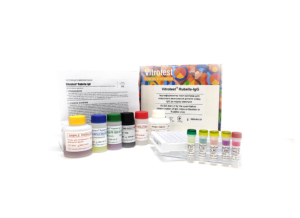
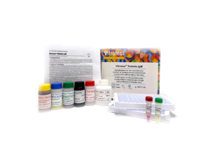
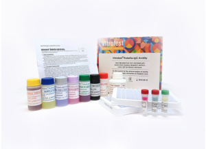
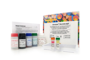
The test kit Vitrotest® Brucella-IgG is an enzyme linked immunosorbent assay (ELISA) for the qualitative and semiquantitative determination of igG class antibodies to Brucella in human serum or plasma. Determination of IgG antibodies to Brucella in the test kit Vitrotest® Brucella-IgG is based on a solid phase, indirect ELISA in a two-step incubation procedure.
- TK153 - 96 tests
- Solid phase: strip ELISA plate pre-coated with antigens of Brucella.
- Conjugate: monoclonal antibodies to human IgG conjugated with horseradish peroxidase.
- Chromogen: ready to use TMB solution.
- Sample volume: 100 μl.
- Assay time: 1 h 15 min.
Brucellosis is a zoonotic infectious disease caused by bacteria of the genus Brucella. Globally, 1.6–2.1 million human cases of brucellosis are reported annually. Among the countries with the highest reported incidence of brucellosis are Iran, Kyrgyzstan, Tajikistan, Kazakhstan, Azerbaijan, Turkmenistan, Armenia, and Uzbekistan. The species most pathogenic to humans include Brucella melitensis, Brucella abortus, Brucella suis, and Brucella canis. Different species of Brucella vary in their reservoir hosts and degree of virulence: B. melitensis is considered the most pathogenic for humans, whereas B. abortus more commonly causes relatively milder forms of the disease. Human infection occurs through contact with infected animals and their biological fluids, as well as through the consumption of animal-derived products (raw milk and insufficiently heat-treated meat), and by the airborne route during occupational exposure. The pathogen enters the human body through mucous membranes or damaged skin. Subsequently, the bacteria are transported by macrophages to lymphoid tissues, spread through the lymphatic system, and may potentially proliferate in multiple organs, causing localized and systemic infections. By persisting within host cells such as macrophages, Brucella spp. employ strategies to evade the host immune response, resulting in prolonged infection. The incubation period of brucellosis usually ranges from 1 to 4 weeks but may extend to several months. Clinical manifestations are characterized by undulating fever, sweating (especially at night), weakness, and enlargement of the lymph nodes, liver, and spleen. In chronic cases, the musculoskeletal, nervous, and cardiovascular systems may also be affected. Due to the nonspecific nature of its symptoms, the diagnosis of brucellosis is challenging and requires a comprehensive approach, including the evaluation of clinical findings, epidemiological history, and laboratory test results (bacterial culture, serological assays, and PCR). Serological methods used for the diagnosis of brucellosis include the agglutination test, complement fixation test, and enzyme-linked immunosorbent assay (ELISA). Using the ELISA method, IgM antibodies are detected during the early stages of infection, whereas IgG antibodies are detected later and may persist for a long period after previous infection or successful treatment. A rapid decline in IgG antibody titers usually indicates a favorable response to antibiotic therapy, while persistently high or increasing titers may indicate treatment failure, residual disease, or an impending clinical relapse. Therefore, the detection of IgG antibodies specific to Brucella is an important marker of current or past infection, particularly in subacute and chronic forms of brucellosis. -
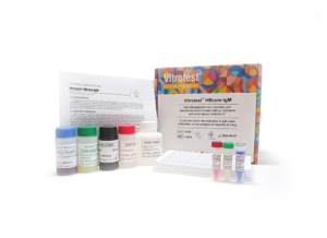
The test kit Vitrotest® HAV-IgM is an enzyme linked immunosorbent assay (ELISA) for the qualitative determination of IgM class antibodies to Hepatitis A Virus in human serum or plasma.
Determination of IgM antibodies to Hepatitis A Virus in the test kit Vitrotest® HAV-IgM is based on a solid phase, «IgM-capture» ELISA in a two-step incubation procedure.
- TK069 - 96 tests
- Solid phase: strip ELISA plate pre-coated with monoclonal anti-IgM antibodies.
- Conjugate: monoclonal antibodies to human HAV conjugated with horseradish peroxidase.
- Chromogen: ready to use TMB solution.
- Sample volume: 100 μl.
- Assay time: 2 h.
Hepatitis A is an acute infectious disease of the liver (also known as Botkin’s disease) caused by the hepatitis A virus (HAV), which belongs to the genus Hepatovirus of the family Picornaviridae. The virus has a single-stranded RNA genome and lacks a lipid envelope, which determines its high stability in the environment, in particular, it is able to remain infectious in water, soil, and on surfaces for prolonged periods and is resistant to low temperatures and the acidic environment of the stomach.
According to the World Health Organization, approximately 100 million HAV infections and 1.5 million clinically apparent cases are reported worldwide each year, of which 15,000–30,000 result in death annually. The main routes of transmission are fecal–oral, including through contaminated water and food, as well as household contact. After infection, HAV penetrates through the intestinal epithelium into the bloodstream and is transported to the liver, where it replicates in hepatocytes and Kupffer cells. Due to its cytolytic effect on liver cells, the virus is excreted with bile into the intestine and released into the environment even before clinical symptoms appear.
The incubation period of hepatitis A usually ranges from 14 to 28 days, although it may extend to 15–50 days depending on immune status, age, concomitant diseases, and other factors. In most cases, the disease is accompanied by weakness, body aches, sore throat, fever, loss of appetite, vomiting, pain, and heaviness in the right upper quadrant of the abdomen. The clinical course varies with age: in young children the infection is often asymptomatic, whereas in adults an icteric form with pronounced intoxication syndrome develops more frequently.
The immune response to HAV is characterized by the sequential appearance of specific antibodies. IgM antibodies appear in the early stages of infection, often before symptoms develop, and are the primary marker of acute infection. Their levels peak during the first weeks of illness and gradually decline over 3–6 months, sometimes persisting for up to 12 months after infection. IgG antibodies appear later and provide long-term, usually lifelong, immunity. They are also produced after vaccination against HAV, which forms the basis of specific disease prevention. The presence of IgG in the absence of IgM indicates past infection or immunization. Therefore, the determination of HAV-specific IgM antibodies by ELISA is a key tool for the early diagnosis of acute hepatitis A, which is essential for timely treatment and epidemiological control of the disease.
-
The test kit Vitrotest® Anti-HIV1/2 is an enzyme linked immunosorbent assay (ELISA) for the detection of total antibodies (IgG, IgA, IgM) to HIV1 (group M and O) and HIV2 in human serum or plasma.
Detection of total antibodies to human immunodeficiency virus types 1 and 2 (HIV1/2) in the Vitrotest® Anti-HIV1/2 test kit is based on a solid-phase sandwich ELISA in a two-step incubation procedure.
- TK129 - 96 tests
- TK130 - 192 tests
- TK131 - 480 tests
- Solid phase: breakable microplate ELISA is coated with recombinant HIV 1/2 antigens.
- Conjugate: recombinant HIV1/2 antigens conjugated to horseradish peroxidase.
- Chromogen: ready to use TMB solution.
- Volume of sample for analysis: 70 μl.
- Assay time: 1 hour 45 minutes.
The complex epidemic situation regarding the human immunodeficiency virus (HIV), which causes acquired immunodeficiency syndrome (AIDS), has remained a global health problem for more than forty years and has led to approximately 40 million deaths during this time. According to WHO data from 2023, approximately 39.9 million people worldwide are infected with HIV.
HIV is an RNA-containing lentivirus of the Retroviridae family. It has a lipid envelope in which the transmembrane glycoprotein gp41 with the surface antigen gp120 (encoded by the viral RNA gene env) is embedded. Under the envelope are matrix, nucleocapsid and core proteins, including the p24 antigen, encoded by the viral gene gag. The virus also has several other proteins with various regulatory or immunomodulatory functions.
Based on their structural and antigenic characteristics, HIV isolates are divided into two main types: the worldwide common HIV type 1 (HIV1) and the less common HIV type 2 (HIV2), the latter occurring predominantly in some regions of West and Central Africa. Based on the identity of the nucleotide sequence of the env and gag genes, HIV1 isolates have been classified into three groups: M (major), O (outlier), and N (non-M/non-O group).
HIV infection occurs by its transmission through infected biological fluids, namely blood, sperm, vaginal discharge, and breast milk. The infection is usually characterized by loss of CD4+ cells and has several stages: an acute phase of intensive viral replication and dissemination in lymphoid tissues; a chronic, often asymptomatic phase of prolonged immune activation and viral replication; and a progressive phase of marked depletion of CD4+ T-cells, leading to AIDS.
Modern laboratory diagnostics of HIV infection is based on the detection of specific markers of infection, namely RNA of the pathogen, core antigen p24 and antibodies to HIV1/2. The choice of markers for testing depends on the purpose of diagnostics and should be consistent with the kinetics and time of their appearance in the patient’s blood. Viral RNA is detected by polymerase chain reaction (PCR) in blood plasma within 10-14 days after infection. Its quantity increases intensively for several months, and after the inclusion of humoral and cellular mechanisms of the immune response, the RNA level drops sharply to a constant level. In the late stages of HIV infection, the RNA level gradually increases to high concentrations with the appearance of symptoms of AIDS-associated diseases. Viral antigen p24 is detected in the blood of an infected person several days later than HIV RNA and remains at the detection level for about 1.5 months. HIV-specific antibodies usually appear 3-4 weeks after infection (the so-called seroconversion “window”), and can be detected in almost all infected individuals within 1-2 months. Detection of total HIV antibodies by enzyme immunoassay is widely used both for diagnosing HIV infection and for screening donor blood. The use of “fourth” generation enzyme-linked immunosorbent test kits, which make it possible to detect not only specific antibodies, but also the HIV p24 core antigen, can increase the sensitivity of the analysis, reducing the seroconversion “window” by 7-10 days.
-
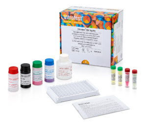
The test kit Vitrotest® HIV Ag/Ab is an enzyme linked immunosorbent assay (ELISA) for the simultaneous detection of HIV p24 antigen and total antibodies (IgG, IgA, IgM) to HIV1 (group M and O) and HIV2 in human serum or plasma.
Simultaneous detection of the HIV1 p24 core antigen and total antibodies to human immunodeficiency virus types 1 and 2 (HIV1/2) in the Vitrotest® HIV Ab/Ag test kit is based on a solid-phase sandwich ELISA in a two-step incubation procedure.
○ TK132 - 96 tests
○ TK133 - 192 tests
○ TK134 - 480 tests- Solid phase: breakable microplate ELISA is coated with monoclonal antibodies specific to p24 HIV1 and recombinant HIV 1/2 antigens.
- Conjugate: solution of streptavidin and recombinant HIV1/2 antigens conjugated to horseradish peroxidase.
- Chromogen: ready to use TMB solution.
- Volume of sample for analysis: 70 μl.
- Assay time: 1 hour 45 minutes.
The complex epidemic situation regarding the human immunodeficiency virus (HIV), which causes acquired immunodeficiency syndrome (AIDS), has remained a global health problem for more than forty years and has led to approximately 40 million deaths during this time. According to WHO data from 2023, approximately 39.9 million people worldwide are infected with HIV.
HIV is an RNA-containing lentivirus of the Retroviridae family. It has a lipid envelope in which the transmembrane glycoprotein gp41 with the surface antigen gp120 (encoded by the viral RNA gene env) is embedded. Under the envelope are matrix, nucleocapsid and core proteins, including the p24 antigen, encoded by the viral gene gag. The virus also has several other proteins with various regulatory or immunomodulatory functions.
Based on their structural and antigenic characteristics, HIV isolates are divided into two main types: the worldwide common HIV type 1 (HIV1) and the less common HIV type 2 (HIV2), the latter occurring predominantly in some regions of West and Central Africa. Based on the identity of the nucleotide sequence of the env and gag genes, HIV1 isolates have been classified into three groups: M (major), O (outlier), and N (non-M/non-O group).
HIV infection occurs by its transmission through infected biological fluids, namely blood, sperm, vaginal discharge, and breast milk. The infection is usually characterized by loss of CD4+ cells and has several stages: an acute phase of intensive viral replication and dissemination in lymphoid tissues; a chronic, often asymptomatic phase of prolonged immune activation and viral replication; and a progressive phase of marked depletion of CD4+ T-cells, leading to AIDS.
Modern laboratory diagnostics of HIV infection is based on the detection of specific markers of infection, namely RNA of the pathogen, core antigen p24 and antibodies to HIV1/2. The choice of markers for testing depends on the purpose of diagnostics and should be consistent with the kinetics and time of their appearance in the patient’s blood. Viral RNA is detected by polymerase chain reaction (PCR) in blood plasma within 10-14 days after infection. Its quantity increases intensively for several months, and after the inclusion of humoral and cellular mechanisms of the immune response, the RNA level drops sharply to a constant level. In the late stages of HIV infection, the RNA level gradually increases to high concentrations with the appearance of symptoms of AIDS-associated diseases. Viral antigen p24 is detected in the blood of an infected person several days later than HIV RNA and remains at the detection level for about 1.5 months. HIV-specific antibodies usually appear 3-4 weeks after infection (the so-called seroconversion “window”), and can be detected in almost all infected individuals within 1-2 months. Detection of total HIV antibodies by enzyme immunoassay is widely used both for diagnosing HIV infection and for screening donor blood. The use of “fourth” generation enzyme-linked immunosorbent test kits, which make it possible to detect not only specific antibodies, but also the HIV p24 core antigen, can increase the sensitivity of the analysis, reducing the seroconversion “window” by 7-10 days.
-
The test kit Vitrotest® Anti-TPO is an enzyme linked immunosorbent assay (ELISA) for the quantitative determination of autoantibodies to thyroid peroxidase (TPO) in human serum or plasma. Determination of autoantibodies to thyroid peroxidase in the test kit Vitrotest® Anti-TPO is based on a solid phase, indirect ELISA in a two-step incubation procedure.
- TK152 - 96 tests
- Solid phase: strip ELISA plate pre-coated with recombinant thyroid peroxidase.
- Conjugate: buffer solution of monoclonal antibodies to human IgG conjugated with horseradish peroxidase.
- Chromogen: ready to use TMB solution.
- Sample volume: 10 μl.
- Assay time: 1 h 20 min.
According to WHO, thyroid diseases are the second most widespread endocrine disorders after diabetes mellitus. Over 200 million people worldwide suffer from various types of thyroid dysfunction. In Ukraine, over the past 5 years, the number of people with thyroid diseases has increased fivefold. Autoimmune thyroid diseases represent a diverse group of organ-specific autoimmune disorders, the most common of which are Hashimoto’s thyroiditis and Graves’ disease. The pathological process is associated with the formation of autoantibodies to thyroid peroxidase and thyroglobulin. Thyroid peroxidase is a membrane-bound enzyme responsible for iodine oxidation and iodination of tyrosyl residues in the thyroglobulin molecule during the synthesis of thyroid hormones T3 and T4. Most anti-TPO antibodies belong to the IgG1 subclass, which activates complement. Additionally, anti-TPO antibodies can bind through their Fc fragment to natural killer cells, which in turn cause cytotoxic damage to thyrocytes. Damage to thyroid cells, as well as direct enzyme inhibition, can lead to insufficient hormone production (hypothyroidism), sometimes preceded by transient hyperthyroidism. Besides thyroid disorders, elevated anti-TPO titers may also occur in a wide range of diseases: pernicious anemia, systemic lupus erythematosus, rheumatoid arthritis, insulin-dependent diabetes mellitus, breast cancer, and others. A correlation has also been established between anti-TPO antibodies and obstetric complications, such as postpartum thyroiditis or postpartum depression. Anti-TPO antibodies may be present in individuals without clinical or laboratory signs of thyroid dysfunction. The presence of anti-TPO without overt disease is associated with a higher risk of developing autoimmune thyroiditis in the future and, together with TSH levels, is used to predict the development of hypo- and hyperthyroidism. In modern laboratory diagnostics, ELISA is widely used for determining the concentration of autoantibodies to thyroid peroxidase due to its simplicity, convenience, high sensitivity, and specificity. Standardization of quantitative determination of anti-TPO in human serum or plasma is ensured by the use of the WHO International Standard with assigned concentration in IU/ml for preparation of internal ELISA calibrators. -
The test kit Vitrotest® Free T3 is an enzyme linked immunosorbent assay (ELISA) for the quantitative determination of free triiodothyronine (FT3) in human serum or plasma. Determination of free triiodothyronine concentration in the test kit Vitrotest® Free T3 is based on a competitive ELISA with a two-step incubation.
- TK150 - 96 tests
- Solid phase: strip ELISA plate pre-coated with triiodothyronine.
- Conjugate: buffer solution of monoclonal antibodies specific to triiodothyronine conjugated with horseradish peroxidase.
- Chromogen: ready to use TMB solution.
- Sample volume: 25 μl.
- Assay time: 1 h 20 min.
According to data from the World Health Organization (WHO), thyroid diseases are the second most common endocrine disorders after diabetes mellitus. More than 200 million people worldwide suffer from various forms of thyroid dysfunction. In Ukraine, over the past five years, the number of people with thyroid diseases has increased fivefold. The main pathological conditions include hyperthyroidism, hypothyroidism, autoimmune thyroid diseases, benign and malignant neoplasms, etc. The main hormones produced by the thyroid gland are thyroxine or tetraiodothyronine (T4, containing four iodine atoms) and triiodothyronine (T3, containing three iodine atoms). These hormones are lipophilic and circulate in the blood mainly bound to transport proteins: thyroxine-binding globulin (TBG), transthyretin (also known as thyroxine-binding prealbumin), and albumin. However, only free triiodothyronine and free thyroxine are functionally active, accounting for just 0.3% and 0.03% of total T3 and T4 in blood serum, respectively. Free triiodothyronine is the biologically active form that directly enters target cells and binds to nuclear receptors to regulate gene expression and cellular function. Free thyroxine has minimal hormonal activity, but its long half-life (8 days) serves as a reservoir or prohormone for free triiodothyronine. All reactions required for the formation and release of T3 and T4 are controlled by thyroid-stimulating hormone (TSH) through a negative feedback mechanism. Although determination of TSH and FT4 concentrations are considered the primary tests of choice for diagnosing thyroid dysfunction, measurement of FT3 levels is useful for detecting T3-thyrotoxicosis, complex or unusual manifestations of hyperthyroidism, monitoring thyroid disease treatment, and assessing metabolism. Accumulated evidence indicates that changes in FT3 levels are also closely associated with a number of systemic disorders, such as cardiovascular diseases, dyslipidemia, type 2 diabetes mellitus, and liver dysfunction, highlighting the clinical importance of FT3 monitoring as a functional indicator of thyroid status. -
The test kit Vitrotest® Free T4 is an enzyme linked immunosorbent assay (ELISA) for the quantitative determination of free thyroxine (FT4) in human serum or plasma. Determination of free thyroxine concentration in the test kit Vitrotest® Free T4 is based on a competitive ELISA with a two-step incubation.
- TK151 - 96 tests
- Solid phase: strip ELISA plate pre-coated with monoclonal antibodies specific to thyroxine.
- Conjugate: buffer solution of thyroxine conjugated with horseradish peroxidase.
- Chromogen: ready to use TMB solution.
- Sample volume: 20 μl.
- Assay time: 1 h 20 min.
According to WHO, thyroid diseases are the second most common endocrine disorders after diabetes mellitus. Over 200 million people worldwide suffer from various forms of thyroid dysfunction. In Ukraine, the number of people with thyroid diseases has increased fivefold over the past 5 years. The main pathological conditions include hyperthyroidism, hypothyroidism, autoimmune thyroid diseases, benign and malignant neoplasms. The main hormones produced by the thyroid gland are thyroxine or tetraiodothyronine (T4, containing four iodine atoms) and triiodothyronine (T3, containing three iodine atoms). They are lipophilic and circulate in the blood mainly bound to transport proteins: thyroxine-binding globulin (TBG), transthyretin (thyroxine-binding prealbumin), and albumin. However, the biologically active forms are free triiodothyronine and free thyroxine, which represent only 0.3% and 0.03% of total T3 and T4 in serum, respectively. Overall, FT4 has minimal hormonal activity, but its long half-life (8 days) serves as a reservoir or prohormone for active free triiodothyronine, which binds to nuclear receptors. All processes required for the synthesis and release of T3 and T4 are controlled by thyroid-stimulating hormone (TSH), secreted by pituitary thyrotropic cells. TSH secretion is regulated through pituitary negative feedback: elevated free T4 and T3 levels suppress TSH synthesis and secretion, while decreased levels increase TSH secretion. Determination of TSH and free thyroxine concentrations are the primary tests for diagnosing thyroid dysfunction. FT4 is an important marker for differentiating subclinical hyperthyroidism from overt hyperthyroidism or hypothyroidism, for investigating suspected abnormal TSH secretion, TSH-secreting pituitary adenomas, and for monitoring treatment of thyroid diseases. -
The test kit Vitrotest® HIV1 p24 is an enzyme linked immunosorbent assay (ELISA) for quantitative determination of p24 core antigen to HIV1 in human serum or plasma.
Determination of the HIV1 p24 core antigen in the Vitrotest® HIV1 p24 test kit is based on a solid-phase sandwich ELISA in a two-step incubation procedure.
- TK138 - 96 tests
- TK139 - 192 tests
- TK140 - 480 tests
- Solid phase: breakable microplate ELISA is coated with monoclonal antibodies specific to p24 HIV1.
- Conjugate: solution of streptavidin conjugated to horseradish peroxidase.
- Chromogen: ready to use TMB solution.
- Volume of sample for analysis: 70 μl.
- Assay time: 1 hour 45 minutes.
The complex epidemic situation regarding the human immunodeficiency virus (HIV), which causes acquired immunodeficiency syndrome (AIDS), has remained a global health problem for more than forty years and has led to approximately 40 million deaths during this time. According to WHO data from 2023, approximately 39.9 million people worldwide are infected with HIV.
HIV is an RNA-containing lentivirus of the Retroviridae family. It has a lipid envelope in which the transmembrane glycoprotein gp41 with the surface antigen gp120 (encoded by the viral RNA gene env) is embedded. Under the envelope are matrix, nucleocapsid and core proteins, including the p24 antigen, encoded by the viral gene gag. The virus also has several other proteins with various regulatory or immunomodulatory functions.
Based on their structural and antigenic characteristics, HIV isolates are divided into two main types: the worldwide common HIV type 1 (HIV1) and the less common HIV type 2 (HIV2), the latter occurring predominantly in some regions of West and Central Africa. Based on the identity of the nucleotide sequence of the env and gag genes, HIV1 isolates have been classified into three groups: M (major), O (outlier), and N (non-M/non-O group).
HIV infection occurs by its transmission through infected biological fluids, namely blood, sperm, vaginal discharge, and breast milk. The infection is usually characterized by loss of CD4+ cells and has several stages: an acute phase of intensive viral replication and dissemination in lymphoid tissues; a chronic, often asymptomatic phase of prolonged immune activation and viral replication; and a progressive phase of marked depletion of CD4+ T-cells, leading to AIDS.
Modern laboratory diagnostics of HIV infection is based on the detection of specific markers of infection, namely RNA of the pathogen, core antigen p24 and antibodies to HIV1/2. The choice of markers for testing depends on the purpose of diagnostics and should be consistent with the kinetics and time of their appearance in the patient’s blood. Viral RNA is detected by polymerase chain reaction (PCR) in blood plasma within 10-14 days after infection. Its quantity increases intensively for several months, and after the inclusion of humoral and cellular mechanisms of the immune response, the RNA level drops sharply to a constant level. In the late stages of HIV infection, the RNA level gradually increases to high concentrations with the appearance of symptoms of AIDS-associated diseases. Viral antigen p24 is detected in the blood of an infected person several days later than HIV RNA and remains at the detection level for about 1.5 months. HIV-specific antibodies usually appear 3-4 weeks after infection (the so-called seroconversion “window”), and can be detected in almost all infected individuals within 1-2 months. Detection of total HIV antibodies by enzyme immunoassay is widely used both for diagnosing HIV infection and for screening donor blood. The use of “fourth” generation enzyme-linked immunosorbent test kits, which make it possible to detect not only specific antibodies, but also the HIV p24 core antigen, can increase the sensitivity of the analysis, reducing the seroconversion “window” by 7-10 days.
-
The test kit Vitrotest® TSH is an enzyme linked immunosorbent assay (ELISA) for the quantitative determination of thyroid-stimulating hormone (TSH) in human serum or plasma. Determination of thyroid-stimulating hormone concentration in the test kit Vitrotest® TSH is based on a solid-phase sandwich ELISA in a two-step incubation procedure.
- TK148 - 96 tests
- TK149 - 192 tests
- Solid phase: strip ELISA plate pre-coated with the first monoclonal antibodies specific to the β-subunit of human thyroid-stimulating hormone.
- Conjugate: monoclonal antibodies to human TSH conjugated with horseradish peroxidase.
- Chromogen: ready to use TMB solution.
- Sample volume: 50 μl.
- Assay time: 2 h.
According to the World Health Organization (WHO), thyroid diseases are the second most common endocrine disorders after diabetes mellitus. Over 200 million people worldwide suffer from various forms of thyroid dysfunction. In Ukraine, over the past 5 years, the number of people with thyroid diseases has increased fivefold. The main pathological conditions include hyperthyroidism, hypothyroidism, autoimmune thyroid diseases, benign and malignant neoplasms. A connection has also been established between thyroid dysfunction and other diseases such as diabetes, cardiovascular diseases, depression, oral diseases, and cancer. A valuable biomarker of thyroid functional status widely used for screening, diagnosis, and monitoring of thyroid diseases is thyroid-stimulating hormone (TSH). TSH is a glycoprotein hormone synthesized by the anterior pituitary and is a key regulator of thyroid function. The TSH molecule consists of two different non-covalently bound subunits: the α-subunit, identical in amino acid sequence to the α-subunit of chorionic gonadotropin, luteinizing hormone, and follicle-stimulating hormone, and the hormone-specific β-subunit, which is unique. The main function of TSH is to stimulate the thyroid gland to synthesize and secrete thyroid hormones – thyroxine (T4) and triiodothyronine (T3). TSH binds to the TSH receptor on thyrocytes and activates intracellular signaling cascades that regulate iodine uptake, thyroid metabolism, thyroid growth, and hormone secretion. Through negative feedback, T3 and T4 inhibit TSH secretion. In healthy adults, TSH serum levels are approximately 0.4 to 4.0 μIU/ml, although narrower ranges may be used to better detect subclinical hypothyroidism. Separate reference intervals for TSH are established for pregnant women, infants, and young children. TSH secretion has pulsatile and circadian patterns, and its concentration depends on factors such as age, sex, ethnicity, iodine intake, reproductive status, and body mass index. Over the last three decades, laboratory methods used to determine TSH levels have significantly improved. Among immunochemical methods, ELISA has gained wide application due to its convenience, simplicity, high reproducibility, and sensitivity for determining thyroid-stimulating hormone in human serum and plasma. The standardization of quantitative determination of TSH in human serum or plasma is ensured by the use of the WHO International Standard with assigned TSH concentration in μIU/ml for preparation of internal ELISA calibrators. -
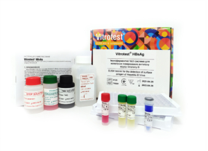
The test kit Vitrotest® HBsAg is an enzyme linked immunosorbent assay (ELISA) for the detection of surface antigen of Hepatitis B Virus (HBsAg) in human serum or plasma.
Detection of the presence of HBsAg in the test kit Vitrotest® HBsAg is based on a solid-phase "sandwich" ELISA.
○ ТК016 - 96 tests
○ ТК059 - 192 tests
○ TK127 - 480 tests- Solid phase: microplate ELISA is coated with monoclonal antibodies to HBsAg.
- Conjugate: monoclonal antibodies to HBsAg conjugated with horseradish peroxidase.
- Chromogen: ready to use TMB solution.
- Volume of sample for analysis: 100 μl.
- Assay time: 2.5 hours.
Hepatitis B virus (HBV) is an enveloped DNA virus of the family Hepadnaviridae. The aetiology of ‘‘serum hepatitis’’, as it was known for many years, was not identified, until the discovery of the so-called Au antigen by Blumberg et al. in 1965 [Blumberg et al., 1965] led to the identification of viral particles by Dane et al. several years later [Dane et al., 1970].
Hepatitis B (HB) has a long incubation period of 45 to 160 days (average: 120 days). The length of incubation period is related to the amount of virus in the inoculum, the mode of transmission and host factors.
The appearance of symptoms under acute HB is inversely related to age: less than 1% of newborns and 30%–50% adults develop symptoms. Those who do get symptoms, which are similar for all types of viral hepatitis, usually suffer from tiredness, loss of appetite, abdominal discomfort, nausea, vomiting, fever and jaundice. In less then 1 % of cases, especially in the elderly, fulminating HB develops, which is mostly fatal due to acute hepatic necrosis.
The acute HB often resolves spontaneously after a 4-8 week illness. Otherwise, the infection can last for six months or more. This condition is known as chronic HB.
More than 90 % of infected infants, 25–50 % of children infected between 1 and 5 years of age, and 6–10 % of acutely infected older children and adults develop chronic infection. As a result, more than 350 million people in the world today are estimated to be persistently infected with HBV.
In a considerable number of patients, chronic HB may lead to liver cirrhosis and hepatocellular carcinoma. Cirrhosis affects around one in five people with chronic hepatitis B. Of all causes of cirrhosis, approximately one third can be attributed to chronic HBV infection.
Transmission occurs by percutaneous and permucosal (through broken skin) exposure to such infective body fluids as blood, vaginal and menstrual fluids, and semen. The main ways of transmission include: vertical - from an infected mother during delivery (rate of transmission around 50%); sexual; horizontal - household contact with an infected person (for example, contact of infected blood with cutaneous scratches), sharing of contaminated injection drug equipment by injection drug users, or unhygienic injection procedures in health-care institutions. -
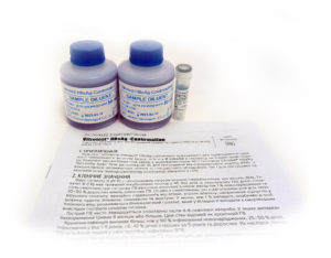
The Vitrotest® HBsAg-Confirmation reagent kit is intended to confirm the presence of the hepatitis B virus surface antigen (HBsAg) in human serum or plasma. The kit is used together with the test kit Vitrotest® HBsAg.
Confirmation of the presence of the hepatitis B virus surface antigen (HBsAg) is based on the solid-phase "sandwich" ELISA using the reagent kit Vitrotest® HBsAg-Confirmation and the test kit Vitrotest® HBsAg .
○ TK017 - 100 testsСomposition of the set:
 sample diluent neutralizing component (monoclonal antibodies specific for HBsAg)Hepatitis B virus (HBV) is an enveloped DNA virus of the family Hepadnaviridae. The aetiology of ‘‘serum hepatitis’’, as it was known for many years, was not identified, until the discovery of the so-called Au antigen by Blumberg et al. in 1965 [Blumberg et al., 1965] led to the identification of viral particles by Dane et al. several years later [Dane et al., 1970].
Hepatitis B (HB) has a long incubation period of 45 to 160 days (average: 120 days). The length of incubation period is related to the amount of virus in the inoculum, the mode of transmission and host factors.
The appearance of symptoms under acute HB is inversely related to age: less than 1% of newborns and 30%–50% adults develop symptoms. Those who do get symptoms, which are similar for all types of viral hepatitis, usually suffer from tiredness, loss of appetite, abdominal discomfort, nausea, vomiting, fever and jaundice. In less then 1 % of cases, especially in the elderly, fulminating HB develops, which is mostly fatal due to acute hepatic necrosis.
The acute HB often resolves spontaneously after a 4-8 week illness. Otherwise, the infection can last for six months or more. This condition is known as chronic HB.
More than 90 % of infected infants, 25–50 % of children infected between 1 and 5 years of age, and 6–10 % of acutely infected older children and adults develop chronic infection. As a result, more than 350 million people in the world today are estimated to be persistently infected with HBV.
In a considerable number of patients, chronic HB may lead to liver cirrhosis and hepatocellular carcinoma. Cirrhosis affects around one in five people with chronic hepatitis B. Of all causes of cirrhosis, approximately one third can be attributed to chronic HBV infection.
Transmission occurs by percutaneous and permucosal (through broken skin) exposure to such infective body fluids as blood, vaginal and menstrual fluids, and semen. The main ways of transmission include: vertical - from an infected mother during delivery (rate of transmission around 50%); sexual; horizontal - household contact with an infected person (for example, contact of infected blood with cutaneous scratches), sharing of contaminated injection drug equipment by injection drug users, or unhygienic injection procedures in health-care institutions. -
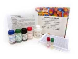
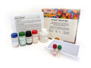
The test kit Vitrotest® Anti-HBcore is an enzyme linked immunosorbent assay (ELISA) for the detection of antibodies to core antigen of Hepatitis B Virus (HBcore antigen) in human serum or
plasma.
Detection of antibodies to HBcore antigen in the test kit Vitrotest® Anti-HBcore is based on a solid phase, indirect ELISA in a two-step incubation procedure.
○ TK018 - 96 tests
○ TK141 - 192 tests- Solid phase: microplate ELISA is coated with recombinant HBcore antigen.
- Conjugate: a mixture of monoclonal antibodies to human IgG and IgM conjugated with horseradish peroxidase.
- Chromogen: ready to use TMB solution.
- Volume of sample for analysis: 20 μl.
- Assay time: 1.5 hours.
Hepatitis B virus (HBV) is an enveloped DNA virus of the family Hepadnaviridae. The aetiology of ‘‘serum hepatitis’’, as it was known for many years, was not identified, until the discovery of the so-called Au antigen by Blumberg et al. in 1965 [Blumberg et al., 1965] led to the identification of viral particles by Dane et al. several years later [Dane et al., 1970].
Hepatitis B (HB) has a long incubation period of 45 to 160 days (average: 120 days). The length of incubation period is related to the amount of virus in the inoculum, the mode of transmission and host factors.
The appearance of symptoms under acute HB is inversely related to age: less than 1% of newborns and 30%–50% adults develop symptoms. Those who do get symptoms, which are similar for all types of viral hepatitis, usually suffer from tiredness, loss of appetite, abdominal discomfort, nausea, vomiting, fever and jaundice. In less then 1 % of cases, especially in the elderly, fulminating HB develops, which is mostly fatal due to acute hepatic necrosis.
The acute HB often resolves spontaneously after a 4-8 week illness. Otherwise, the infection can last for six months or more. This condition is known as chronic HB.
More than 90 % of infected infants, 25–50 % of children infected between 1 and 5 years of age, and 6–10 % of acutely infected older children and adults develop chronic infection. As a result, more than 350 million people in the world today are estimated to be persistently infected with HBV.
In a considerable number of patients, chronic HB may lead to liver cirrhosis and hepatocellular carcinoma. Cirrhosis affects around one in five people with chronic hepatitis B. Of all causes of cirrhosis, approximately one third can be attributed to chronic HBV infection.
Transmission occurs by percutaneous and permucosal (through broken skin) exposure to such infective body fluids as blood, vaginal and menstrual fluids, and semen. The main ways of transmission include: vertical - from an infected mother during delivery (rate of transmission around 50%); sexual; horizontal - household contact with an infected person (for example, contact of infected blood with cutaneous scratches), sharing of contaminated injection drug equipment by injection drug users, or unhygienic injection procedures in health-care institutions. -
The test kit Vitrotest® HBcore-IgG is an enzyme linked immunosorbent assay (ELISA) for the detection of IgG class antibodies to core antigen of Hepatitis B Virus (HBcore antigen) in human serum or plasma.
Detection of IgG class antibodies to HBcore antigen in the test kit Vitrotest® HBcore-IgG is based on a solid phase, indirect ELISA in a two-step incubation procedure.
○ TK050 - 96 tests
○ TK142 - 192 tests- Solid phase: microplate ELISA is coated with recombinant HBcore antigen.
- Conjugate: monoclonal antibodies to human IgG conjugated to horseradish peroxidase.
- Chromogen: ready to use TMB solution.
- Volume of sample for analysis: 20 μl.
- Assay time: 1.5 hours.
Hepatitis B virus (HBV) is an enveloped DNA virus of the family Hepadnaviridae. The aetiology of ‘‘serum hepatitis’’, as it was known for many years, was not identified, until the discovery of the so-called Au antigen by Blumberg et al. in 1965 [Blumberg et al., 1965] led to the identification of viral particles by Dane et al. several years later [Dane et al., 1970].
Hepatitis B (HB) has a long incubation period of 45 to 160 days (average: 120 days). The length of incubation period is related to the amount of virus in the inoculum, the mode of transmission and host factors.
The appearance of symptoms under acute HB is inversely related to age: less than 1% of newborns and 30%–50% adults develop symptoms. Those who do get symptoms, which are similar for all types of viral hepatitis, usually suffer from tiredness, loss of appetite, abdominal discomfort, nausea, vomiting, fever and jaundice. In less then 1 % of cases, especially in the elderly, fulminating HB develops, which is mostly fatal due to acute hepatic necrosis.
The acute HB often resolves spontaneously after a 4-8 week illness. Otherwise, the infection can last for six months or more. This condition is known as chronic HB.
More than 90 % of infected infants, 25–50 % of children infected between 1 and 5 years of age, and 6–10 % of acutely infected older children and adults develop chronic infection. As a result, more than 350 million people in the world today are estimated to be persistently infected with HBV.
In a considerable number of patients, chronic HB may lead to liver cirrhosis and hepatocellular carcinoma. Cirrhosis affects around one in five people with chronic hepatitis B. Of all causes of cirrhosis, approximately one third can be attributed to chronic HBV infection.
Transmission occurs by percutaneous and permucosal (through broken skin) exposure to such infective body fluids as blood, vaginal and menstrual fluids, and semen. The main ways of transmission include: vertical - from an infected mother during delivery (rate of transmission around 50%); sexual; horizontal - household contact with an infected person (for example, contact of infected blood with cutaneous scratches), sharing of contaminated injection drug equipment by injection drug users, or unhygienic injection procedures in health-care institutions. -
The test kit Vitrotest® HBcore-IgM is an enzyme linked immunosorbent assay (ELISA) for the detection of IgM class antibodies to the core antigen of the Hepatitis B Virus (HBcore) in human serum or plasma.
Detection of IgM class antibodies to the core antigen of the Hepatitis B Virus in the test kit Vitrotest® HBcore-IgM is based on the principle of "IgM-capture" of solid-phase ELISA in two-stage incubation.
○ TK019 - 96 tests
○ TK143 - 192 tests- Solid phase: microplate ELISA is coated with monoclonal antibodies specific for human immunoglobulin M.
- Conjugate: HBcore recombinant antigen conjugated with horseradish peroxidase.
- Chromogen: ready to use TMB solution.
- Volume of sample for analysis: 10 μl.
- Assay time: 1.5 hours.
Hepatitis B virus (HBV) is an enveloped DNA virus of the family Hepadnaviridae. The aetiology of ‘‘serum hepatitis’’, as it was known for many years, was not identified, until the discovery of the so-called Au antigen by Blumberg et al. in 1965 [Blumberg et al., 1965] led to the identification of viral particles by Dane et al. several years later [Dane et al., 1970].
Hepatitis B (HB) has a long incubation period of 45 to 160 days (average: 120 days). The length of incubation period is related to the amount of virus in the inoculum, the mode of transmission and host factors.
The appearance of symptoms under acute HB is inversely related to age: less than 1% of newborns and 30%–50% adults develop symptoms. Those who do get symptoms, which are similar for all types of viral hepatitis, usually suffer from tiredness, loss of appetite, abdominal discomfort, nausea, vomiting, fever and jaundice. In less then 1 % of cases, especially in the elderly, fulminating HB develops, which is mostly fatal due to acute hepatic necrosis.
The acute HB often resolves spontaneously after a 4-8 week illness. Otherwise, the infection can last for six months or more. This condition is known as chronic HB.
More than 90 % of infected infants, 25–50 % of children infected between 1 and 5 years of age, and 6–10 % of acutely infected older children and adults develop chronic infection. As a result, more than 350 million people in the world today are estimated to be persistently infected with HBV.
In a considerable number of patients, chronic HB may lead to liver cirrhosis and hepatocellular carcinoma. Cirrhosis affects around one in five people with chronic hepatitis B. Of all causes of cirrhosis, approximately one third can be attributed to chronic HBV infection.
Transmission occurs by percutaneous and permucosal (through broken skin) exposure to such infective body fluids as blood, vaginal and menstrual fluids, and semen. The main ways of transmission include: vertical - from an infected mother during delivery (rate of transmission around 50%); sexual; horizontal - household contact with an infected person (for example, contact of infected blood with cutaneous scratches), sharing of contaminated injection drug equipment by injection drug users, or unhygienic injection procedures in health-care institutions. -
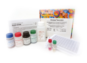
The test kit Vitrotest® Anti-HCV is an enzyme linked immunosorbent assay (ELISA) for the detection of total antibodies to Hepatitis C Virus (HCV) in human serum or plasma.
Determination of antibodies to HCV in the test kit Vitrotest® Anti-HCV is based on a solid phase indirect ELISA in a two-step incubation procedure.
○ TK022 - 96 tests
○ TK060 - 192 tests
○ TK128 - 480 tests- Solid phase: breakable microplate ELISA is coated with recombinant antigens (core, NS3, NS4 and NS5).
- Conjugate: monoclonal antibodies to human IgG and IgM conjugated to horseradish peroxidase.
- Chromogen: ready to use TMB solution.
- Volume of sample for analysis: 40 μl.
- Assay time: 2 hours.
According to the World Health Organization, approximately 150 million people are chronically infected with hepatitis C, and each year more than 350 thousand people die of hepatitis C-related liver disease. The disease may be either acute or chronic, and it often occurs without symptoms. However, chronic infection leads to liver cirrhosis and hepatocellular carcinoma development.
The causative agent of the disease is Hepatitis C Virus (HCV), a small enveloped single-stranded RNA virus (50 nm in diameter), belong to the Flaviviridae family. HCV genome has sequences encoding structural and nonstructural proteins. The structural antigens are nucleocapsid protein (core) and two envelope proteins (E1 and E2). Nonstructural proteins are complex proteins with enzymatic activity (NS2, NS3, NS4a, NS4b, NS5a and NS5b). In response to virus infection specific antibodies to all viral proteins are produced in human bodies.
Incubation period of hepatitis C is 14-180 (average 45) days. After this period, symptoms could arise. They include fever, fatigue, loss of appetite, nausea, vomiting, abdominal pain, joint pain, jaundice. Yet, most (70-80%) people infected with HCV are asymptomatic. Roughly 20% of infected patients clear the virus spontaneously; the rest develop chronic infection. HCV is a leading cause of chronic hepatitis, which progresses into cirrhosis in 5-20% of cases over a period of 20-30 years.
HCV is spread mainly through blood-to-blood contact. Therefore, in developed countries virus infects primarily persons who have injected illicit drugs and recipients of blood transfusions before introduction of regular blood screening for HCV. In developing countries many HCV infections occur in the health-care institutions as a result of unhygienic injections and various surgical manipulations such as tattooing or circumcision. Of other routes of transmission, the most important are sexual and vertical, from mother to fetus. Sexual transmission is regarded as a minor risk factor. Virus transmission from HCV-infected mother to unborn child is possible, with rates of transmission of around several percent.
HCV is divided into six major genotypes that can be further divided into several subtypes from A to L. The amino acid sequences of the major HCV genotypes differ approximately 30% from each other. The genotypes 1, 2 and 3 are found throughout the world whereas the distribution of the other genotypes is much more restricted. The immunity after cleared infection does not result in reliable protection against reinfections.
The overall worldwide prevalence of HCV is approximately 3%. The highest HCV prevalence figures (up to 10–20%) are found in Egypt. The prevalence of HCV infection varies remarkably and, for instance, in different European countries it ranges from 0,1% to 4%.
Adaptive immune responses are typically delayed during acute HCV infection. HCV RNA can be detected 1–3 weeks following infection, but neither HCV-specific T-cells nor HCV-specific antibodies are observed until 1–2 months after infection. The titre of IgG antibodies during the acute phase is relatively low in comparison with other virus infections in the majority of patients, gradually increasing during transformation to chronicity. In patients with resolved infection the titers of IgG after cure are low and often not detectable.
The IgM response in acute HCV infection also does not follow the classical pattern when IgM antibodies precede IgG response. Firstly, it was shown that HCV-specific IgM is more readily detected in chronically than in acutely infected patients (80% and 50% respectively); besides, the IgM titers under chronic infections are higher. Secondly, HCV-specific IgM and IgG are both almost simultaneously detected in acute infection. In individuals recovered from the infection no anti-HCV IgM antibodies are detectable.
A number of diagnostically relevant antigenic epitopes have been found within the C region, E2, NS3, NS4A/B and NS5 proteins, while E1, NS2 and NS5B are less immunogenic. In one study on chronic HCV patients, the following data on prevalence of antibodies were obtained: E2 - 98%, core-97%, NS3-88%, NS5A-68%, and NS4-48%. These data were similar to those observed by other investigators. Antibody titers were highest for core protein while titers for other proteins were considerably lower.
Antibody response against different HCV proteins is temporarily regulated. After infection, relatively early in the acute phase anticore antibodies are produced whereas significant levels of anti-E2 and anti-NS antibodies are detected only during the chronic phase. In recovering patients, anti-core antibodies persist longer than anti-NS antibodies, which often disappear. -
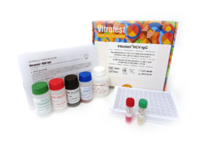
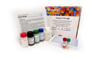
The test kit Vitrotest® HCV-IgG is an enzyme linked immunosorbent assay (ELISA) for the detection of IgG class antibodies to Hepatitis C Virus (HCV) in human serum or plasma.
Determination of IgG class antibodies to HCV in the test kit Vitrotest® HCV-IgG is based on a solid phase indirect ELISA in a two-step incubation procedure.
○ ТК055 - 96 tests
○ TK144 - 192 tests- Solid phase: breakable microplate ELISA is coated with recombinant antigens (core, NS3, NS4 and NS5).
- Conjugate: monoclonal antibodies to human IgG conjugated to horseradish peroxidase.
- Chromogen: ready to use TMB solution.
- Volume of sample for analysis: 40 μl.
- Assay time: 2 hours.
According to the World Health Organization, approximately 150 million people are chronically infected with hepatitis C, and each year more than 350 thousand people die of hepatitis C-related liver disease. The disease may be either acute or chronic, and it often occurs without symptoms. However, chronic infection leads to liver cirrhosis and hepatocellular carcinoma development.
The causative agent of the disease is Hepatitis C Virus (HCV), a small enveloped single-stranded RNA virus (50 nm in diameter), belong to the Flaviviridae family. HCV genome has sequences encoding structural and nonstructural proteins. The structural antigens are nucleocapsid protein (core) and two envelope proteins (E1 and E2). Nonstructural proteins are complex proteins with enzymatic activity (NS2, NS3, NS4a, NS4b, NS5a and NS5b). In response to virus infection specific antibodies to all viral proteins are produced in human bodies.
Incubation period of hepatitis C is 14-180 (average 45) days. After this period, symptoms could arise. They include fever, fatigue, loss of appetite, nausea, vomiting, abdominal pain, joint pain, jaundice. Yet, most (70-80%) people infected with HCV are asymptomatic. Roughly 20% of infected patients clear the virus spontaneously; the rest develop chronic infection. HCV is a leading cause of chronic hepatitis, which progresses into cirrhosis in 5-20% of cases over a period of 20-30 years.
HCV is spread mainly through blood-to-blood contact. Therefore, in developed countries virus infects primarily persons who have injected illicit drugs and recipients of blood transfusions before introduction of regular blood screening for HCV. In developing countries many HCV infections occur in the health-care institutions as a result of unhygienic injections and various surgical manipulations such as tattooing or circumcision. Of other routes of transmission, the most important are sexual and vertical, from mother to fetus. Sexual transmission is regarded as a minor risk factor. Virus transmission from HCV-infected mother to unborn child is possible, with rates of transmission of around several percent.
HCV is divided into six major genotypes that can be further divided into several subtypes from A to L. The amino acid sequences of the major HCV genotypes differ approximately 30% from each other. The genotypes 1, 2 and 3 are found throughout the world whereas the distribution of the other genotypes is much more restricted. The immunity after cleared infection does not result in reliable protection against reinfections.
The overall worldwide prevalence of HCV is approximately 3%. The highest HCV prevalence figures (up to 10–20%) are found in Egypt. The prevalence of HCV infection varies remarkably and, for instance, in different European countries it ranges from 0,1% to 4%.
Adaptive immune responses are typically delayed during acute HCV infection. HCV RNA can be detected 1–3 weeks following infection, but neither HCV-specific T-cells nor HCV-specific antibodies are observed until 1–2 months after infection. The titre of IgG antibodies during the acute phase is relatively low in comparison with other virus infections in the majority of patients, gradually increasing during transformation to chronicity. In patients with resolved infection the titers of IgG after cure are low and often not detectable.
The IgM response in acute HCV infection also does not follow the classical pattern when IgM antibodies precede IgG response. Firstly, it was shown that HCV-specific IgM is more readily detected in chronically than in acutely infected patients (80% and 50% respectively); besides, the IgM titers under chronic infections are higher. Secondly, HCV-specific IgM and IgG are both almost simultaneously detected in acute infection. In individuals recovered from the infection no anti-HCV IgM antibodies are detectable.
A number of diagnostically relevant antigenic epitopes have been found within the C region, E2, NS3, NS4A/B and NS5 proteins, while E1, NS2 and NS5B are less immunogenic. In one study on chronic HCV patients, the following data on prevalence of antibodies were obtained: E2 - 98%, core-97%, NS3-88%, NS5A-68%, and NS4-48%. These data were similar to those observed by other investigators. Antibody titers were highest for core protein while titers for other proteins were considerably lower.
Antibody response against different HCV proteins is temporarily regulated. After infection, relatively early in the acute phase anticore antibodies are produced whereas significant levels of anti-E2 and anti-NS antibodies are detected only during the chronic phase. In recovering patients, anti-core antibodies persist longer than anti-NS antibodies, which often disappear. -
The test kit Vitrotest® HCV-IgМ is an enzyme linked immunosorbent assay (ELISA) for the detection of IgМ class antibodies to Hepatitis C Virus (HCV) in human serum or plasma.
Determination of IgМ class antibodies to HCV in the test kit Vitrotest® HCV-IgМ is based on a solid phase indirect ELISA in a two-step incubation procedure.
○ ТК043 - 96 tests
○ TK145 - 192 tests- Solid phase: breakable microplate ELISA is coated with recombinant antigens (core, NS3 and NS4).
- Conjugate: monoclonal antibodies to human IgМ conjugated to horseradish peroxidase.
- Chromogen: ready to use TMB solution.
- Volume of sample for analysis: 40 μl.
- Assay time: 1 hour 30 minutes.
According to the World Health Organization, approximately 150 million people are chronically infected with hepatitis C, and each year more than 350 thousand people die of hepatitis C-related liver disease. The disease may be either acute or chronic, and it often occurs without symptoms. However, chronic infection leads to liver cirrhosis and hepatocellular carcinoma development.
The causative agent of the disease is Hepatitis C Virus (HCV), a small enveloped single-stranded RNA virus (50 nm in diameter), belong to the Flaviviridae family. HCV genome has sequences encoding structural and nonstructural proteins. The structural antigens are nucleocapsid protein (core) and two envelope proteins (E1 and E2). Nonstructural proteins are complex proteins with enzymatic activity (NS2, NS3, NS4a, NS4b, NS5a and NS5b). In response to virus infection specific antibodies to all viral proteins are produced in human bodies.
Incubation period of hepatitis C is 14-180 (average 45) days. After this period, symptoms could arise. They include fever, fatigue, loss of appetite, nausea, vomiting, abdominal pain, joint pain, jaundice. Yet, most (70-80%) people infected with HCV are asymptomatic. Roughly 20% of infected patients clear the virus spontaneously; the rest develop chronic infection. HCV is a leading cause of chronic hepatitis, which progresses into cirrhosis in 5-20% of cases over a period of 20-30 years.
HCV is spread mainly through blood-to-blood contact. Therefore, in developed countries virus infects primarily persons who have injected illicit drugs and recipients of blood transfusions before introduction of regular blood screening for HCV. In developing countries many HCV infections occur in the health-care institutions as a result of unhygienic injections and various surgical manipulations such as tattooing or circumcision. Of other routes of transmission, the most important are sexual and vertical, from mother to fetus. Sexual transmission is regarded as a minor risk factor. Virus transmission from HCV-infected mother to unborn child is possible, with rates of transmission of around several percent.
HCV is divided into six major genotypes that can be further divided into several subtypes from A to L. The amino acid sequences of the major HCV genotypes differ approximately 30% from each other. The genotypes 1, 2 and 3 are found throughout the world whereas the distribution of the other genotypes is much more restricted. The immunity after cleared infection does not result in reliable protection against reinfections.
The overall worldwide prevalence of HCV is approximately 3%. The highest HCV prevalence figures (up to 10–20%) are found in Egypt. The prevalence of HCV infection varies remarkably and, for instance, in different European countries it ranges from 0,1% to 4%.
Adaptive immune responses are typically delayed during acute HCV infection. HCV RNA can be detected 1–3 weeks following infection, but neither HCV-specific T-cells nor HCV-specific antibodies are observed until 1–2 months after infection. The titre of IgG antibodies during the acute phase is relatively low in comparison with other virus infections in the majority of patients, gradually increasing during transformation to chronicity. In patients with resolved infection the titers of IgG after cure are low and often not detectable.
The IgM response in acute HCV infection also does not follow the classical pattern when IgM antibodies precede IgG response. Firstly, it was shown that HCV-specific IgM is more readily detected in chronically than in acutely infected patients (80% and 50% respectively); besides, the IgM titers under chronic infections are higher. Secondly, HCV-specific IgM and IgG are both almost simultaneously detected in acute infection. In individuals recovered from the infection no anti-HCV IgM antibodies are detectable.
A number of diagnostically relevant antigenic epitopes have been found within the C region, E2, NS3, NS4A/B and NS5 proteins, while E1, NS2 and NS5B are less immunogenic. In one study on chronic HCV patients, the following data on prevalence of antibodies were obtained: E2 - 98%, core-97%, NS3-88%, NS5A-68%, and NS4-48%. These data were similar to those observed by other investigators. Antibody titers were highest for core protein while titers for other proteins were considerably lower.
Antibody response against different HCV proteins is temporarily regulated. After infection, relatively early in the acute phase anticore antibodies are produced whereas significant levels of anti-E2 and anti-NS antibodies are detected only during the chronic phase. In recovering patients, anti-core antibodies persist longer than anti-NS antibodies, which often disappear.
-
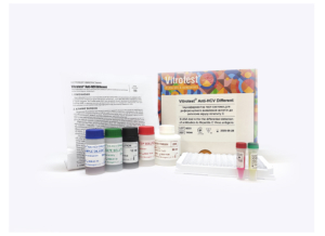
The test kit Vitrotest® Anti-HCV Different is an enzyme linked immunosorbent assay (ELISA) for the differential detection of antibodies to antigens of hepatitis C - core, NS3, NS4, NS5 in human serum or plasma.
The differential detection of antibodies to specific antigens of the hepatitis C virus in the test kit Vitrotest® Anti-HCV Different is based on a solid phase indirect ELISA in a two-step incubation procedure.
○ TK044 - 24 tests- Solid phase: breakable microplate ELISA is coated with recombinant HCV antigens in separate wells: core (strips 1, 5, and 9, marked in black), NS3 (strips 2, 6, and 10, marked in green), NS4 (strips 3, 7, and 11, marked in blue), and NS5 (strips 4, 8, and 12, marked in red).
- Conjugate: monoclonal antibodies to human IgG and IgM conjugated to horseradish peroxidase.
- Chromogen: ready to use TMB solution.
- Volume of sample for analysis: 40 μl.
- Assay time: 2 hours.
According to the World Health Organization, approximately 150 million people are chronically infected with hepatitis C, and each year more than 350 thousand people die of hepatitis C-related liver disease. The disease may be either acute or chronic, and it often occurs without symptoms. However, chronic infection leads to liver cirrhosis and hepatocellular carcinoma development.
The causative agent of the disease is Hepatitis C Virus (HCV), a small enveloped single-stranded RNA virus (50 nm in diameter), belong to the Flaviviridae family. HCV genome has sequences encoding structural and nonstructural proteins. The structural antigens are nucleocapsid protein (core) and two envelope proteins (E1 and E2). Nonstructural proteins are complex proteins with enzymatic activity (NS2, NS3, NS4a, NS4b, NS5a and NS5b). In response to virus infection specific antibodies to all viral proteins are produced in human bodies.
Incubation period of hepatitis C is 14-180 (average 45) days. After this period, symptoms could arise. They include fever, fatigue, loss of appetite, nausea, vomiting, abdominal pain, joint pain, jaundice. Yet, most (70-80%) people infected with HCV are asymptomatic. Roughly 20% of infected patients clear the virus spontaneously; the rest develop chronic infection. HCV is a leading cause of chronic hepatitis, which progresses into cirrhosis in 5-20% of cases over a period of 20-30 years.
HCV is spread mainly through blood-to-blood contact. Therefore, in developed countries virus infects primarily persons who have injected illicit drugs and recipients of blood transfusions before introduction of regular blood screening for HCV. In developing countries many HCV infections occur in the health-care institutions as a result of unhygienic injections and various surgical manipulations such as tattooing or circumcision. Of other routes of transmission, the most important are sexual and vertical, from mother to fetus. Sexual transmission is regarded as a minor risk factor. Virus transmission from HCV-infected mother to unborn child is possible, with rates of transmission of around several percent.
HCV is divided into six major genotypes that can be further divided into several subtypes from A to L. The amino acid sequences of the major HCV genotypes differ approximately 30% from each other. The genotypes 1, 2 and 3 are found throughout the world whereas the distribution of the other genotypes is much more restricted. The immunity after cleared infection does not result in reliable protection against reinfections.
The overall worldwide prevalence of HCV is approximately 3%. The highest HCV prevalence figures (up to 10–20%) are found in Egypt. The prevalence of HCV infection varies remarkably and, for instance, in different European countries it ranges from 0,1% to 4%.
Adaptive immune responses are typically delayed during acute HCV infection. HCV RNA can be detected 1–3 weeks following infection, but neither HCV-specific T-cells nor HCV-specific antibodies are observed until 1–2 months after infection. The titre of IgG antibodies during the acute phase is relatively low in comparison with other virus infections in the majority of patients, gradually increasing during transformation to chronicity. In patients with resolved infection the titers of IgG after cure are low and often not detectable.
The IgM response in acute HCV infection also does not follow the classical pattern when IgM antibodies precede IgG response. Firstly, it was shown that HCV-specific IgM is more readily detected in chronically than in acutely infected patients (80% and 50% respectively); besides, the IgM titers under chronic infections are higher. Secondly, HCV-specific IgM and IgG are both almost simultaneously detected in acute infection. In individuals recovered from the infection no anti-HCV IgM antibodies are detectable.
A number of diagnostically relevant antigenic epitopes have been found within the C region, E2, NS3, NS4A/B and NS5 proteins, while E1, NS2 and NS5B are less immunogenic. In one study on chronic HCV patients, the following data on prevalence of antibodies were obtained: E2 - 98%, core-97%, NS3-88%, NS5A-68%, and NS4-48%. These data were similar to those observed by other investigators. Antibody titers were highest for core protein while titers for other proteins were considerably lower.
Antibody response against different HCV proteins is temporarily regulated. After infection, relatively early in the acute phase anticore antibodies are produced whereas significant levels of anti-E2 and anti-NS antibodies are detected only during the chronic phase. In recovering patients, anti-core antibodies persist longer than anti-NS antibodies, which often disappear. -
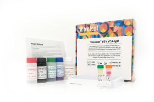
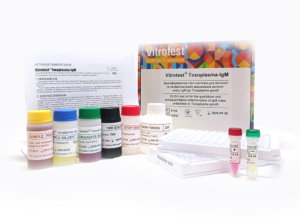
The test kit Vitrotest® Anti-Treponema is an enzyme linked immunosorbent assay (ELISA) for the detection of IgG and IgM class antibodies to Treponema pallidum in human serum or plasma.
Detection of antibodies to T. pallidum in the test kit Vitrotest® Anti-Treponema is based on a solid phase, indirect ELISA in a two-step incubation procedure.
○ ТК063 - 192 tests- Solid phase: breakable microplate ELISA is coated with recombinant Treponema pallidum antigens.
- Conjugate: monoclonal antibodies to human IgG and IgM conjugated to horseradish peroxidase.
- Chromogen: ready to use TMB solution.
- Volume of sample for analysis: 20 μl.
- Assay time: 1h 15 min.
Syphilis is a sexually transmitted infection (STI) caused by the bacterium Treponema pallidum. Syphilis develops in four successive stages, primary, secondary, latent, and tertiary. The primary ulcer at entry site ("primary chancre", 10-90 days postinfection), is followed by spread of the treponemes to the regional lymph nodes and haematogenous dissemination to other parts of the body. The ulcer is classically indurated and painless. While the local immunity leads to ulcer healing in approximately 3-6 weeks, systemic dissemination results in intense immune response to the deposited treponemes, leading to secondary syphilis soon after healing of primary chancre. Rash is the presenting complaint in majority of patients with secondary syphilis, and it is found on physical examination in more than 90% of patients. Rash frequently covers palms and soles, it is usually maculopapular and never vesicular. Other symptoms include fever, sore throat, malaise, headache, and lymphadenopathy. Neurological symptoms (meningitis, ocular complaints), although more characteristic for tertiary syphilis, could arise already at secondary stage. Secondary syphilis resolves without treatment, followed by latent stage. Latent stage is asymptomatic, the bacteria remain dormant and are undetected by traditional methods in blood and issues. Tertiary syphilis represents an activation of dormant infection, occuring in about one-third of affected individuals several decades after primary infection. Tertiary syphilis can affect multiple organ systems, including brain, nerves, eyes, heart, blood vessels, liver, bones, and joints, and is often fatal. Symptoms of tertiary syphilis vary depending on the organ system affected.
The fundamental histological changes at all stages are vasculitis and its consequences, necrosis and fibrosis.
Syphilis could be transmitted:
1) by direct contact of skin/mucosa with infective treponema-rich ulcers;
2) vertically from infected mother to her unborn child;
3) by blood sharing.
The main routes of transmission are sexual and congenital. Sexual transmission occurs by inoculation of bacteria into tiny abrasions resulting from sexual trauma. Minor routes of transmission are by blood transfusion, by needle sharing, and by fomites among medical personnel .
Persons with syphilis are infective for sexual transmission only during primary and secondary stage. On the contrary, women with syphilis could transmit the disease congenitally also during the latent stage.
T. pallidum gains access to the fetal compartment as early as 9–10 weeks after conception. Pregnancy in women with syphilis could result in spontaneous abortion, newborn death, and congenital syphilis. For women with syphilis, the probability of delivering a healthy infant is only 1/3. -
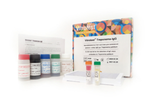
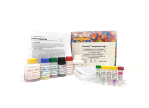
The test kit Vitrotest® Treponema-IgG is an enzyme linked immunosorbent assay (ELISA) for the detection of IgG class antibodies to Treponema pallidum in human serum or plasma.
Detection of IgG antibodies to T. pallidum in the test kit Vitrotest® Treponema-IgG is based on a solid phase, indirect ELISA in a two-step incubation procedure.
○ ТК056 - 96 tests- Solid phase: breakable microplate ELISA is coated with recombinant Treponema pallidum antigens.
- Conjugate: monoclonal antibodies to human IgG conjugated to horseradish peroxidase.
- Chromogen: ready to use TMB solution.
- Volume of sample for analysis: 20 μl.
- Assay time: 1h 15 min.
Syphilis is a sexually transmitted infection (STI) caused by the bacterium Treponema pallidum. Syphilis develops in four successive stages, primary, secondary, latent, and tertiary. The primary ulcer at entry site ("primary chancre", 10-90 days postinfection), is followed by spread of the treponemes to the regional lymph nodes and haematogenous dissemination to other parts of the body. The ulcer is classically indurated and painless. While the local immunity leads to ulcer healing in approximately 3-6 weeks, systemic dissemination results in intense immune response to the deposited treponemes, leading to secondary syphilis soon after healing of primary chancre. Rash is the presenting complaint in majority of patients with secondary syphilis, and it is found on physical examination in more than 90% of patients. Rash frequently covers palms and soles, it is usually maculopapular and never vesicular. Other symptoms include fever, sore throat, malaise, headache, and lymphadenopathy. Neurological symptoms (meningitis, ocular complaints), although more characteristic for tertiary syphilis, could arise already at secondary stage. Secondary syphilis resolves without treatment, followed by latent stage. Latent stage is asymptomatic, the bacteria remain dormant and are undetected by traditional methods in blood and issues. Tertiary syphilis represents an activation of dormant infection, occuring in about one-third of affected individuals several decades after primary infection. Tertiary syphilis can affect multiple organ systems, including brain, nerves, eyes, heart, blood vessels, liver, bones, and joints, and is often fatal. Symptoms of tertiary syphilis vary depending on the organ system affected.
The fundamental histological changes at all stages are vasculitis and its consequences, necrosis and fibrosis.
Syphilis could be transmitted:
1) by direct contact of skin/mucosa with infective treponema-rich ulcers;
2) vertically from infected mother to her unborn child;
3) by blood sharing.
The main routes of transmission are sexual and congenital. Sexual transmission occurs by inoculation of bacteria into tiny abrasions resulting from sexual trauma. Minor routes of transmission are by blood transfusion, by needle sharing, and by fomites among medical personnel .
Persons with syphilis are infective for sexual transmission only during primary and secondary stage. On the contrary, women with syphilis could transmit the disease congenitally also during the latent stage.
T. pallidum gains access to the fetal compartment as early as 9–10 weeks after conception. Pregnancy in women with syphilis could result in spontaneous abortion, newborn death, and congenital syphilis. For women with syphilis, the probability of delivering a healthy infant is only 1/3. -
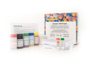
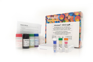
The test kit Vitrotest® HSV1/2-IgG is an enzyme linked immunosorbent assay (ELISA) for the qualitative and semiquantitative determination of IgG class antibodies to Herpes Simplex Virus types 1 and 2 (HSV1/2) in human serum or plasma.
Determination of IgG antibodies to HSV1/2 in the test kit Vitrotest® HSV1/2-IgG is based on a solid phase, indirect ELISA in a two-step incubation procedure.
○ ТК007 - 96 tests- Solid phase: breakable microplate ELISA is coated with mixture of purified antigens of herpes simplex virus type 1 and type 2.
- Conjugate: a monoclonal antibodies to human IgG conjugated to horseradish peroxidase.
- Chromogen: ready to use TMB solution.
- Volume of sample for analysis: 10 μl.
- Assay time: 1h 15 min.
Herpes simplex virus (HSV) is an etiological agent of 2 widespread diseases in humans: orofacial infection, the so called “cold sores”, and genital herpes (GH).
There are two closely related species of HSV, HSV-1 and HSV-2. Both are large enveloped viruses containing double-stranded DNA. HSV-1 is normally (but not always) associated with orofacial infections whereas HSV-2 causes mainly GH (though it could also cause orofacial infections, clinically indistinguishable from those caused by HSV-1). Both viruses could be transmitted from infected mothers to neonates.
HSV infects its host at mucosal or epithelial surfaces and causes primary infection of the epithelium. Then the virus enters sensory neurons and is transported by retrograde flow along axons that connect the point of entry into the body to nuclei of sensory neurons. Viral multiplication occurs in a small number of sensory neurons; the viral genome then remains in a latent state for the life of the host.
HSV frequently reactivates from latency and infects epithelial cells adjacent to neuron where it resides. This process results in lesions with viral shedding or with asymptomatic shedding alone. Reactivation is triggered by another infection, psychological stress, tissue damage, etc.
HSV causes benign vesicular and ulcerative lesions in immunocompetent adults and may cause severe systemic disease in neonates and immunosuppressed hosts. In children >5 years old and immunocompetent adults duration of the first attack is 2-4 weeks. Symptoms of recurrent outbreaks are typically shorter in duration (7-10 days) and even less severe than the first outbreak of genital herpes.
The course of disease is more severe in small children (the so-called neonatal herpes (NH) and immunocompromised persons. Complications of herpes in these patients could remain localized and affect only eyes, mouth and skin; infection could spread to the central nervous system causing meningitis, myelitis, and encephalitis; in the last, disseminated, stage, infection involves multiple organs.
To initiate infection, HSV must contact either mucosal surfaces or abraded skin. Transmission usually occurs by direct oral-oral, oral-genital or genital-genital contact. Orofacial herpes is transmitted mainly by household contact during childhood whereas GH is a disease mainly transmitted by sex. Neonatal HSV infection most commonly results from contact between the newborn and HSV that is present in the birth canal of the mother during delivery.
HSV can be transmitted to child under primary or recurrent herpes, both in the presence or absence of symptoms. However, under recurrent herpes, the risk of transmission to a newborn is low even if open sores are present at the time of birth (the risk is 3/100). On the contrary, the risk is considerably higher if the primary infection is acquired in the third trimester, and especially in the last 6 weeks of pregnancy (the risk increases to around 50%). As a result, seronegative women in the third trimester of pregnancy are the main group at risk, especially, if their partner is seropositive. Intrauterine infection, contrary to neonatal one, is a relatively rare event.
Infection with HHV-6 usually occurs during the first or second year of life, and accordingly, about 95% of adults have antibodies to HHV-6. At birth, most children are seropositive due to maternal antibodies, the titer of which decreases by 5 months. However, by the end of the first year of life, the percentage of seropositive infants is similar to that among older children and adults. -
The test kit Vitrotest® HSV1/2-IgM is an enzyme linked immunosorbent assay (ELISA) for the qualitative determination of IgM class antibodies to Herpes Simplex Virus types 1 and 2 (HSV1/2) in human serum or plasma.
Determination of IgM antibodies to HSV1 and HSV2 in the test kit Vitrotest® HSV1/2-IgM is based on a solid phase, «IgM-capture» ELISA in a two-step incubation procedure.
○ ТК008 - 96 tests- Solid phase: breakable microplate ELISA is coated with monoclonal anti-IgM antibodies.
- Conjugate: mixture of recombinant antigens of herpes simplex virus type 1 and type 2 gG1 and gG2 conjugated to horseradish peroxidase.
- Chromogen: ready to use TMB solution.
- Volume of sample for analysis: 10 μl.
- Assay time: 2 hours.
Herpes simplex virus (HSV) is an etiological agent of 2 widespread diseases in humans: orofacial infection, the so called “cold sores”, and genital herpes (GH).
There are two closely related species of HSV, HSV-1 and HSV-2. Both are large enveloped viruses containing double-stranded DNA. HSV-1 is normally (but not always) associated with orofacial infections whereas HSV-2 causes mainly GH (though it could also cause orofacial infections, clinically indistinguishable from those caused by HSV-1). Both viruses could be transmitted from infected mothers to neonates.
HSV infects its host at mucosal or epithelial surfaces and causes primary infection of the epithelium. Then the virus enters sensory neurons and is transported by retrograde flow along axons that connect the point of entry into the body to nuclei of sensory neurons. Viral multiplication occurs in a small number of sensory neurons; the viral genome then remains in a latent state for the life of the host.
HSV frequently reactivates from latency and infects epithelial cells adjacent to neuron where it resides. This process results in lesions with viral shedding or with asymptomatic shedding alone. Reactivation is triggered by another infection, psychological stress, tissue damage, etc.
HSV causes benign vesicular and ulcerative lesions in immunocompetent adults and may cause severe systemic disease in neonates and immunosuppressed hosts. In children >5 years old and immunocompetent adults duration of the first attack is 2-4 weeks. Symptoms of recurrent outbreaks are typically shorter in duration (7-10 days) and even less severe than the first outbreak of genital herpes.
The course of disease is more severe in small children (the so-called neonatal herpes (NH) and immunocompromised persons. Complications of herpes in these patients could remain localized and affect only eyes, mouth and skin; infection could spread to the central nervous system causing meningitis, myelitis, and encephalitis; in the last, disseminated, stage, infection involves multiple organs.
To initiate infection, HSV must contact either mucosal surfaces or abraded skin. Transmission usually occurs by direct oral-oral, oral-genital or genital-genital contact. Orofacial herpes is transmitted mainly by household contact during childhood whereas GH is a disease mainly transmitted by sex. Neonatal HSV infection most commonly results from contact between the newborn and HSV that is present in the birth canal of the mother during delivery.
HSV can be transmitted to child under primary or recurrent herpes, both in the presence or absence of symptoms. However, under recurrent herpes, the risk of transmission to a newborn is low even if open sores are present at the time of birth (the risk is 3/100). On the contrary, the risk is considerably higher if the primary infection is acquired in the third trimester, and especially in the last 6 weeks of pregnancy (the risk increases to around 50%). As a result, seronegative women in the third trimester of pregnancy are the main group at risk, especially, if their partner is seropositive. Intrauterine infection, contrary to neonatal one, is a relatively rare event.
Infection with HHV-6 usually occurs during the first or second year of life, and accordingly, about 95% of adults have antibodies to HHV-6. At birth, most children are seropositive due to maternal antibodies, the titer of which decreases by 5 months. However, by the end of the first year of life, the percentage of seropositive infants is similar to that among older children and adults. -
The test kit Vitrotest® HSV1-IgG is an enzyme linked immunosorbent assay (ELISA) for the qualitative and semiquantitative determination of IgG class antibodies to Herpes Simplex Virus type 1 (HSV-1) in human serum or plasma.
Determination of IgG antibodies to HSV-1 in the test kit Vitrotest® HSV1-IgG is based on a solid phase, indirect ELISA in a two-step incubation procedure.
○ ТК026 - 96 tests- Solid phase: breakable microplate ELISA is coated with recombinant antigens of herpes simplex virus type 1.
- Conjugate: a monoclonal antibodies to human IgG conjugated to horseradish peroxidase.
- Chromogen: ready to use TMB solution.
- Volume of sample for analysis: 10 μl.
- Assay time: 1h 15 min.
Herpes simplex virus (HSV) is an etiological agent of 2 widespread diseases in humans: orofacial infection, the so called “cold sores”, and genital herpes (GH).
There are two closely related species of HSV, HSV-1 and HSV-2. Both are large enveloped viruses containing double-stranded DNA. HSV-1 is normally (but not always) associated with orofacial infections whereas HSV-2 causes mainly GH (though it could also cause orofacial infections, clinically indistinguishable from those caused by HSV-1). Both viruses could be transmitted from infected mothers to neonates.
HSV infects its host at mucosal or epithelial surfaces and causes primary infection of the epithelium. Then the virus enters sensory neurons and is transported by retrograde flow along axons that connect the point of entry into the body to nuclei of sensory neurons. Viral multiplication occurs in a small number of sensory neurons; the viral genome then remains in a latent state for the life of the host.
HSV frequently reactivates from latency and infects epithelial cells adjacent to neuron where it resides. This process results in lesions with viral shedding or with asymptomatic shedding alone. Reactivation is triggered by another infection, psychological stress, tissue damage, etc.
HSV causes benign vesicular and ulcerative lesions in immunocompetent adults and may cause severe systemic disease in neonates and immunosuppressed hosts. In children >5 years old and immunocompetent adults duration of the first attack is 2-4 weeks. Symptoms of recurrent outbreaks are typically shorter in duration (7-10 days) and even less severe than the first outbreak of genital herpes.
The course of disease is more severe in small children (the so-called neonatal herpes (NH) and immunocompromised persons. Complications of herpes in these patients could remain localized and affect only eyes, mouth and skin; infection could spread to the central nervous system causing meningitis, myelitis, and encephalitis; in the last, disseminated, stage, infection involves multiple organs.
To initiate infection, HSV must contact either mucosal surfaces or abraded skin. Transmission usually occurs by direct oral-oral, oral-genital or genital-genital contact. Orofacial herpes is transmitted mainly by household contact during childhood whereas GH is a disease mainly transmitted by sex. Neonatal HSV infection most commonly results from contact between the newborn and HSV that is present in the birth canal of the mother during delivery.
HSV can be transmitted to child under primary or recurrent herpes, both in the presence or absence of symptoms. However, under recurrent herpes, the risk of transmission to a newborn is low even if open sores are present at the time of birth (the risk is 3/100). On the contrary, the risk is considerably higher if the primary infection is acquired in the third trimester, and especially in the last 6 weeks of pregnancy (the risk increases to around 50%). As a result, seronegative women in the third trimester of pregnancy are the main group at risk, especially, if their partner is seropositive. Intrauterine infection, contrary to neonatal one, is a relatively rare event.
Infection with HHV-6 usually occurs during the first or second year of life, and accordingly, about 95% of adults have antibodies to HHV-6. At birth, most children are seropositive due to maternal antibodies, the titer of which decreases by 5 months. However, by the end of the first year of life, the percentage of seropositive infants is similar to that among older children and adults. -
The test kit Vitrotest® HSV2-IgG is an enzyme linked immunosorbent assay (ELISA) for the qualitative and semiquantitative determination of IgG class antibodies to Herpes Simplex Virus type 2 (HSV-2) in human serum or plasma.
Determination of IgG antibodies to HSV-2 in the test kit Vitrotest® HSV2-IgG is based on a solid phase, indirect ELISA in a two-step incubation procedure.
○ ТК011 - 96 tests- Solid phase: breakable microplate ELISA is coated with recombinant antigens of herpes simplex virus type 2.
- Conjugate: a monoclonal antibodies to human IgG conjugated to horseradish peroxidase.
- Chromogen: ready to use TMB solution.
- Volume of sample for analysis: 10 μl.
- Assay time: 1h 15 min.
Herpes simplex virus (HSV) is an etiological agent of 2 widespread diseases in humans: orofacial infection, the so called “cold sores”, and genital herpes (GH).
There are two closely related species of HSV, HSV-1 and HSV-2. Both are large enveloped viruses containing double-stranded DNA. HSV-1 is normally (but not always) associated with orofacial infections whereas HSV-2 causes mainly GH (though it could also cause orofacial infections, clinically indistinguishable from those caused by HSV-1). Both viruses could be transmitted from infected mothers to neonates.
HSV infects its host at mucosal or epithelial surfaces and causes primary infection of the epithelium. Then the virus enters sensory neurons and is transported by retrograde flow along axons that connect the point of entry into the body to nuclei of sensory neurons. Viral multiplication occurs in a small number of sensory neurons; the viral genome then remains in a latent state for the life of the host.
HSV frequently reactivates from latency and infects epithelial cells adjacent to neuron where it resides. This process results in lesions with viral shedding or with asymptomatic shedding alone. Reactivation is triggered by another infection, psychological stress, tissue damage, etc.
HSV causes benign vesicular and ulcerative lesions in immunocompetent adults and may cause severe systemic disease in neonates and immunosuppressed hosts. In children >5 years old and immunocompetent adults duration of the first attack is 2-4 weeks. Symptoms of recurrent outbreaks are typically shorter in duration (7-10 days) and even less severe than the first outbreak of genital herpes.
The course of disease is more severe in small children (the so-called neonatal herpes (NH) and immunocompromised persons. Complications of herpes in these patients could remain localized and affect only eyes, mouth and skin; infection could spread to the central nervous system causing meningitis, myelitis, and encephalitis; in the last, disseminated, stage, infection involves multiple organs.
To initiate infection, HSV must contact either mucosal surfaces or abraded skin. Transmission usually occurs by direct oral-oral, oral-genital or genital-genital contact. Orofacial herpes is transmitted mainly by household contact during childhood whereas GH is a disease mainly transmitted by sex. Neonatal HSV infection most commonly results from contact between the newborn and HSV that is present in the birth canal of the mother during delivery.
HSV can be transmitted to child under primary or recurrent herpes, both in the presence or absence of symptoms. However, under recurrent herpes, the risk of transmission to a newborn is low even if open sores are present at the time of birth (the risk is 3/100). On the contrary, the risk is considerably higher if the primary infection is acquired in the third trimester, and especially in the last 6 weeks of pregnancy (the risk increases to around 50%). As a result, seronegative women in the third trimester of pregnancy are the main group at risk, especially, if their partner is seropositive. Intrauterine infection, contrary to neonatal one, is a relatively rare event.
Infection with HHV-6 usually occurs during the first or second year of life, and accordingly, about 95% of adults have antibodies to HHV-6. At birth, most children are seropositive due to maternal antibodies, the titer of which decreases by 5 months. However, by the end of the first year of life, the percentage of seropositive infants is similar to that among older children and adults. -
The test kit Vitrotest® HSV1-IgM is an enzyme linked immunosorbent assay (ELISA) for the qualitative determination of IgM class antibodies to Herpes Simplex Virus type 1 (HSV-1) in human serum or plasma.
Determination of IgM antibodies to HSV-1 in the test kit Vitrotest® HSV1-IgM is based on a solid phase, «IgM-capture» ELISA in a two-step incubation procedure.
○ ТК009 - 96 tests- Solid phase: breakable microplate ELISA is coated with monoclonal anti-IgM antibodies.
- Conjugate: recombinant HSV1 antigen gG1 conjugated to horseradish peroxidase.
- Chromogen: ready to use TMB solution.
- Volume of sample for analysis: 10 μl.
- Assay time: 2 hours.
Herpes simplex virus (HSV) is an etiological agent of 2 widespread diseases in humans: orofacial infection, the so called “cold sores”, and genital herpes (GH).
There are two closely related species of HSV, HSV-1 and HSV-2. Both are large enveloped viruses containing double-stranded DNA. HSV-1 is normally (but not always) associated with orofacial infections whereas HSV-2 causes mainly GH (though it could also cause orofacial infections, clinically indistinguishable from those caused by HSV-1). Both viruses could be transmitted from infected mothers to neonates.
HSV infects its host at mucosal or epithelial surfaces and causes primary infection of the epithelium. Then the virus enters sensory neurons and is transported by retrograde flow along axons that connect the point of entry into the body to nuclei of sensory neurons. Viral multiplication occurs in a small number of sensory neurons; the viral genome then remains in a latent state for the life of the host.
HSV frequently reactivates from latency and infects epithelial cells adjacent to neuron where it resides. This process results in lesions with viral shedding or with asymptomatic shedding alone. Reactivation is triggered by another infection, psychological stress, tissue damage, etc.
HSV causes benign vesicular and ulcerative lesions in immunocompetent adults and may cause severe systemic disease in neonates and immunosuppressed hosts. In children >5 years old and immunocompetent adults duration of the first attack is 2-4 weeks. Symptoms of recurrent outbreaks are typically shorter in duration (7-10 days) and even less severe than the first outbreak of genital herpes.
The course of disease is more severe in small children (the so-called neonatal herpes (NH) and immunocompromised persons. Complications of herpes in these patients could remain localized and affect only eyes, mouth and skin; infection could spread to the central nervous system causing meningitis, myelitis, and encephalitis; in the last, disseminated, stage, infection involves multiple organs.
To initiate infection, HSV must contact either mucosal surfaces or abraded skin. Transmission usually occurs by direct oral-oral, oral-genital or genital-genital contact. Orofacial herpes is transmitted mainly by household contact during childhood whereas GH is a disease mainly transmitted by sex. Neonatal HSV infection most commonly results from contact between the newborn and HSV that is present in the birth canal of the mother during delivery.
HSV can be transmitted to child under primary or recurrent herpes, both in the presence or absence of symptoms. However, under recurrent herpes, the risk of transmission to a newborn is low even if open sores are present at the time of birth (the risk is 3/100). On the contrary, the risk is considerably higher if the primary infection is acquired in the third trimester, and especially in the last 6 weeks of pregnancy (the risk increases to around 50%). As a result, seronegative women in the third trimester of pregnancy are the main group at risk, especially, if their partner is seropositive. Intrauterine infection, contrary to neonatal one, is a relatively rare event.
Infection with HHV-6 usually occurs during the first or second year of life, and accordingly, about 95% of adults have antibodies to HHV-6. At birth, most children are seropositive due to maternal antibodies, the titer of which decreases by 5 months. However, by the end of the first year of life, the percentage of seropositive infants is similar to that among older children and adults. -
The test kit Vitrotest® HSV2-IgM is an enzyme linked immunosorbent assay (ELISA) for the qualitative determination of IgM class antibodies to Herpes Simplex Virus type 2 (HSV-2) in human serum or plasma.
Determination of IgM antibodies to HSV-2 in the test kit Vitrotest® HSV2-IgM is based on a solid phase, «IgM-capture» ELISA in a two-step incubation procedure.
○ ТК010 - 96 tests- Solid phase: breakable microplate ELISA is coated with monoclonal anti-IgM antibodies.
- Conjugate: recombinant HSV2 antigen gG2 conjugated to horseradish peroxidase.
- Chromogen: ready to use TMB solution.
- Volume of sample for analysis: 10 μl.
- Assay time: 2 hours.
Herpes simplex virus (HSV) is an etiological agent of 2 widespread diseases in humans: orofacial infection, the so called “cold sores”, and genital herpes (GH).
There are two closely related species of HSV, HSV-1 and HSV-2. Both are large enveloped viruses containing double-stranded DNA. HSV-1 is normally (but not always) associated with orofacial infections whereas HSV-2 causes mainly GH (though it could also cause orofacial infections, clinically indistinguishable from those caused by HSV-1). Both viruses could be transmitted from infected mothers to neonates.
HSV infects its host at mucosal or epithelial surfaces and causes primary infection of the epithelium. Then the virus enters sensory neurons and is transported by retrograde flow along axons that connect the point of entry into the body to nuclei of sensory neurons. Viral multiplication occurs in a small number of sensory neurons; the viral genome then remains in a latent state for the life of the host.
HSV frequently reactivates from latency and infects epithelial cells adjacent to neuron where it resides. This process results in lesions with viral shedding or with asymptomatic shedding alone. Reactivation is triggered by another infection, psychological stress, tissue damage, etc.
HSV causes benign vesicular and ulcerative lesions in immunocompetent adults and may cause severe systemic disease in neonates and immunosuppressed hosts. In children >5 years old and immunocompetent adults duration of the first attack is 2-4 weeks. Symptoms of recurrent outbreaks are typically shorter in duration (7-10 days) and even less severe than the first outbreak of genital herpes.
The course of disease is more severe in small children (the so-called neonatal herpes (NH) and immunocompromised persons. Complications of herpes in these patients could remain localized and affect only eyes, mouth and skin; infection could spread to the central nervous system causing meningitis, myelitis, and encephalitis; in the last, disseminated, stage, infection involves multiple organs.
To initiate infection, HSV must contact either mucosal surfaces or abraded skin. Transmission usually occurs by direct oral-oral, oral-genital or genital-genital contact. Orofacial herpes is transmitted mainly by household contact during childhood whereas GH is a disease mainly transmitted by sex. Neonatal HSV infection most commonly results from contact between the newborn and HSV that is present in the birth canal of the mother during delivery.
HSV can be transmitted to child under primary or recurrent herpes, both in the presence or absence of symptoms. However, under recurrent herpes, the risk of transmission to a newborn is low even if open sores are present at the time of birth (the risk is 3/100). On the contrary, the risk is considerably higher if the primary infection is acquired in the third trimester, and especially in the last 6 weeks of pregnancy (the risk increases to around 50%). As a result, seronegative women in the third trimester of pregnancy are the main group at risk, especially, if their partner is seropositive. Intrauterine infection, contrary to neonatal one, is a relatively rare event.
Infection with HHV-6 usually occurs during the first or second year of life, and accordingly, about 95% of adults have antibodies to HHV-6. At birth, most children are seropositive due to maternal antibodies, the titer of which decreases by 5 months. However, by the end of the first year of life, the percentage of seropositive infants is similar to that among older children and adults. -
The test kit Vitrotest® HHV6-IgG is an enzyme linked immunosorbent assay (ELISA) for the qualitative and semiquantitative determination of IgG class antibodies to Human Herpes Virus type 6 (HHV-6) in human serum or plasma.
Determination of IgG antibodies to HHV6 in the test kit Vitrotest® HHV6-IgG is based on a solid phase, indirect ELISA in a two-step incubation procedure.
○ ТК090 - 96 tests- Solid phase: breakable microplate ELISA is coated with recombinant antigens of Human Herpes Virus type 6.
- Conjugate: a monoclonal antibodies to human IgG conjugated to horseradish peroxidase.
- Chromogen: ready to use TMB solution.
- Volume of sample for analysis: 10 μl.
- Assay time: 1h 15 min.
Herpes simplex virus (HSV) is an etiological agent of 2 widespread diseases in humans: orofacial infection, the so called “cold sores”, and genital herpes (GH).
There are two closely related species of HSV, HSV-1 and HSV-2. Both are large enveloped viruses containing double-stranded DNA. HSV-1 is normally (but not always) associated with orofacial infections whereas HSV-2 causes mainly GH (though it could also cause orofacial infections, clinically indistinguishable from those caused by HSV-1). Both viruses could be transmitted from infected mothers to neonates.
HSV infects its host at mucosal or epithelial surfaces and causes primary infection of the epithelium. Then the virus enters sensory neurons and is transported by retrograde flow along axons that connect the point of entry into the body to nuclei of sensory neurons. Viral multiplication occurs in a small number of sensory neurons; the viral genome then remains in a latent state for the life of the host.
HSV frequently reactivates from latency and infects epithelial cells adjacent to neuron where it resides. This process results in lesions with viral shedding or with asymptomatic shedding alone. Reactivation is triggered by another infection, psychological stress, tissue damage, etc.
HSV causes benign vesicular and ulcerative lesions in immunocompetent adults and may cause severe systemic disease in neonates and immunosuppressed hosts. In children >5 years old and immunocompetent adults duration of the first attack is 2-4 weeks. Symptoms of recurrent outbreaks are typically shorter in duration (7-10 days) and even less severe than the first outbreak of genital herpes.
The course of disease is more severe in small children (the so-called neonatal herpes (NH) and immunocompromised persons. Complications of herpes in these patients could remain localized and affect only eyes, mouth and skin; infection could spread to the central nervous system causing meningitis, myelitis, and encephalitis; in the last, disseminated, stage, infection involves multiple organs.
To initiate infection, HSV must contact either mucosal surfaces or abraded skin. Transmission usually occurs by direct oral-oral, oral-genital or genital-genital contact. Orofacial herpes is transmitted mainly by household contact during childhood whereas GH is a disease mainly transmitted by sex. Neonatal HSV infection most commonly results from contact between the newborn and HSV that is present in the birth canal of the mother during delivery.
HSV can be transmitted to child under primary or recurrent herpes, both in the presence or absence of symptoms. However, under recurrent herpes, the risk of transmission to a newborn is low even if open sores are present at the time of birth (the risk is 3/100). On the contrary, the risk is considerably higher if the primary infection is acquired in the third trimester, and especially in the last 6 weeks of pregnancy (the risk increases to around 50%). As a result, seronegative women in the third trimester of pregnancy are the main group at risk, especially, if their partner is seropositive. Intrauterine infection, contrary to neonatal one, is a relatively rare event.
Infection with HHV-6 usually occurs during the first or second year of life, and accordingly, about 95% of adults have antibodies to HHV-6. At birth, most children are seropositive due to maternal antibodies, the titer of which decreases by 5 months. However, by the end of the first year of life, the percentage of seropositive infants is similar to that among older children and adults. -
The test kit Vitrotest® EBNA-IgG is an enzyme linked immunosorbent assay (ELISA) for the qualitative and semiquantitative determination of IgG class antibodies to nuclear antigen (EBNA) of Epstein-Barr virus (EBV) in human serum or plasma.
Determination of IgG antibodies to EBV nuclear antigen (EBNA) in the test kit Vitrotest® EBNA-IgG is based on a solid phase, indirect ELISA in a two-step incubation procedure.
○ ТК054 - 96 tests- Solid phase: breakable microplate ELISA is coated with EBV recombinant nuclear antigen.
- Conjugate: a monoclonal antibodies to human IgG conjugated to horseradish peroxidase.
- Chromogen: ready to use TMB solution.
- Volume of sample for analysis: 10 μl.
- Assay time: 1h 15 min.
Epstein-Barr virus (EBV), also known as human herpesvirus 4, is the causative agent of diseases such as infectious mononucleosis (IM), Burkitt’s lymphoma (BL), nasopharyngeal carcinoma.
EBV acquired in early childhood usually does not cause symptoms. Infection in adolescence or early adulthood often results in clinical IM. Typically, the disease presents as pharyngitis, lymphadenopathy, and fever. In most cases, symptoms resolve within 2–4 weeks, but more than 90% of adults develop a latent B-lymphocyte infection. Approximately 1% of immunocompetent individuals may experience severe complications (hepatitis, myocarditis, splenic rupture, neurological complications). In immunosuppressed individuals, primary EBV infection leads to severe disorders (eg, BL).
The clinical symptoms range from mild fever to prolonged debilitating illness. Typically, the disease presents as pharyngitis, lymphadenopathy, and fever. Enlarged spleen and a swollen liver are less common symptoms. Microscopic blood observation reveals lymphocytosis; most lymphocytes have atypical morphology.
EBV spreads most commonly through bodily fluids, especially saliva, sporadically also through blood and semen. Humans are the most infectious during acute IM, but the virus could be shed into saliva even during latency. Transmission occurs mainly via household and sexual routes; however, transmission via blood transfusion and organ transplantation was also demonstrated.
Risk groups for IM are adolescents, senior school students, blood transfusion recipients, as well as people with immunodeficiencies (risk group for complications). Up to 50% of adolescents and young adults in developed countries are seronegative and hence at risk of developing IM.
EBV is a genetically stable virus. EBV is one of the most common human viruses. EBV is found all over the world; virus is able to infect more than 95% of all individuals within the first four decades of life. The current incidence of IM in USA is estimated at approximately 125 000 reported cases/year. EBV DNA is surrounded by a nucleocapsid; protein tegument is situated between the nucleocapsid and envelope; outer envelope has glycoprotein spikes typical of all herpesviruses. The viral genome encodes around 90 proteins. The most of these proteins (80) are expressed only during EBV lytic cycle.
On the basis of their time of expression, EBV genes are designated early or late. Early genes encode mostly non-structural proteins, the most important among them is viral DNA polymerase. Late genes encode mostly structural viral proteins which are required for successful packaging of the viral DNA and formation of the virion. The viral capsids have several polypeptide components ranging in size from 18 to 155 kDa, with a 155-kDa polypeptide being the major component. The membrane antigen (MA) complex is found on the virion envelope and consists of at least three major EBV glycoproteins: gp350/220 (the most abundant protein), gp110, and gp85.
The presence of EBV in epithelial and B cells provokes an intense immune response consisting of antibodies to a large variety of virally encoded antigens. During the acute phase of IM, the humoral response is directed toward viral antigens of the lytic cycle, notably membrane antigen complex, several early antigens (p 54 and p138), and VCA. A strong IgM response (the so-called heterophile antibodies) is directed towards a complex glycoprotein structure on the surface of EBV infected cells. As IM patients recover from clinical symptoms, the IgM response reduces significantly while the IgG response in serum plateaus at a reduced level and is maintained throughout the persistent infection.
The main purpose of the diagnostics of IM is to rule out less common but more serious causes of IM-like symptoms, requiring immediate intervention, such as hepatitis or various leukemic malignancies. The mainstay in the diagnostics of IM are still clinical symptoms (classic triad-fever, lymphoadenopathy, and pharyngytis) in combination with anamnestic aspects.
The specificity of the “classic triad” is around 90%, since IM-like symptoms may be caused by a variety of other pathogens and malignancies, such as cytomegalovirus, human herpesvirus 6, adenovirus, rubella virus, mumps virus, human immunodeficiency virus, hepatitis A virus, influenza A and B viruses, and Toxoplasma gondii, lymphoma and leukemia. Therefore, EBV infection should be confirmed with a blood antibody test.
It is well known for many years that acute IM is characterized by the appearance of heterophile antibodies (i.e. antibodies to EBV antigen described above) in the serum. This peculiarity is used in the so-called “monospot tests”, detecting agglutination of sheep erythrocytes in the presence of serum of patients with acute-phase IM. This test is now not recommended for general use by the Centers for Disease Prevention and Control, USA, since it produces both false positive and false negative results.
Only three serological parameters are essential for the detection of EBV-specific antibodies in immunocompetent individuals: VCA IgG (marker of disease, present or past), VCA IgM (marker of recent or present disease), and EBNA-1 IgG (marker of past disease).
The most widely used serological methods are immunofluorescence assay (IFA) and enzyme immunoassay (EIA). IFA has always been used as a reference method. However, some investigators found EIA to be more sensitive than IFA.
On the contrary, in immunosuppressed individuals serological assays are not recommended for many reasons, such as dysfunctions in the production of antibodies. To date, only detection of viral load by PCR is an established marker for immunosuppressed patients. -
The test kit Vitrotest® EBV VCA-IgG is an enzyme linked immunosorbent assay (ELISA) for the qualitative and semiquantitative determination of IgG class antibodies to viral capsid antigen (VCA) of Epstein-Barr virus (EBV) in human serum or plasma.
Determination of IgG antibodies to EBV capsid antigen (VCA) in the test kit Vitrotest® EBV VCA-IgG is based on a solid phase, indirect ELISA in a two-step incubation procedure.
○ ТК053 - 96 tests- Solid phase: breakable microplate ELISA is coated with EBV recombinant capsid antigen.
- Conjugate: a monoclonal antibodies to human IgG conjugated to horseradish peroxidase.
- Chromogen: ready to use TMB solution.
- Volume of sample for analysis: 10 μl.
- Assay time: 1h 15 min.
Epstein-Barr virus (EBV), also known as human herpesvirus 4, is the causative agent of diseases such as infectious mononucleosis (IM), Burkitt’s lymphoma (BL), nasopharyngeal carcinoma.
EBV acquired in early childhood usually does not cause symptoms. Infection in adolescence or early adulthood often results in clinical IM. Typically, the disease presents as pharyngitis, lymphadenopathy, and fever. In most cases, symptoms resolve within 2–4 weeks, but more than 90% of adults develop a latent B-lymphocyte infection. Approximately 1% of immunocompetent individuals may experience severe complications (hepatitis, myocarditis, splenic rupture, neurological complications). In immunosuppressed individuals, primary EBV infection leads to severe disorders (eg, BL).
The clinical symptoms range from mild fever to prolonged debilitating illness. Typically, the disease presents as pharyngitis, lymphadenopathy, and fever. Enlarged spleen and a swollen liver are less common symptoms. Microscopic blood observation reveals lymphocytosis; most lymphocytes have atypical morphology.
EBV spreads most commonly through bodily fluids, especially saliva, sporadically also through blood and semen. Humans are the most infectious during acute IM, but the virus could be shed into saliva even during latency. Transmission occurs mainly via household and sexual routes; however, transmission via blood transfusion and organ transplantation was also demonstrated.
Risk groups for IM are adolescents, senior school students, blood transfusion recipients, as well as people with immunodeficiencies (risk group for complications). Up to 50% of adolescents and young adults in developed countries are seronegative and hence at risk of developing IM.
EBV is a genetically stable virus. EBV is one of the most common human viruses. EBV is found all over the world; virus is able to infect more than 95% of all individuals within the first four decades of life. The current incidence of IM in USA is estimated at approximately 125 000 reported cases/year. EBV DNA is surrounded by a nucleocapsid; protein tegument is situated between the nucleocapsid and envelope; outer envelope has glycoprotein spikes typical of all herpesviruses. The viral genome encodes around 90 proteins. The most of these proteins (80) are expressed only during EBV lytic cycle.
On the basis of their time of expression, EBV genes are designated early or late. Early genes encode mostly non-structural proteins, the most important among them is viral DNA polymerase. Late genes encode mostly structural viral proteins which are required for successful packaging of the viral DNA and formation of the virion. The viral capsids have several polypeptide components ranging in size from 18 to 155 kDa, with a 155-kDa polypeptide being the major component. The membrane antigen (MA) complex is found on the virion envelope and consists of at least three major EBV glycoproteins: gp350/220 (the most abundant protein), gp110, and gp85.
The presence of EBV in epithelial and B cells provokes an intense immune response consisting of antibodies to a large variety of virally encoded antigens. During the acute phase of IM, the humoral response is directed toward viral antigens of the lytic cycle, notably membrane antigen complex, several early antigens (p 54 and p138), and VCA. A strong IgM response (the so-called heterophile antibodies) is directed towards a complex glycoprotein structure on the surface of EBV infected cells. As IM patients recover from clinical symptoms, the IgM response reduces significantly while the IgG response in serum plateaus at a reduced level and is maintained throughout the persistent infection.
The main purpose of the diagnostics of IM is to rule out less common but more serious causes of IM-like symptoms, requiring immediate intervention, such as hepatitis or various leukemic malignancies. The mainstay in the diagnostics of IM are still clinical symptoms (classic triad-fever, lymphoadenopathy, and pharyngytis) in combination with anamnestic aspects.
The specificity of the “classic triad” is around 90%, since IM-like symptoms may be caused by a variety of other pathogens and malignancies, such as cytomegalovirus, human herpesvirus 6, adenovirus, rubella virus, mumps virus, human immunodeficiency virus, hepatitis A virus, influenza A and B viruses, and Toxoplasma gondii, lymphoma and leukemia. Therefore, EBV infection should be confirmed with a blood antibody test.
It is well known for many years that acute IM is characterized by the appearance of heterophile antibodies (i.e. antibodies to EBV antigen described above) in the serum. This peculiarity is used in the so-called “monospot tests”, detecting agglutination of sheep erythrocytes in the presence of serum of patients with acute-phase IM. This test is now not recommended for general use by the Centers for Disease Prevention and Control, USA, since it produces both false positive and false negative results.
Only three serological parameters are essential for the detection of EBV-specific antibodies in immunocompetent individuals: VCA IgG (marker of disease, present or past), VCA IgM (marker of recent or present disease), and EBNA-1 IgG (marker of past disease).
The most widely used serological methods are immunofluorescence assay (IFA) and enzyme immunoassay (EIA). IFA has always been used as a reference method. However, some investigators found EIA to be more sensitive than IFA.
On the contrary, in immunosuppressed individuals serological assays are not recommended for many reasons, such as dysfunctions in the production of antibodies. To date, only detection of viral load by PCR is an established marker for immunosuppressed patients. -
The test kit Vitrotest® EBV VCA-IgА is an enzyme linked immunosorbent assay (ELISA) for the qualitative and semiquantitative determination of IgА class antibodies to viral capsid antigen (VCA) of Epstein-Barr virus (EBV) in human serum or plasma.
Determination of IgА antibodies to EBV capsid antigen (VCA) in the test kit Vitrotest® EBV VCA-IgА is based on a solid phase, indirect ELISA in a two-step incubation procedure.
○ ТК123 - 96 tests- Solid phase: breakable microplate ELISA is coated with EBV recombinant capsid antigen.
- Conjugate: a monoclonal antibodies to human IgА conjugated to horseradish peroxidase.
- Chromogen: ready to use TMB solution.
- Volume of sample for analysis: 10 μl.
- Assay time: 1h 15 min.
Epstein-Barr virus (EBV), also known as human herpesvirus 4, is the causative agent of diseases such as infectious mononucleosis (IM), Burkitt’s lymphoma (BL), nasopharyngeal carcinoma.
EBV acquired in early childhood usually does not cause symptoms. Infection in adolescence or early adulthood often results in clinical IM. Typically, the disease presents as pharyngitis, lymphadenopathy, and fever. In most cases, symptoms resolve within 2–4 weeks, but more than 90% of adults develop a latent B-lymphocyte infection. Approximately 1% of immunocompetent individuals may experience severe complications (hepatitis, myocarditis, splenic rupture, neurological complications). In immunosuppressed individuals, primary EBV infection leads to severe disorders (eg, BL).
The clinical symptoms range from mild fever to prolonged debilitating illness. Typically, the disease presents as pharyngitis, lymphadenopathy, and fever. Enlarged spleen and a swollen liver are less common symptoms. Microscopic blood observation reveals lymphocytosis; most lymphocytes have atypical morphology.
EBV spreads most commonly through bodily fluids, especially saliva, sporadically also through blood and semen. Humans are the most infectious during acute IM, but the virus could be shed into saliva even during latency. Transmission occurs mainly via household and sexual routes; however, transmission via blood transfusion and organ transplantation was also demonstrated.
Risk groups for IM are adolescents, senior school students, blood transfusion recipients, as well as people with immunodeficiencies (risk group for complications). Up to 50% of adolescents and young adults in developed countries are seronegative and hence at risk of developing IM.
EBV is a genetically stable virus. EBV is one of the most common human viruses. EBV is found all over the world; virus is able to infect more than 95% of all individuals within the first four decades of life. The current incidence of IM in USA is estimated at approximately 125 000 reported cases/year. EBV DNA is surrounded by a nucleocapsid; protein tegument is situated between the nucleocapsid and envelope; outer envelope has glycoprotein spikes typical of all herpesviruses. The viral genome encodes around 90 proteins. The most of these proteins (80) are expressed only during EBV lytic cycle.
On the basis of their time of expression, EBV genes are designated early or late. Early genes encode mostly non-structural proteins, the most important among them is viral DNA polymerase. Late genes encode mostly structural viral proteins which are required for successful packaging of the viral DNA and formation of the virion. The viral capsids have several polypeptide components ranging in size from 18 to 155 kDa, with a 155-kDa polypeptide being the major component. The membrane antigen (MA) complex is found on the virion envelope and consists of at least three major EBV glycoproteins: gp350/220 (the most abundant protein), gp110, and gp85.
The presence of EBV in epithelial and B cells provokes an intense immune response consisting of antibodies to a large variety of virally encoded antigens. During the acute phase of IM, the humoral response is directed toward viral antigens of the lytic cycle, notably membrane antigen complex, several early antigens (p 54 and p138), and VCA. A strong IgM response (the so-called heterophile antibodies) is directed towards a complex glycoprotein structure on the surface of EBV infected cells. As IM patients recover from clinical symptoms, the IgM response reduces significantly while the IgG response in serum plateaus at a reduced level and is maintained throughout the persistent infection.
The main purpose of the diagnostics of IM is to rule out less common but more serious causes of IM-like symptoms, requiring immediate intervention, such as hepatitis or various leukemic malignancies. The mainstay in the diagnostics of IM are still clinical symptoms (classic triad-fever, lymphoadenopathy, and pharyngytis) in combination with anamnestic aspects.
The specificity of the “classic triad” is around 90%, since IM-like symptoms may be caused by a variety of other pathogens and malignancies, such as cytomegalovirus, human herpesvirus 6, adenovirus, rubella virus, mumps virus, human immunodeficiency virus, hepatitis A virus, influenza A and B viruses, and Toxoplasma gondii, lymphoma and leukemia. Therefore, EBV infection should be confirmed with a blood antibody test.
It is well known for many years that acute IM is characterized by the appearance of heterophile antibodies (i.e. antibodies to EBV antigen described above) in the serum. This peculiarity is used in the so-called “monospot tests”, detecting agglutination of sheep erythrocytes in the presence of serum of patients with acute-phase IM. This test is now not recommended for general use by the Centers for Disease Prevention and Control, USA, since it produces both false positive and false negative results.
Only three serological parameters are essential for the detection of EBV-specific antibodies in immunocompetent individuals: VCA IgG (marker of disease, present or past), VCA IgM (marker of recent or present disease), and EBNA-1 IgG (marker of past disease).
The most widely used serological methods are immunofluorescence assay (IFA) and enzyme immunoassay (EIA). IFA has always been used as a reference method. However, some investigators found EIA to be more sensitive than IFA.
On the contrary, in immunosuppressed individuals serological assays are not recommended for many reasons, such as dysfunctions in the production of antibodies. To date, only detection of viral load by PCR is an established marker for immunosuppressed patients. -
The test kit Vitrotest® EBV VCA-IgM is an enzyme linked immunosorbent assay (ELISA) for the qualitative and semiquantitative determination of IgM class antibodies to viral capsid antigen (VCA) of Epstein-Barr virus (EBV) in human serum or plasma.
Determination of IgM antibodies to EBV VCA in the test kit Vitrotest® EBV VCA-IgM is based on a solid phase «IgM-capture» ELISA in a two-step incubation procedure.
○ ТК052 - 96 tests- Solid phase: breakable microplate ELISA is coated with monoclonal anti-IgM antibodies.
- Conjugate: recombinant Epstein-Barr virus capsid antigen (VCA) conjugated to horseradish peroxidase.
- Chromogen: ready to use TMB solution.
- Volume of sample for analysis: 10 μl.
- Assay time: 1h 15 min.
Epstein-Barr virus (EBV), also known as human herpesvirus 4, is the causative agent of diseases such as infectious mononucleosis (IM), Burkitt’s lymphoma (BL), nasopharyngeal carcinoma.
EBV acquired in early childhood usually does not cause symptoms. Infection in adolescence or early adulthood often results in clinical IM. Typically, the disease presents as pharyngitis, lymphadenopathy, and fever. In most cases, symptoms resolve within 2–4 weeks, but more than 90% of adults develop a latent B-lymphocyte infection. Approximately 1% of immunocompetent individuals may experience severe complications (hepatitis, myocarditis, splenic rupture, neurological complications). In immunosuppressed individuals, primary EBV infection leads to severe disorders (eg, BL).
The clinical symptoms range from mild fever to prolonged debilitating illness. Typically, the disease presents as pharyngitis, lymphadenopathy, and fever. Enlarged spleen and a swollen liver are less common symptoms. Microscopic blood observation reveals lymphocytosis; most lymphocytes have atypical morphology.
EBV spreads most commonly through bodily fluids, especially saliva, sporadically also through blood and semen. Humans are the most infectious during acute IM, but the virus could be shed into saliva even during latency. Transmission occurs mainly via household and sexual routes; however, transmission via blood transfusion and organ transplantation was also demonstrated.
Risk groups for IM are adolescents, senior school students, blood transfusion recipients, as well as people with immunodeficiencies (risk group for complications). Up to 50% of adolescents and young adults in developed countries are seronegative and hence at risk of developing IM.
EBV is a genetically stable virus. EBV is one of the most common human viruses. EBV is found all over the world; virus is able to infect more than 95% of all individuals within the first four decades of life. The current incidence of IM in USA is estimated at approximately 125 000 reported cases/year. EBV DNA is surrounded by a nucleocapsid; protein tegument is situated between the nucleocapsid and envelope; outer envelope has glycoprotein spikes typical of all herpesviruses. The viral genome encodes around 90 proteins. The most of these proteins (80) are expressed only during EBV lytic cycle.
On the basis of their time of expression, EBV genes are designated early or late. Early genes encode mostly non-structural proteins, the most important among them is viral DNA polymerase. Late genes encode mostly structural viral proteins which are required for successful packaging of the viral DNA and formation of the virion. The viral capsids have several polypeptide components ranging in size from 18 to 155 kDa, with a 155-kDa polypeptide being the major component. The membrane antigen (MA) complex is found on the virion envelope and consists of at least three major EBV glycoproteins: gp350/220 (the most abundant protein), gp110, and gp85.
The presence of EBV in epithelial and B cells provokes an intense immune response consisting of antibodies to a large variety of virally encoded antigens. During the acute phase of IM, the humoral response is directed toward viral antigens of the lytic cycle, notably membrane antigen complex, several early antigens (p 54 and p138), and VCA. A strong IgM response (the so-called heterophile antibodies) is directed towards a complex glycoprotein structure on the surface of EBV infected cells. As IM patients recover from clinical symptoms, the IgM response reduces significantly while the IgG response in serum plateaus at a reduced level and is maintained throughout the persistent infection.
The main purpose of the diagnostics of IM is to rule out less common but more serious causes of IM-like symptoms, requiring immediate intervention, such as hepatitis or various leukemic malignancies. The mainstay in the diagnostics of IM are still clinical symptoms (classic triad-fever, lymphoadenopathy, and pharyngytis) in combination with anamnestic aspects.
The specificity of the “classic triad” is around 90%, since IM-like symptoms may be caused by a variety of other pathogens and malignancies, such as cytomegalovirus, human herpesvirus 6, adenovirus, rubella virus, mumps virus, human immunodeficiency virus, hepatitis A virus, influenza A and B viruses, and Toxoplasma gondii, lymphoma and leukemia. Therefore, EBV infection should be confirmed with a blood antibody test.
It is well known for many years that acute IM is characterized by the appearance of heterophile antibodies (i.e. antibodies to EBV antigen described above) in the serum. This peculiarity is used in the so-called “monospot tests”, detecting agglutination of sheep erythrocytes in the presence of serum of patients with acute-phase IM. This test is now not recommended for general use by the Centers for Disease Prevention and Control, USA, since it produces both false positive and false negative results.
Only three serological parameters are essential for the detection of EBV-specific antibodies in immunocompetent individuals: VCA IgG (marker of disease, present or past), VCA IgM (marker of recent or present disease), and EBNA-1 IgG (marker of past disease).
The most widely used serological methods are immunofluorescence assay (IFA) and enzyme immunoassay (EIA). IFA has always been used as a reference method. However, some investigators found EIA to be more sensitive than IFA.
On the contrary, in immunosuppressed individuals serological assays are not recommended for many reasons, such as dysfunctions in the production of antibodies. To date, only detection of viral load by PCR is an established marker for immunosuppressed patients. -
The test kit Vitrotest® Toxoplasma-IgG is an enzyme linked immunosorbent assay (ELISA) for the quantitative determination of IgG class antibodies to Toxoplasma gondii in human serum or plasma.
Determination of IgG antibodies to Toxoplasma gondii in the test kit Vitrotest® Toxoplasma-IgG is based on a solid phase, indirect ELISA in a two-step incubation procedure.
○ ТК001 - 96 tests- Solid phase: breakable microplate ELISA is coated Toxoplasma gondii antigens.
- Conjugate: a monoclonal antibodies to human IgG conjugated to horseradish peroxidase.
- Chromogen: ready to use TMB solution.
- Volume of sample for analysis: 10 μl.
- Assay time: 1h 15 min.
Toxoplasma gondii is a protozoan parasite firstly described in 1909 by Charles Nicolle and Louis Manceaux. T. gondii is an intracellular parasite, which has a complex life cycle consisting of three stages:
a) tachyzoite - this form of the parasite invades and replicates within cells during the acute stage of infection;
b) bradyzoite - present in tissue cysts;
c) sporozoite - is found in environmentally resistant oocysts excreted by the members of the family Felidae (including domestic and feral cats), which are the only definitive hosts of T. gondii.
Incubation period of primary toxoplasmosis is 5–23 days. Central nervous system (CNS), retina and cardiac/skeletal muscles are the main and ultimate target organs for tachyzoites. Symptoms, if they arise, in otherwise healthy people include: fever, mild flu-like symptoms, enlarged lymph nodes in the head and neck, muscle pain, pneumonia, and disturbances of CNS. Tissue cysts in immunocompetent patients usually remain latent for life. In immunosuppressed individuals (patients with AIDS or lymphoproliferative disorders, organ transplant patients) the course of toxoplasmosis is less favourable, often resulting in meningoencephalitis. In this group of patients toxoplasmosis is mostly a result of reactivation of pre-existing latent T. gondii infections. Another group at risk are pregnant females. Damage to the unborn child is sometimes severe and pregnancy can result in miscarriage, stillbirth, or a child born with signs of toxoplasmosis. The classic triad of signs suggestive of congenital toxoplasmosis (CT) include chorioretinitis, intracranial calcifications, and hydrocephalus. Although most severe cases are diagnosed during the first month of life, severe disease can sometimes only become obvious in the second or third month of life. Visual impairment is the most common long-term sequelae, and can greatly impact the quality of life of congenitally infected children.
Women infected with T. gondii before conception, with rare exceptions, do not transmit the infection to their fetuses. Women infected with T. gondii less than 6 months before conception or after it can transmit the infection across the placenta to their fetuses. The overall risk of transmission of CT from mother acutely ill with toxoplasmosis to fetus could be as high as 50-60% in untreated cases. The frequency of vertical transmission increases with the gestational age. In contrast, more severe clinical signs in the infected infant are more commonly observed in offspring of women whose infection was acquired early in gestation.
Toxoplasma can be transmitted to humans via three principal routes:
a) foodborne-by ingestion of raw or inadequately cooked infected meat;
b)catborne- due to ingestion of oocysts that cats pass in their feces through exposure to cat litter or soil;
c) vertical-an infected pregnant woman passing the infection to her fetus.
The infection leaves a long-lasting immunity except for cases of infection with more virulent strain. It is estimated that more than a third of the world’s population has been infected with the parasite, but infection is unevenly distributed across countries, being mostly in the range of 10-80% .The incidence of CT per 10,000 live births also varies significantly across countries. In the USA, it is estimated in one case per 10,000 live births. In France, it was reported in 2.9 per 10,000 live births. In certain countries, such as Brazil, rates as high as nine per 10,000 live births have been reported, while in other areas, such as the UK, CT was estimated in 0.33-1 per 10,000.
A well-orchestrated cellular, humoral and innate immune response must be triggered upon parasite invasion, in order to prevent the uncontrolled proliferation of tachyzoites. IgM are considered as the earliest immunoglobulines in acute infection, since they are produced during the first week after infection, and become undetectable after several months. IgA antibodies show kinetics similar to IgM. IgG can usually be detected 2–3 weeks after IgM, then decreases to reach a plateau in 2–3 months, leading to persistent detectability.
Acute toxoplasmosis in immunocompetent patients is rarely diagnosed by directly detecting the parasite in body fluids, tissue, or secretions; the clinical symptoms are also not specific enough. Therefore, the most common methods of diagnosis are based on antibody detection. A number of serological methods are used to detect antibodies to T. gondii (dye test, indirect immunofluorescence assays, agglutination assays, such as the immunosorbent agglutination assay (ISAGA), immunoenzyme assays). IgG and IgM ELISA are the methods most often used in non-specialized laboratories, whereas many other serological methods should be used by trained personnel of reference laboratories.
In immunosuppressed patients, antibody production can be affected. Therefore, serological tests may not be as efficient as in immunocompetent people. Hence, direct evidence of the parasites must be sought by PCR or parasite isolation.
Serological methods are mostly used in following clinical situations:
1) In pregnant women-to determine serological status before conception/in the first trimester of pregnancy.
2) In pregnant seronegative women-to carry out systematic screening for possible CT during pregnancy.
3) In newborns and infants-to diagnose possible CT (IgG at 12 months is a ‘gold standard’ for ultimate and definite CT diagnosis).
4) In organ donors and recipients-to determine serological status before transplantation.
In the case of CT the early diagnosis and treatment has proved to be efficient. In countries where serological screening and prenatal treatment is systematically offered to pregnant women, such as France, the majority of the cases of congenital T. gondii infection do not have overt clinical disease during gestation or the neonatal period. In Austria, nearly all women who become pregnant are serologically screened early in pregnancy and, if found to be negative initially, are tested again during the second and third trimesters. Women with T. gondii infections are treated as soon as infection is detected. These measures helped reduce the incidence of CT from 50-70 cases per 10,000 births before the program to 1 per 10,000 births thereafter.
It needs to be emphasized that a positive IgM antibody test result does not necessarily mean a recently acquired infection. IgM antibodies may persist for 1 year following acute infection, and most positive IgM antibody test results ( 60%) are obtained in pregnant women with past infection. Therefore, IgM results should not be used by alone; the greatest value of a positive IgM antibody test result is that it raises the question of a recently acquired infection, thereby necessitating confirmatory testing, for example, IgG avidity test.
High-avidity IgG antibodies develop at least 12–16 weeks after infection. Therefore, the presence of high-avidity antibodies in the serum taken during the first trimester indicates that infection was acquired before pregnancy, and the risk for fetus is negligible. Low-avidity IgG results indicate the necessity for further tests, since the peculiarity of T. gondii infection is the persistence of low-avidity antibodies in some individuals for many months-year or more after the primary infection. -
The test kit Vitrotest® Toxoplasma-IgM is an enzyme linked immunosorbent assay (ELISA) for the qualitative and semiquantitative determination of IgM class antibodies to Toxoplasma gondii in human serum or plasma.
Determination of IgM antibodies to Toxoplasma gondii in the test kit Vitrotest® Toxoplasma-IgM is based on a solid phase, indirect ELISA in a two-step incubation procedure.
○ ТК002 - 96 tests- Solid phase: breakable microplate ELISA is coated Toxoplasma gondii antigens.
- Conjugate: a monoclonal antibodies to human IgM conjugated to horseradish peroxidase.
- Chromogen: ready to use TMB solution.
- Volume of sample for analysis: 10 μl.
- Assay time: 1h 15 min.
Toxoplasma gondii is a protozoan parasite firstly described in 1909 by Charles Nicolle and Louis Manceaux. T. gondii is an intracellular parasite, which has a complex life cycle consisting of three stages:
a) tachyzoite - this form of the parasite invades and replicates within cells during the acute stage of infection;
b) bradyzoite - present in tissue cysts;
c) sporozoite - is found in environmentally resistant oocysts excreted by the members of the family Felidae (including domestic and feral cats), which are the only definitive hosts of T. gondii.
Incubation period of primary toxoplasmosis is 5–23 days. Central nervous system (CNS), retina and cardiac/skeletal muscles are the main and ultimate target organs for tachyzoites. Symptoms, if they arise, in otherwise healthy people include: fever, mild flu-like symptoms, enlarged lymph nodes in the head and neck, muscle pain, pneumonia, and disturbances of CNS. Tissue cysts in immunocompetent patients usually remain latent for life. In immunosuppressed individuals (patients with AIDS or lymphoproliferative disorders, organ transplant patients) the course of toxoplasmosis is less favourable, often resulting in meningoencephalitis. In this group of patients toxoplasmosis is mostly a result of reactivation of pre-existing latent T. gondii infections. Another group at risk are pregnant females. Damage to the unborn child is sometimes severe and pregnancy can result in miscarriage, stillbirth, or a child born with signs of toxoplasmosis. The classic triad of signs suggestive of congenital toxoplasmosis (CT) include chorioretinitis, intracranial calcifications, and hydrocephalus. Although most severe cases are diagnosed during the first month of life, severe disease can sometimes only become obvious in the second or third month of life. Visual impairment is the most common long-term sequelae, and can greatly impact the quality of life of congenitally infected children.
Women infected with T. gondii before conception, with rare exceptions, do not transmit the infection to their fetuses. Women infected with T. gondii less than 6 months before conception or after it can transmit the infection across the placenta to their fetuses. The overall risk of transmission of CT from mother acutely ill with toxoplasmosis to fetus could be as high as 50-60% in untreated cases. The frequency of vertical transmission increases with the gestational age. In contrast, more severe clinical signs in the infected infant are more commonly observed in offspring of women whose infection was acquired early in gestation.
Toxoplasma can be transmitted to humans via three principal routes:
a) foodborne-by ingestion of raw or inadequately cooked infected meat;
b)catborne- due to ingestion of oocysts that cats pass in their feces through exposure to cat litter or soil;
c) vertical-an infected pregnant woman passing the infection to her fetus.
The infection leaves a long-lasting immunity except for cases of infection with more virulent strain. It is estimated that more than a third of the world’s population has been infected with the parasite, but infection is unevenly distributed across countries, being mostly in the range of 10-80% .The incidence of CT per 10,000 live births also varies significantly across countries. In the USA, it is estimated in one case per 10,000 live births. In France, it was reported in 2.9 per 10,000 live births. In certain countries, such as Brazil, rates as high as nine per 10,000 live births have been reported, while in other areas, such as the UK, CT was estimated in 0.33-1 per 10,000.
A well-orchestrated cellular, humoral and innate immune response must be triggered upon parasite invasion, in order to prevent the uncontrolled proliferation of tachyzoites. IgM are considered as the earliest immunoglobulines in acute infection, since they are produced during the first week after infection, and become undetectable after several months. IgA antibodies show kinetics similar to IgM. IgG can usually be detected 2–3 weeks after IgM, then decreases to reach a plateau in 2–3 months, leading to persistent detectability.
Acute toxoplasmosis in immunocompetent patients is rarely diagnosed by directly detecting the parasite in body fluids, tissue, or secretions; the clinical symptoms are also not specific enough. Therefore, the most common methods of diagnosis are based on antibody detection. A number of serological methods are used to detect antibodies to T. gondii (dye test, indirect immunofluorescence assays, agglutination assays, such as the immunosorbent agglutination assay (ISAGA), immunoenzyme assays). IgG and IgM ELISA are the methods most often used in non-specialized laboratories, whereas many other serological methods should be used by trained personnel of reference laboratories.
In immunosuppressed patients, antibody production can be affected. Therefore, serological tests may not be as efficient as in immunocompetent people. Hence, direct evidence of the parasites must be sought by PCR or parasite isolation.
Serological methods are mostly used in following clinical situations:
1) In pregnant women-to determine serological status before conception/in the first trimester of pregnancy.
2) In pregnant seronegative women-to carry out systematic screening for possible CT during pregnancy.
3) In newborns and infants-to diagnose possible CT (IgG at 12 months is a ‘gold standard’ for ultimate and definite CT diagnosis).
4) In organ donors and recipients-to determine serological status before transplantation.
In the case of CT the early diagnosis and treatment has proved to be efficient. In countries where serological screening and prenatal treatment is systematically offered to pregnant women, such as France, the majority of the cases of congenital T. gondii infection do not have overt clinical disease during gestation or the neonatal period. In Austria, nearly all women who become pregnant are serologically screened early in pregnancy and, if found to be negative initially, are tested again during the second and third trimesters. Women with T. gondii infections are treated as soon as infection is detected. These measures helped reduce the incidence of CT from 50-70 cases per 10,000 births before the program to 1 per 10,000 births thereafter.
It needs to be emphasized that a positive IgM antibody test result does not necessarily mean a recently acquired infection. IgM antibodies may persist for 1 year following acute infection, and most positive IgM antibody test results ( 60%) are obtained in pregnant women with past infection. Therefore, IgM results should not be used by alone; the greatest value of a positive IgM antibody test result is that it raises the question of a recently acquired infection, thereby necessitating confirmatory testing, for example, IgG avidity test.
High-avidity IgG antibodies develop at least 12–16 weeks after infection. Therefore, the presence of high-avidity antibodies in the serum taken during the first trimester indicates that infection was acquired before pregnancy, and the risk for fetus is negligible. Low-avidity IgG results indicate the necessity for further tests, since the peculiarity of T. gondii infection is the persistence of low-avidity antibodies in some individuals for many months-year or more after the primary infection. -
The test kit Vitrotest® Toxoplasma-IgG Avidity is an enzyme linked immunosorbent assay (ELISA) for the determination of avidity index of IgG class antibodies to Toxoplasma gondii in human serum or plasma.
Determination of avidity index of IgG antibodies to Toxoplasma gondii in the test kit Vitrotest® Toxoplasma-IgG Avidity is based on a solid phase, indirect ELISA.
○ ТК064 - 48 tests- Solid phase: breakable microplate ELISA is coated Toxoplasma gondii antigens.
- Conjugate: a monoclonal antibodies to human IgG conjugated to horseradish peroxidase.
- Chromogen: ready to use TMB solution.
- Volume of sample for analysis: 10 μl.
- Assay time: 1h 30 min.
Toxoplasma gondii is a protozoan parasite firstly described in 1909 by Charles Nicolle and Louis Manceaux. T. gondii is an intracellular parasite, which has a complex life cycle consisting of three stages:
a) tachyzoite - this form of the parasite invades and replicates within cells during the acute stage of infection;
b) bradyzoite - present in tissue cysts;
c) sporozoite - is found in environmentally resistant oocysts excreted by the members of the family Felidae (including domestic and feral cats), which are the only definitive hosts of T. gondii.
Incubation period of primary toxoplasmosis is 5–23 days. Central nervous system (CNS), retina and cardiac/skeletal muscles are the main and ultimate target organs for tachyzoites. Symptoms, if they arise, in otherwise healthy people include: fever, mild flu-like symptoms, enlarged lymph nodes in the head and neck, muscle pain, pneumonia, and disturbances of CNS. Tissue cysts in immunocompetent patients usually remain latent for life. In immunosuppressed individuals (patients with AIDS or lymphoproliferative disorders, organ transplant patients) the course of toxoplasmosis is less favourable, often resulting in meningoencephalitis. In this group of patients toxoplasmosis is mostly a result of reactivation of pre-existing latent T. gondii infections. Another group at risk are pregnant females. Damage to the unborn child is sometimes severe and pregnancy can result in miscarriage, stillbirth, or a child born with signs of toxoplasmosis. The classic triad of signs suggestive of congenital toxoplasmosis (CT) include chorioretinitis, intracranial calcifications, and hydrocephalus. Although most severe cases are diagnosed during the first month of life, severe disease can sometimes only become obvious in the second or third month of life. Visual impairment is the most common long-term sequelae, and can greatly impact the quality of life of congenitally infected children.
Women infected with T. gondii before conception, with rare exceptions, do not transmit the infection to their fetuses. Women infected with T. gondii less than 6 months before conception or after it can transmit the infection across the placenta to their fetuses. The overall risk of transmission of CT from mother acutely ill with toxoplasmosis to fetus could be as high as 50-60% in untreated cases. The frequency of vertical transmission increases with the gestational age. In contrast, more severe clinical signs in the infected infant are more commonly observed in offspring of women whose infection was acquired early in gestation.
Toxoplasma can be transmitted to humans via three principal routes:
a) foodborne-by ingestion of raw or inadequately cooked infected meat;
b)catborne- due to ingestion of oocysts that cats pass in their feces through exposure to cat litter or soil;
c) vertical-an infected pregnant woman passing the infection to her fetus.
The infection leaves a long-lasting immunity except for cases of infection with more virulent strain. It is estimated that more than a third of the world’s population has been infected with the parasite, but infection is unevenly distributed across countries, being mostly in the range of 10-80% .The incidence of CT per 10,000 live births also varies significantly across countries. In the USA, it is estimated in one case per 10,000 live births. In France, it was reported in 2.9 per 10,000 live births. In certain countries, such as Brazil, rates as high as nine per 10,000 live births have been reported, while in other areas, such as the UK, CT was estimated in 0.33-1 per 10,000.
A well-orchestrated cellular, humoral and innate immune response must be triggered upon parasite invasion, in order to prevent the uncontrolled proliferation of tachyzoites. IgM are considered as the earliest immunoglobulines in acute infection, since they are produced during the first week after infection, and become undetectable after several months. IgA antibodies show kinetics similar to IgM. IgG can usually be detected 2–3 weeks after IgM, then decreases to reach a plateau in 2–3 months, leading to persistent detectability.
Acute toxoplasmosis in immunocompetent patients is rarely diagnosed by directly detecting the parasite in body fluids, tissue, or secretions; the clinical symptoms are also not specific enough. Therefore, the most common methods of diagnosis are based on antibody detection. A number of serological methods are used to detect antibodies to T. gondii (dye test, indirect immunofluorescence assays, agglutination assays, such as the immunosorbent agglutination assay (ISAGA), immunoenzyme assays). IgG and IgM ELISA are the methods most often used in non-specialized laboratories, whereas many other serological methods should be used by trained personnel of reference laboratories.
In immunosuppressed patients, antibody production can be affected. Therefore, serological tests may not be as efficient as in immunocompetent people. Hence, direct evidence of the parasites must be sought by PCR or parasite isolation.
Serological methods are mostly used in following clinical situations:
1) In pregnant women-to determine serological status before conception/in the first trimester of pregnancy.
2) In pregnant seronegative women-to carry out systematic screening for possible CT during pregnancy.
3) In newborns and infants-to diagnose possible CT (IgG at 12 months is a ‘gold standard’ for ultimate and definite CT diagnosis).
4) In organ donors and recipients-to determine serological status before transplantation.
In the case of CT the early diagnosis and treatment has proved to be efficient. In countries where serological screening and prenatal treatment is systematically offered to pregnant women, such as France, the majority of the cases of congenital T. gondii infection do not have overt clinical disease during gestation or the neonatal period. In Austria, nearly all women who become pregnant are serologically screened early in pregnancy and, if found to be negative initially, are tested again during the second and third trimesters. Women with T. gondii infections are treated as soon as infection is detected. These measures helped reduce the incidence of CT from 50-70 cases per 10,000 births before the program to 1 per 10,000 births thereafter.
It needs to be emphasized that a positive IgM antibody test result does not necessarily mean a recently acquired infection. IgM antibodies may persist for 1 year following acute infection, and most positive IgM antibody test results ( 60%) are obtained in pregnant women with past infection. Therefore, IgM results should not be used by alone; the greatest value of a positive IgM antibody test result is that it raises the question of a recently acquired infection, thereby necessitating confirmatory testing, for example, IgG avidity test.
High-avidity IgG antibodies develop at least 12–16 weeks after infection. Therefore, the presence of high-avidity antibodies in the serum taken during the first trimester indicates that infection was acquired before pregnancy, and the risk for fetus is negligible. Low-avidity IgG results indicate the necessity for further tests, since the peculiarity of T. gondii infection is the persistence of low-avidity antibodies in some individuals for many months-year or more after the primary infection. -
The test kit Vitrotest® Rubella-IgG is an enzyme linked immunosorbent assay (ELISA) for the quantitative determination of IgG class antibodies to Rubella virus in human serum or plasma.
Determination of IgG antibodies to Rubella virus in the test kit Vitrotest® Rubella-IgG is based on a solid phase, indirect ELISA in a two-step incubation procedure.
○ ТК003 - 96 tests- Solid phase: breakable microplate ELISA is coated with Rubella virus antigens.
- Conjugate: a monoclonal antibodies to human IgG conjugated to horseradish peroxidase.
- Chromogen: ready to use TMB solution.
- Volume of sample for analysis: 10 μl.
- Assay time: 1h 15 min.
Rubella is an acute viral infectious disease with significant teratogenic effects. The causative agent of rubella is an RNA virus Rubivirus genus Togaviridae family and was first isolated in 1962. Rubella is an air-bourne disease, takes few days to pass without a special treatment in most patients and does not cause health problems later. The main danger occurs during the first contact of a pregnant woman with the Rubella virus during the first trimester of pregnancy when the foetus is the most vulnerable to rubella. If the virus is being passed on from mother to foetus, it can be cause of a misbirth, a stillbirth or congenital rubella syndrome (CRS) - all of which comprise a group of serious deficiencies that can cause a developmental delay, mental retardation, deafness, cataracts, microcephaly, liver problems and a newborn heart disease. Because of the strong teratogenic effect of the Rubella virus a large-scale vaccination that provides permanent immunity to infection is conducted in most countries.
Extensive vaccination against rubella over the past decade has led to the near eradication of rubella and CRS in many developed countries and some developing countries. Before the introduction of vaccination, up to 4 children with CRS were born per 1,000 live births worldwide. Even now, more than 100,000 children are born with CRS annually in developing countries. In Ukraine, the incidence of rubella is about 4 cases per 100,000 population per year.
Many studies have shown that viral protection is mostly induced by neutralizing antibodies. The rubella specific IgM antibodies are usually detected within 4 days after the onset of rash and for 4–8 weeks thereafter, but in some cases these antibodies can persist for over a year. IgG antibodies appear during the acute phase (7 to 30 days postonset) and persist at varying levels for life. During viral infection, antibodies specific to three structural proteins of the rubella virus develop; the protective immune response is predominantly directed toward the glycoproteins, mainly against the glycoprotein E1.
Since the symptoms of rubella are often not specific, and many cases of rubella are asymptomatic, the diagnosis of rubella is rarely based upon clinical symptoms. The presence of rubella virus in nasal, throat, urine, blood, and cerebrospinal fluid specimens from persons with suspected rubella should be proven by virus isolation or alternatively viral nucleic acid should be detected by polymerase chain reaction. Immunological tests are by far the most popular in the diagnostics of rubella.
Serum assays are used for 3 main purposes:
1) for determining seroconversion after RV vaccination by the level of IgG;
2) for evaluation of the immunity to virus by the level of IgG, determining the need to vaccinate women at reproductive age;
3) to diagnose possible infection in pregnant women by the level of IgM and by the avidity of IgG.
Detection of rubella-specific IgM alone cannot be considered absolute proof of a recent primary infection for several reasons. IgM response after primary infection may be prolonged, lasting up to several years. Furthermore, sometimes rubella IgM is detectable in the case of mild secondary infection occuring despite a vaccination. This secondary infection is regarded as safe for the fetus. False-positive IgM results may also be artifacts due to various reasons.
This issue of doubtful IgM results is especially important when investigating suspected rubella in pregnant women because of the risk of CRS, so additional diagnostic tests should be used in such situations. Аvidity of IgG, which in most cases begins to increase 3 months following rubella infection, is another independent parameter allowing to differentiate between recent and past infection. For example, in one study low avidity specific IgG was detected in 91% of sera taken at 3–4 months after exposure to rubella virus; at 5–7 months after exposure only 21% of sera remained low avidity.
The IgG avidity assay is gaining popularity as a diagnostic method for the assessment of the time of infection. According to the recommendations of the Center for Disease Control and Prevention, USA, if IgM of the first probe is positive, avidity of IgG of the second probe, collected in 5-10 days, should be also measured. If the IgM and IgG of this second probe are positive, but the IgG avidity is high, this may indicate either a false-positive IgM result or a benign secondary infection. -
The test kit Vitrotest® Rubella-IgM is an enzyme linked immunosorbent assay (ELISA) for the qualitative and semiquantitative determination of IgM class antibodies to Rubella virus in human serum or plasma.
Determination of IgM antibodies to Rubella virus in the test kit Vitrotest® Rubella-IgM is based on a solid phase, indirect ELISA in a two-step incubation procedure.
○ ТК004 - 96 tests- Solid phase: breakable microplate ELISA is coated with Rubella virus antigens.
- Conjugate: a monoclonal antibodies to human IgM conjugated to horseradish peroxidase.
- Chromogen: ready to use TMB solution.
- Volume of sample for analysis: 10 μl.
- Assay time: 1h 15 min.
Rubella is an acute viral infectious disease with significant teratogenic effects. The causative agent of rubella is an RNA virus Rubivirus genus Togaviridae family and was first isolated in 1962. Rubella is an air-bourne disease, takes few days to pass without a special treatment in most patients and does not cause health problems later. The main danger occurs during the first contact of a pregnant woman with the Rubella virus during the first trimester of pregnancy when the foetus is the most vulnerable to rubella. If the virus is being passed on from mother to foetus, it can be cause of a misbirth, a stillbirth or congenital rubella syndrome (CRS) - all of which comprise a group of serious deficiencies that can cause a developmental delay, mental retardation, deafness, cataracts, microcephaly, liver problems and a newborn heart disease. Because of the strong teratogenic effect of the Rubella virus a large-scale vaccination that provides permanent immunity to infection is conducted in most countries.
Extensive vaccination against rubella over the past decade has led to the near eradication of rubella and CRS in many developed countries and some developing countries. Before the introduction of vaccination, up to 4 children with CRS were born per 1,000 live births worldwide. Even now, more than 100,000 children are born with CRS annually in developing countries. In Ukraine, the incidence of rubella is about 4 cases per 100,000 population per year.
Many studies have shown that viral protection is mostly induced by neutralizing antibodies. The rubella specific IgM antibodies are usually detected within 4 days after the onset of rash and for 4–8 weeks thereafter, but in some cases these antibodies can persist for over a year. IgG antibodies appear during the acute phase (7 to 30 days postonset) and persist at varying levels for life. During viral infection, antibodies specific to three structural proteins of the rubella virus develop; the protective immune response is predominantly directed toward the glycoproteins, mainly against the glycoprotein E1.
Since the symptoms of rubella are often not specific, and many cases of rubella are asymptomatic, the diagnosis of rubella is rarely based upon clinical symptoms. The presence of rubella virus in nasal, throat, urine, blood, and cerebrospinal fluid specimens from persons with suspected rubella should be proven by virus isolation or alternatively viral nucleic acid should be detected by polymerase chain reaction. Immunological tests are by far the most popular in the diagnostics of rubella.
Serum assays are used for 3 main purposes:
1) for determining seroconversion after RV vaccination by the level of IgG;
2) for evaluation of the immunity to virus by the level of IgG, determining the need to vaccinate women at reproductive age;
3) to diagnose possible infection in pregnant women by the level of IgM and by the avidity of IgG.
Detection of rubella-specific IgM alone cannot be considered absolute proof of a recent primary infection for several reasons. IgM response after primary infection may be prolonged, lasting up to several years. Furthermore, sometimes rubella IgM is detectable in the case of mild secondary infection occuring despite a vaccination. This secondary infection is regarded as safe for the fetus. False-positive IgM results may also be artifacts due to various reasons.
This issue of doubtful IgM results is especially important when investigating suspected rubella in pregnant women because of the risk of CRS, so additional diagnostic tests should be used in such situations. Аvidity of IgG, which in most cases begins to increase 3 months following rubella infection, is another independent parameter allowing to differentiate between recent and past infection. For example, in one study low avidity specific IgG was detected in 91% of sera taken at 3–4 months after exposure to rubella virus; at 5–7 months after exposure only 21% of sera remained low avidity.
The IgG avidity assay is gaining popularity as a diagnostic method for the assessment of the time of infection. According to the recommendations of the Center for Disease Control and Prevention, USA, if IgM of the first probe is positive, avidity of IgG of the second probe, collected in 5-10 days, should be also measured. If the IgM and IgG of this second probe are positive, but the IgG avidity is high, this may indicate either a false-positive IgM result or a benign secondary infection. -
The test kit Vitrotest® Rubella-IgG Avidity is an enzyme linked immunosorbent assay (ELISA) for the determination of avidity index of IgG class antibodies to Rubella virus in human serum or plasma.
Determination of avidity index of IgG antibodies to Rubella virus in the test kit Vitrotest® Rubella-IgG Avidity is based on a solid phase, indirect ELISA.
○ ТК065 - 48 tests- Solid phase: breakable microplate ELISA is coated with Rubella virus antigens.
- Conjugate: a monoclonal antibodies to human IgG conjugated to horseradish peroxidase.
- Chromogen: ready to use TMB solution.
- Volume of sample for analysis: 10 μl.
- Assay time: 1h 25 min.
Rubella is an acute viral infectious disease with significant teratogenic effects. The causative agent of rubella is an RNA virus Rubivirus genus Togaviridae family and was first isolated in 1962. Rubella is an air-bourne disease, takes few days to pass without a special treatment in most patients and does not cause health problems later. The main danger occurs during the first contact of a pregnant woman with the Rubella virus during the first trimester of pregnancy when the foetus is the most vulnerable to rubella. If the virus is being passed on from mother to foetus, it can be cause of a misbirth, a stillbirth or congenital rubella syndrome (CRS) - all of which comprise a group of serious deficiencies that can cause a developmental delay, mental retardation, deafness, cataracts, microcephaly, liver problems and a newborn heart disease. Because of the strong teratogenic effect of the Rubella virus a large-scale vaccination that provides permanent immunity to infection is conducted in most countries.
Extensive vaccination against rubella over the past decade has led to the near eradication of rubella and CRS in many developed countries and some developing countries. Before the introduction of vaccination, up to 4 children with CRS were born per 1,000 live births worldwide. Even now, more than 100,000 children are born with CRS annually in developing countries. In Ukraine, the incidence of rubella is about 4 cases per 100,000 population per year.
Many studies have shown that viral protection is mostly induced by neutralizing antibodies. The rubella specific IgM antibodies are usually detected within 4 days after the onset of rash and for 4–8 weeks thereafter, but in some cases these antibodies can persist for over a year. IgG antibodies appear during the acute phase (7 to 30 days postonset) and persist at varying levels for life. During viral infection, antibodies specific to three structural proteins of the rubella virus develop; the protective immune response is predominantly directed toward the glycoproteins, mainly against the glycoprotein E1.
Since the symptoms of rubella are often not specific, and many cases of rubella are asymptomatic, the diagnosis of rubella is rarely based upon clinical symptoms. The presence of rubella virus in nasal, throat, urine, blood, and cerebrospinal fluid specimens from persons with suspected rubella should be proven by virus isolation or alternatively viral nucleic acid should be detected by polymerase chain reaction. Immunological tests are by far the most popular in the diagnostics of rubella.
Serum assays are used for 3 main purposes:
1) for determining seroconversion after RV vaccination by the level of IgG;
2) for evaluation of the immunity to virus by the level of IgG, determining the need to vaccinate women at reproductive age;
3) to diagnose possible infection in pregnant women by the level of IgM and by the avidity of IgG.
Detection of rubella-specific IgM alone cannot be considered absolute proof of a recent primary infection for several reasons. IgM response after primary infection may be prolonged, lasting up to several years. Furthermore, sometimes rubella IgM is detectable in the case of mild secondary infection occuring despite a vaccination. This secondary infection is regarded as safe for the fetus. False-positive IgM results may also be artifacts due to various reasons.
This issue of doubtful IgM results is especially important when investigating suspected rubella in pregnant women because of the risk of CRS, so additional diagnostic tests should be used in such situations. Аvidity of IgG, which in most cases begins to increase 3 months following rubella infection, is another independent parameter allowing to differentiate between recent and past infection. For example, in one study low avidity specific IgG was detected in 91% of sera taken at 3–4 months after exposure to rubella virus; at 5–7 months after exposure only 21% of sera remained low avidity.
The IgG avidity assay is gaining popularity as a diagnostic method for the assessment of the time of infection. According to the recommendations of the Center for Disease Control and Prevention, USA, if IgM of the first probe is positive, avidity of IgG of the second probe, collected in 5-10 days, should be also measured. If the IgM and IgG of this second probe are positive, but the IgG avidity is high, this may indicate either a false-positive IgM result or a benign secondary infection. -
The test kit Vitrotest® CMV-IgG is an enzyme linked immunosorbent assay (ELISA) for the quantitative and semiquantitative determination of IgG class antibodies to Human Cytomegalovirus in human serum or plasma.
Determination of IgG antibodies to human CMV in the test kit Vitrotest® CMV-IgG is based on a solid phase, indirect ELISA in a two-step incubation procedure.
○ ТК005 - 96 tests- Solid phase: breakable microplate ELISA is coated with CMV antigens.
- Conjugate: a monoclonal antibodies to human IgG conjugated to horseradish peroxidase.
- Chromogen: ready to use TMB solution.
- Volume of sample for analysis: 10 μl.
- Assay time: 1h 15 min.
Human cytomegalovirus (CMV, herpesvirus 5) is a member of the herpesvirus family. This group includes herpes simplex virus types 1 and 2, varicella-zoster virus, and Epstein-Barr virus. CMV is a large enveloped virus; its double-stranded DNA genome codes for over 200 proteins. Viral proteins are regulatory and structural. Among structural proteins, glycoproteins and phosphoproteins are described.
CMV, similarly to other herpes viruses, shares a characteristic ability to remain dormant within the body for life. After initial infection, which may cause few symptoms, CMV becomes latent, residing in cells without causing detectable damage or clinical illness. Severe impairment of the body’s immune system by medication or disease may allow the virus to reactivate from the latent or dormant state and become symptomatic.
CMV infects most humans without harm. However infection with CMV causes serious, life-threatening disease in two circumstances: in immunosuppressed adults and in congenital infections of developing fetuses. In immunocompromised patients (organ transplant recipients, patients with lymphoid cancers, and HIV-infected patients) CMV is a major cause of disease and death. The common manifestations of disease in those patients are pneumonia, retinitis, and gastrointestinal diseases.
Congenital CMV (CCMV) infection is mostly (86% of all cases) asymptomatic. Symptoms, if they develop, include jaundice, pneumonia, a rash, an enlarged liver and spleen, low birth weight, seizures, small head. Mortality of children under symptomatic CCMV is around 20%. Around a half of symptomatic infants and around 12% of asymptomatic ones later develop physical or mental problems. These can include hearing loss, visual impairment or blindness, learning difficulties, epilepsy. -
The test kit Vitrotest® CMV-IgM is an enzyme linked immunosorbent assay (ELISA) for the qualitative and semiquantitative determination of IgM class antibodies to Human Cytomegalovirus in human serum or plasma.
Determination of IgM antibodies to human CMV in the test kit Vitrotest® CMV-IgM is based on a solid phase, indirect ELISA in a two-step incubation procedure.
○ ТК006 - 96 tests- Solid phase: breakable microplate ELISA is coated with recombinant CMV antigen.
- Conjugate: a monoclonal antibodies to human IgM conjugated to horseradish peroxidase.
- Chromogen: ready to use TMB solution.
- Volume of sample for analysis: 10 μl.
- Assay time: 1h 15 min.
Human cytomegalovirus (CMV, herpesvirus 5) is a member of the herpesvirus family. This group includes herpes simplex virus types 1 and 2, varicella-zoster virus, and Epstein-Barr virus. CMV is a large enveloped virus; its double-stranded DNA genome codes for over 200 proteins. Viral proteins are regulatory and structural. Among structural proteins, glycoproteins and phosphoproteins are described.
CMV, similarly to other herpes viruses, shares a characteristic ability to remain dormant within the body for life. After initial infection, which may cause few symptoms, CMV becomes latent, residing in cells without causing detectable damage or clinical illness. Severe impairment of the body’s immune system by medication or disease may allow the virus to reactivate from the latent or dormant state and become symptomatic.
CMV infects most humans without harm. However infection with CMV causes serious, life-threatening disease in two circumstances: in immunosuppressed adults and in congenital infections of developing fetuses. In immunocompromised patients (organ transplant recipients, patients with lymphoid cancers, and HIV-infected patients) CMV is a major cause of disease and death. The common manifestations of disease in those patients are pneumonia, retinitis, and gastrointestinal diseases.
Congenital CMV (CCMV) infection is mostly (86% of all cases) asymptomatic. Symptoms, if they develop, include jaundice, pneumonia, a rash, an enlarged liver and spleen, low birth weight, seizures, small head. Mortality of children under symptomatic CCMV is around 20%. Around a half of symptomatic infants and around 12% of asymptomatic ones later develop physical or mental problems. These can include hearing loss, visual impairment or blindness, learning difficulties, epilepsy. -
The test kit Vitrotest® CMV-IgG Avidity is an enzyme linked immunosorbent assay (ELISA) for the determination of avidity index of IgG class antibodies to Human Cytomegalovirus in human serum or plasma.
Determination of avidity index of IgG antibodies to Human Cytomegalovirus in the test kit Vitrotest® CMV-IgG Avidity is based on a solid phase, indirect ELISA.
○ ТК092 - 48 tests- Solid phase: breakable microplate ELISA is coated with CMV antigens.
- Conjugate: a monoclonal antibodies to human IgG conjugated to horseradish peroxidase.
- Chromogen: ready to use TMB solution.
- Volume of sample for analysis: 10 μl.
- Assay time: 1h 30 min.
Human cytomegalovirus (CMV, herpesvirus 5) is a member of the herpesvirus family. This group includes herpes simplex virus types 1 and 2, varicella-zoster virus, and Epstein-Barr virus. CMV is a large enveloped virus; its double-stranded DNA genome codes for over 200 proteins. Viral proteins are regulatory and structural. Among structural proteins, glycoproteins and phosphoproteins are described.
CMV, similarly to other herpes viruses, shares a characteristic ability to remain dormant within the body for life. After initial infection, which may cause few symptoms, CMV becomes latent, residing in cells without causing detectable damage or clinical illness. Severe impairment of the body’s immune system by medication or disease may allow the virus to reactivate from the latent or dormant state and become symptomatic.
CMV infects most humans without harm. However infection with CMV causes serious, life-threatening disease in two circumstances: in immunosuppressed adults and in congenital infections of developing fetuses. In immunocompromised patients (organ transplant recipients, patients with lymphoid cancers, and HIV-infected patients) CMV is a major cause of disease and death. The common manifestations of disease in those patients are pneumonia, retinitis, and gastrointestinal diseases.
Congenital CMV (CCMV) infection is mostly (86% of all cases) asymptomatic. Symptoms, if they develop, include jaundice, pneumonia, a rash, an enlarged liver and spleen, low birth weight, seizures, small head. Mortality of children under symptomatic CCMV is around 20%. Around a half of symptomatic infants and around 12% of asymptomatic ones later develop physical or mental problems. These can include hearing loss, visual impairment or blindness, learning difficulties, epilepsy. -
The test kit Vitrotest® Measles-IgG is an enzyme linked immunosorbent assay (ELISA) for the quantitative and semiquantitative determination of IgG class antibodies to measles virus in human serum or plasma.
Determination of IgG antibodies to measles virus in the test kit Vitrotest® Measles-IgG is based on a solid phase, indirect ELISA in a two-step incubation procedure.
○ ТК080 - 96 tests- Solid phase: breakable microplate ELISA is coated with purified antigens of measles virus.
- Conjugate: a monoclonal antibodies to human IgG conjugated to horseradish peroxidase.
- Chromogen: ready to use TMB solution.
- Volume of sample for analysis: 10 μl.
- Assay time: 1h 15 min.
Measles is a dangerous, rapidly spreading, highly contagious viral disease that can lead to severe complications and death. The causative agent of measles (eng. Measles virus) is an RNA-containing virus of the Paramyxoviruses family.
The measles virus is usually transmitted through direct contact, as well as through the air, infects the mucous membrane of a person, and then spreads throughout the body. The first sign of measles is usually a significant fever, runny nose, cough, red eyes and watery eyes, and small white spots on the inside of the cheeks. After a few days, a rash appears, first on the face and upper neck. After about three days, the rash spreads all over the body, including the arms and legs, lasts for 5 to 6 days, and then disappears.
Most measles deaths are due to complications associated with the disease. Complications are more common in children under the age of five or in adults over 30 years old.
Introduced in 1963, measures to vaccinate against measles have significantly reduced mortality from this disease.
Screening of pregnant women, young people and patients at risk to determine the level of protective immunity is extremely important during periods of epidemic outbreaks. For this purpose, the most common quantitative determination of IgG antibodies specific to the measles virus is by enzyme immunoassay. -
The test kit Vitrotest® Measles-IgM is an enzyme linked immunosorbent assay (ELISA) for the qualitative and semiquantitative determination of IgM class antibodies to measles virus in human serum or plasma.
Determination of IgM antibodies to measles virus in the test kit Vitrotest® Measles-IgM is based on a solid phase, indirect ELISA in a two-step incubation procedure.
○ ТК079 - 96 tests- Solid phase: breakable microplate ELISA is coated with purified antigens of measles virus.
- Conjugate: a monoclonal antibodies to human IgM conjugated to horseradish peroxidase.
- Chromogen: ready to use TMB solution.
- Volume of sample for analysis: 10 μl.
- Assay time: 1h 15 min.
Measles is a dangerous, rapidly spreading, highly contagious viral disease that can lead to severe complications and death. The causative agent of measles (eng. Measles virus) is an RNA-containing virus of the Paramyxoviruses family.
The measles virus is usually transmitted through direct contact, as well as through the air, infects the mucous membrane of a person, and then spreads throughout the body. The first sign of measles is usually a significant fever, runny nose, cough, red eyes and watery eyes, and small white spots on the inside of the cheeks. After a few days, a rash appears, first on the face and upper neck. After about three days, the rash spreads all over the body, including the arms and legs, lasts for 5 to 6 days, and then disappears.
Most measles deaths are due to complications associated with the disease. Complications are more common in children under the age of five or in adults over 30 years old.
Introduced in 1963, measures to vaccinate against measles have significantly reduced mortality from this disease.
Screening of pregnant women, young people and patients at risk to determine the level of protective immunity is extremely important during periods of epidemic outbreaks. For this purpose, the most common quantitative determination of IgG antibodies specific to the measles virus is by enzyme immunoassay. -
The test kit Vitrotest® Measles-IgG Avidity is an enzyme linked immunosorbent assay (ELISA) for the determination of avidity index of IgG class antibodies to to measles virus in human serum or plasma.
Determination of avidity index of IgG antibodies to to measles virus in the test kit Vitrotest® Measles-IgG Avidity is based on a solid phase, indirect ELISA.
○ ТК103 - 48 tests- Solid phase: breakable microplate ELISA is coated with purified antigens of measles virus.
- Conjugate: a monoclonal antibodies to human IgG conjugated to horseradish peroxidase.
- Chromogen: ready to use TMB solution.
- Volume of sample for analysis: 10 μl.
- Assay time: 1h 25 min.
Measles is a dangerous, rapidly spreading, highly contagious viral disease that can lead to severe complications and death. The causative agent of measles (eng. Measles virus) is an RNA-containing virus of the Paramyxoviruses family.
The measles virus is usually transmitted through direct contact, as well as through the air, infects the mucous membrane of a person, and then spreads throughout the body. The first sign of measles is usually a significant fever, runny nose, cough, red eyes and watery eyes, and small white spots on the inside of the cheeks. After a few days, a rash appears, first on the face and upper neck. After about three days, the rash spreads all over the body, including the arms and legs, lasts for 5 to 6 days, and then disappears.
Most measles deaths are due to complications associated with the disease. Complications are more common in children under the age of five or in adults over 30 years old.
Introduced in 1963, measures to vaccinate against measles have significantly reduced mortality from this disease.
Screening of pregnant women, young people and patients at risk to determine the level of protective immunity is extremely important during periods of epidemic outbreaks. For this purpose, the most common quantitative determination of IgG antibodies specific to the measles virus is by enzyme immunoassay. -
The test kit Vitrotest® Diphtheria-IgG is an enzyme linked immunosorbent assay (ELISA) for the quantitative determination of IgG class antibodies to diphtheria toxoid in human serum or plasma.
Determination of IgG antibodies to diphtheria toxoid in the test kit Vitrotest® Diphtheria-IgG is based on a solid phase, indirect ELISA.
○ ТК027 - 96 tests- Solid phase: breakable microplate ELISA is coated with diphtheria toxoid.
- Conjugate: a monoclonal antibodies to human IgG conjugated to horseradish peroxidase.
- Chromogen: ready to use TMB solution.
- Volume of sample for analysis: 10 μl.
- Assay time: 1h 45 min.
Diphtheria is an infectious disease that primarily affects children. The causative agent of diphtheria Corynebacterium diphtheriae enters the human body, primarily infecting the throat and upper respiratory tract, and produces a toxin that can affect other organs. The disease is characterized by an acute onset, with the main symptoms being a sore throat, fever, and swollen glands in the neck. In severe cases, the bacteria produce a toxin that causes a thick gray or white film to form on the back of the throat.
The most important virulent factor of C. diphtheriae is the diphtheria toxin (exotoxin), which is secreted by the bacteria, enters the bloodstream, and damages the myocardium, kidneys, and central nervous system. Damage to the heart muscle can lead to arrhythmias, and nerve inflammation can cause paralysis.
Vaccination against diphtheria is a highly effective means of preventing the disease. According to WHO recommendations, all children worldwide should be vaccinated against diphtheria. The primary vaccination with diphtheria toxoid, consisting of three doses of the vaccine, should be administered to infants at the ages of 2, 4, and 6 months (with at least 4-week intervals between vaccinations). This should be followed by three booster immunizations with combined vaccines at the ages of 1-2, 4-7, and 9-15 years. At any age, those who are unvaccinated or incompletely vaccinated against diphtheria should receive the necessary doses to complete the vaccination series.
After the 3-dose primary series of the toxoid-containing vaccine, 94-100% of children show diphtheria antitoxin levels > 0.01 IU/ml, but booster doses of the vaccine are necessary to ensure long-term protection. Quantitative enzyme-linked immunosorbent assay (ELISA) for the determination of IgG antibodies to diphtheria toxoid is widely used to assess the level of protective immunity. -
The test kit Vitrotest® Bordetella pertussis Toxin IgG is an enzyme linked immunosorbent assay (ELISA) for the quantitative determination of IgG class antibodies to Bordetella pertussis toxin in human serum or plasma.
Determination of IgG antibodies to Bordetella pertussis toxin in the test kit Vitrotest® Bordetella pertussis Toxin IgG is based on a solid phase, indirect ELISA in a two-step incubation procedure.
○ ТК125 - 96 tests- Solid phase: breakable microplate ELISA is coated with purified Bordetella pertussis toxin.
- Conjugate: a monoclonal antibodies to human IgG conjugated to horseradish peroxidase.
- Chromogen: ready to use TMB solution.
- Volume of sample for analysis: 10 μl.
- Assay time: 1h 15 min.
Whooping cough is a dangerous respiratory infection caused by the bacterium Bordetella pertussis. Characteristic symptoms of the disease include paroxysmal spasmodic coughing, a high-pitched "whoop" sound when inhaling, post-cough vomiting, and more. Whooping cough is most severe in infants and young children, often leading to apnea (breathing pauses) and, in some cases, death.
Whooping cough is a globally widespread disease with a cyclic pattern, with peaks in incidence every 3-5 years. Due to the severity of its clinical manifestations, many countries have implemented vaccination programs against whooping cough. As a result of widespread vaccination campaigns conducted in the 1950s and 1960s in developed countries, there was a sharp decrease in both incidence (by more than 90%) and mortality from whooping cough.
However, despite high vaccination coverage, whooping cough remains a public health issue worldwide, with approximately 140,000 cases reported annually.
In Ukraine, according to the National Immunization Schedule, children should be vaccinated against whooping cough at the ages of 2, 4, 6, and 18 months. For vaccinating children against whooping cough during the first year of life, both acellular (aP) and whole-cell (wP) pertussis vaccines can be used. The immunity formed as a result of the full course of vaccination against whooping cough lasts for 5-7 years.
Laboratory methods are used to diagnose whooping cough, with the most common being polymerase chain reaction (PCR) (to detect the pathogen during the first 2-3 weeks of the illness) and enzyme-linked immunosorbent assay (ELISA) (to detect specific antibodies). The presence of IgM antibodies specific to Bordetella pertussis indicates an acute infection or recent vaccination. The detection of IgG antibodies to the pertussis toxin (PT) in the serum of an unvaccinated individual allows for the diagnosis of a past or current infection and provides information about specific immunity after vaccination. -
The test kit Vitrotest® Bordetella pertussis IgМ is an enzyme linked immunosorbent assay (ELISA) for the qualitative and semiquantitative determination of IgM class antibodies to Bordetella pertussis in human serum or plasma.
Determination of IgM antibodies to Bordetella pertussis in the test kit Vitrotest® Bordetella pertussis IgM is based on a solid phase, indirect ELISA in a two-step incubation procedure.
○ ТК126 - 96 tests- Solid phase: breakable microplate ELISA is coated with Bordetella pertussis antigens.
- Conjugate: a monoclonal antibodies to human IgM conjugated to horseradish peroxidase.
- Chromogen: ready to use TMB solution.
- Volume of sample for analysis: 10 μl.
- Assay time: 1h 15 min.
Whooping cough is a dangerous respiratory infection caused by the bacterium Bordetella pertussis. Characteristic symptoms of the disease include paroxysmal spasmodic coughing, a high-pitched "whoop" sound when inhaling, post-cough vomiting, and more. Whooping cough is most severe in infants and young children, often leading to apnea (breathing pauses) and, in some cases, death.
Whooping cough is a globally widespread disease with a cyclic pattern, with peaks in incidence every 3-5 years. Due to the severity of its clinical manifestations, many countries have implemented vaccination programs against whooping cough. As a result of widespread vaccination campaigns conducted in the 1950s and 1960s in developed countries, there was a sharp decrease in both incidence (by more than 90%) and mortality from whooping cough.
However, despite high vaccination coverage, whooping cough remains a public health issue worldwide, with approximately 140,000 cases reported annually.
In Ukraine, according to the National Immunization Schedule, children should be vaccinated against whooping cough at the ages of 2, 4, 6, and 18 months. For vaccinating children against whooping cough during the first year of life, both acellular (aP) and whole-cell (wP) pertussis vaccines can be used. The immunity formed as a result of the full course of vaccination against whooping cough lasts for 5-7 years.
Laboratory methods are used to diagnose whooping cough, with the most common being polymerase chain reaction (PCR) (to detect the pathogen during the first 2-3 weeks of the illness) and enzyme-linked immunosorbent assay (ELISA) (to detect specific antibodies). The presence of IgM antibodies specific to Bordetella pertussis indicates an acute infection or recent vaccination. The detection of IgG antibodies to the pertussis toxin (PT) in the serum of an unvaccinated individual allows for the diagnosis of a past or current infection and provides information about specific immunity after vaccination. -
The test kit Vitrotest® SARS-CoV-2 IgG is an enzyme linked immunosorbent assay (ELISA) for the qualitative determination of IgG class antibodies to the nucleocapsid antigen of the SARS-CoV-2 coronavirus, as well as the spike protein (S1 and S2 antigen domains) of the SARS-CoV-2 in human serum or plasma.
Determination of IgG class antibodies specific to SARS-CoV-2 in the test kit Vitrotest® SARS-CoV-2 IgG is based on a solid phase indirect ELISA in a two-step incubation procedure.
○ ТК039 - 96 tests- Solid phase: breakable microplate ELISA is coated recombinant antigens SARS-CoV-2.
- Conjugate: a monoclonal antibodies to human IgG conjugated to horseradish peroxidase.
- Chromogen: ready to use TMB solution.
- Volume of sample for analysis: 10 μl.
- Assay time: 1h 15 min.
COVID-19 is an infectious disease caused by a new SARS-CoV-2 coronavirus which had not previously been detected in humans.
The viral infection leads to the development of a respiratory flu-like disease with symptoms such as cough and fever. In more severe cases pneumonia can develop. The average incubation period of the COVID-19 is 6.5 days, but it can range from 3 to 21 days.
SARS-CoV-2 is an RNA-virus with a specific envelope with spikes in the form of a “corona”. The main structural proteins of the virus include envelope protein (E), membrane protein (M), spike (S) glycoprotein, and nucleocapsid (N) protein. S protein on the surface of the SARS-CoV-2 virion mediates the receptor recognition and cell membrane fusion with ACE2 molecules, which are mainly expressed on type II pneumocytes, colon and kidney epithelial cells. It contains three fragments, namely the ectodomain, the transmembrane domain and the short intracellular segment. The ectodomain consists of a receptor-binding subunit S1 containing the RBD domain and a fusion subunit (S2). During viral infection, S1 C-terminal domain binds to the extracellular peptidase (PD) domain of ACE2 to ensure that the virus attaches to the surface of the target cell. The S1 N-terminal domain binds to glycans causing the cleavage of S protein between S1 and S2 fragments by cellular proteases, which, in turn, initiates the fusion of viral and cell membranes by the S2 subunit.
Although most viral proteins are able to induce the production of specific antibodies after SARS-CoV-2 infection, and antibodies to N- and S-protein are widely used in the serological diagnosis of COVID-19, antibodies targeting viral S-protein are more noteworthy because they can block SARS-CoV-2 entry into the host cells. And since most vaccines induce antibodies to the spike protein the determination of IgG specific to this antigen also makes it possible to assess the presence of protective antibodies after the disease or vaccination against COVID-19 -
The test kit Vitrotest® SARS-CoV-2 IgМ is an enzyme linked immunosorbent assay (ELISA) for the qualitative determination of IgМ class antibodies to the nucleocapsid antigen and also spike-protein of coronavirus SARS-CoV-2 in human serum or plasma.
Determination of IgM class antibodies to SARS-CoV-2 in the test kit Vitrotest® SARS-CoV-2 IgМ is based on a solid phase, «IgM-capture» ELISA in a two-step incubation procedure.
○ ТК034 - 96 tests
○ ТК042 - 192 tests- Solid phase: breakable microplate ELISA is coated with monoclonal anti-IgM antibodies.
- Conjugate: recombinant antigens of the SARS-CoV-2 conjugated to horseradish peroxidase.
- Chromogen: ready to use TMB solution.
- Volume of sample for analysis: 10 μl.
- Assay time: 1h 30 min.
COVID-19 is an infectious disease caused by a new SARS-CoV-2 coronavirus which had not previously been detected in humans.
The viral infection leads to the development of a respiratory flu-like disease with symptoms such as cough and fever. In more severe cases pneumonia can develop. The average incubation period of the COVID-19 is 6.5 days, but it can range from 3 to 21 days.
SARS-CoV-2 is an RNA-virus with a specific envelope with spikes in the form of a “corona”. The main structural proteins of the virus include envelope protein (E), membrane protein (M), spike (S) glycoprotein, and nucleocapsid (N) protein. S protein on the surface of the SARS-CoV-2 virion mediates the receptor recognition and cell membrane fusion with ACE2 molecules, which are mainly expressed on type II pneumocytes, colon and kidney epithelial cells. It contains three fragments, namely the ectodomain, the transmembrane domain and the short intracellular segment. The ectodomain consists of a receptor-binding subunit S1 containing the RBD domain and a fusion subunit (S2). During viral infection, S1 C-terminal domain binds to the extracellular peptidase (PD) domain of ACE2 to ensure that the virus attaches to the surface of the target cell. The S1 N-terminal domain binds to glycans causing the cleavage of S protein between S1 and S2 fragments by cellular proteases, which, in turn, initiates the fusion of viral and cell membranes by the S2 subunit.
Although most viral proteins are able to induce the production of specific antibodies after SARS-CoV-2 infection, and antibodies to N- and S-protein are widely used in the serological diagnosis of COVID-19, antibodies targeting viral S-protein are more noteworthy because they can block SARS-CoV-2 entry into the host cells. And since most vaccines induce antibodies to the spike protein the determination of IgG specific to this antigen also makes it possible to assess the presence of protective antibodies after the disease or vaccination against COVID-19 -
The test kit Vitrotest® SARS-CoV-2 IgG QuantiSpikeTM is an enzyme linked immunosorbent assay (ELISA) for the quantitative determination of IgG class antibodies to SARS-CoV-2 Spike protein synthesized in humans due to the disease or vaccination in serum or plasma.
Determination of IgG class antibodies to S-protein of SARS-CoV-2 in the test kit Vitrotest® SARS-CoV-2 IgG QuantiSpikeTM is based on a solid phase, indirect ELISA in a two-step incubation procedure.
○ ТК040 - 96 tests- Solid phase: breakable microplate ELISA is coated recombinant antigens, SARS-CoV-2 S-protein analogues.
- Conjugate: a monoclonal antibodies to human IgG conjugated to horseradish peroxidase.
- Chromogen: ready to use TMB solution.
- Volume of sample for analysis: 10 μl.
- Assay time: 1h 15 min.
COVID-19 is an infectious disease caused by a new SARS-CoV-2 coronavirus which had not previously been detected in humans.
The viral infection leads to the development of a respiratory flu-like disease with symptoms such as cough and fever. In more severe cases pneumonia can develop. The average incubation period of the COVID-19 is 6.5 days, but it can range from 3 to 21 days.
SARS-CoV-2 is an RNA-virus with a specific envelope with spikes in the form of a “corona”. The main structural proteins of the virus include envelope protein (E), membrane protein (M), spike (S) glycoprotein, and nucleocapsid (N) protein. S protein on the surface of the SARS-CoV-2 virion mediates the receptor recognition and cell membrane fusion with ACE2 molecules, which are mainly expressed on type II pneumocytes, colon and kidney epithelial cells. It contains three fragments, namely the ectodomain, the transmembrane domain and the short intracellular segment. The ectodomain consists of a receptor-binding subunit S1 containing the RBD domain and a fusion subunit (S2). During viral infection, S1 C-terminal domain binds to the extracellular peptidase (PD) domain of ACE2 to ensure that the virus attaches to the surface of the target cell. The S1 N-terminal domain binds to glycans causing the cleavage of S protein between S1 and S2 fragments by cellular proteases, which, in turn, initiates the fusion of viral and cell membranes by the S2 subunit.
Although most viral proteins are able to induce the production of specific antibodies after SARS-CoV-2 infection, and antibodies to N- and S-protein are widely used in the serological diagnosis of COVID-19, antibodies targeting viral S-protein are more noteworthy because they can block SARS-CoV-2 entry into the host cells. And since most vaccines induce antibodies to the spike protein the determination of IgG specific to this antigen also makes it possible to assess the presence of protective antibodies after the disease or vaccination against COVID-19 -
The test kit Vitrotest® Borrelia-IgG is an enzyme linked immunosorbent assay (ELISA) for the qualitative and semiquantitative determination of IgG class antibodies to Borrelia burgdorferi sensu lato in human serum or plasma.
Determination of IgG antibodies to B. burgdorferi in the test kit Vitrotest® Borrelia-IgG is based on a solid phase indirect ELISA in a two-step incubation procedure.
○ ТК084 - 96 tests- Solid phase: breakable microplate ELISA is coated with Borrelia burgdorferi sensu lato antigens.
- Conjugate: a monoclonal antibodies to human IgG conjugated to horseradish peroxidase.
- Chromogen: ready to use TMB solution.
- Volume of sample for analysis: 10 μl.
- Assay time: 1h 15 min.
Lyme disease (tick-borne borreliosis) is the most common tick-borne disease in the Northern hemisphere. The causative agent of borreliosis is spirochetes of the Borrelia burgdorferi group - were first identified in 1982 by the American microbiologist Willie Burgdorfer. Today, the most pathogenic spirochetes belong to the Borrelia burgdorferi sensu lato group, which includes Borrelia burgdorferi sensu stricto, Borrelia afzelii and Borrelia garinii.
Lyme borreliosis is a progressing disease which encompasses several clinical syndromes (erythema migrans (EM), neuroborreliosis, Lyme arthritis, and acrodermatitis chronica atrophicans). The most common clinical manifestation of early localized LD is EM, which appears in approximately 70% of patients 3-30 (average 7) days after bite. EM is a rash which appears at the site of the tick bite and gradually expands to a ring with a central clear zone. If the infection remains unnoticed and untreated in this early localized stage, B. burgdorferi can spread to other tissues and organs. The second, so-called early disseminated stage of the disease causes more severe manifestations that can involve the skin, nervous system, joints, or heart. This stage is mainly characterized by neurological signs (neuroborreliosis) or joint aches (Lyme arthritis). The third stadium is late disseminated LD. It’s typical presentations are acrodermatitis chronica atrophicans and arthritis. Manifestations of LD are similar to such immune-mediated disorders as rheumatoid arthritis, multiple sclerosis and systemic lupus.
Ticks most frequently acquire spirochetes from infected rodents during their larval feeding. Once infected, a tick can transmit infection throughout its life.
Not all ticks of competent species are carriers of an infectious agent. The prevalence of B. burgdorferi among ticks was measured to be as different as 19% and 60%. The probability to be infected is regarded as high if the tick remains attached to skin for more than 24 hours.
People living in or visiting rural areas, particularly campers and hikers, are most at risk. The infection risk appears not only in forests but more and more in parks, which attract more attention from the public. Ixodes ticks are the only natural agents through which humans have been shown to become infected. There is no evidence that LD is transmitted from person-to-person.
B.burgdorferi infection is known to induce strong humoral immune response in the host. IgM titres are typically highest between 3rd and 6th week after disease onset and then decline, however in many patients IgM remains high during later manifestations of illness, reflecting disease activity. IgG titres start increasing 1-2 weeks after onset, but reach highest titers only months later when arthritis is already present.
If the patient is cured, antibody levels in the first weeks after convalescence increase, and only then begin to decrease. If LD reaches an advanced stage, IgM and especially IgG are still detectable after cure for many years in the majority of patients. On the contrary, prompt antibiotic treatment at localized stage often aborts the development of a sustained humoral response, resulting in an absence of IgG and IgM after cure. Hence, reinfection is possible after successfully treated early, but not late LD.
B. burgdorferi genome encodes around 1500 proteins; almost half of them are plasmid encoded. Of these proteins, around 100 are immunogenic. Significant serological cross-reactivity exists between B.burdhoferi and causative agents of human spirochetal infections, particularly relapsing fever and syphilis.
Only several proteins are proven specific antigens and therefore valuable for serodiagnosis. These proteins include FlaB (BB0147), the P66 outer membrane protein (BB0603), OspA and OspB (BBA15 and BBA16), decorinbinding protein B (BBA25), OspC (BBB19), fibronectin-binding protein (BBK32), and VlsE (BBF33). FlaB is a part of flagellar apparatus; the rest are outer membrane proteins.
Currently, there is no “gold standard” in the diagnostics of LD. LD diagnosis is based upon:
a) signs and symptoms;
b) history of exposure to infectious ticks;
c) laboratory diagnosis.
The only manifestation characteristic to LD is EM, whereas clinical features of later stage presentations are not unique to B. burgdorferi infection. Unfortunately, only around 70% of patients develop EM; many patients present to the clinics at disseminated stage. Exposure to ticks may easily left unnoticed as many humans are bitten by ticks at their juvenile stage, called nymphs, which are much smaller in size. Therefore, laboratory support of LD must be sought by isolation of B. burgdorferi in culture, PCR or immunoassays.
Assays available for serology are ELISAs (or EIAs) and indirect fluorescent antibody (IFA) assays. Their performance is best at the late stages of LB. ELISAs are available as first, second or third generation tests. First generation ELISAs use whole cell lysates as antigens; second generation ELISAs use native purified antigens; and third generation ELISAs use recombinant or synthetic antigens. Immunoblots are mainly used as confirmatory tests. Apart from testing for antibody response in serum, antibody response may also be measured in cerebrospinal fluid.