-
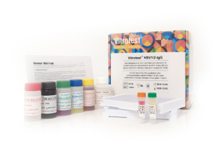
The test kit Vitrotest® HSV1/2-IgG is an enzyme linked immunosorbent assay (ELISA) for the qualitative and semiquantitative determination of IgG class antibodies to Herpes Simplex Virus types 1 and 2 (HSV1/2) in human serum or plasma.
Determination of IgG antibodies to HSV1/2 in the test kit Vitrotest® HSV1/2-IgG is based on a solid phase, indirect ELISA in a two-step incubation procedure.
○ ТК007 - 96 tests- Solid phase: breakable microplate ELISA is coated with mixture of purified antigens of herpes simplex virus type 1 and type 2.
- Conjugate: a monoclonal antibodies to human IgG conjugated to horseradish peroxidase.
- Chromogen: ready to use TMB solution.
- Volume of sample for analysis: 10 μl.
- Assay time: 1h 15 min.
Herpes simplex virus (HSV) is an etiological agent of 2 widespread diseases in humans: orofacial infection, the so called “cold sores”, and genital herpes (GH).
There are two closely related species of HSV, HSV-1 and HSV-2. Both are large enveloped viruses containing double-stranded DNA. HSV-1 is normally (but not always) associated with orofacial infections whereas HSV-2 causes mainly GH (though it could also cause orofacial infections, clinically indistinguishable from those caused by HSV-1). Both viruses could be transmitted from infected mothers to neonates.
HSV infects its host at mucosal or epithelial surfaces and causes primary infection of the epithelium. Then the virus enters sensory neurons and is transported by retrograde flow along axons that connect the point of entry into the body to nuclei of sensory neurons. Viral multiplication occurs in a small number of sensory neurons; the viral genome then remains in a latent state for the life of the host.
HSV frequently reactivates from latency and infects epithelial cells adjacent to neuron where it resides. This process results in lesions with viral shedding or with asymptomatic shedding alone. Reactivation is triggered by another infection, psychological stress, tissue damage, etc.
HSV causes benign vesicular and ulcerative lesions in immunocompetent adults and may cause severe systemic disease in neonates and immunosuppressed hosts. In children >5 years old and immunocompetent adults duration of the first attack is 2-4 weeks. Symptoms of recurrent outbreaks are typically shorter in duration (7-10 days) and even less severe than the first outbreak of genital herpes.
The course of disease is more severe in small children (the so-called neonatal herpes (NH) and immunocompromised persons. Complications of herpes in these patients could remain localized and affect only eyes, mouth and skin; infection could spread to the central nervous system causing meningitis, myelitis, and encephalitis; in the last, disseminated, stage, infection involves multiple organs.
To initiate infection, HSV must contact either mucosal surfaces or abraded skin. Transmission usually occurs by direct oral-oral, oral-genital or genital-genital contact. Orofacial herpes is transmitted mainly by household contact during childhood whereas GH is a disease mainly transmitted by sex. Neonatal HSV infection most commonly results from contact between the newborn and HSV that is present in the birth canal of the mother during delivery.
HSV can be transmitted to child under primary or recurrent herpes, both in the presence or absence of symptoms. However, under recurrent herpes, the risk of transmission to a newborn is low even if open sores are present at the time of birth (the risk is 3/100). On the contrary, the risk is considerably higher if the primary infection is acquired in the third trimester, and especially in the last 6 weeks of pregnancy (the risk increases to around 50%). As a result, seronegative women in the third trimester of pregnancy are the main group at risk, especially, if their partner is seropositive. Intrauterine infection, contrary to neonatal one, is a relatively rare event.
Infection with HHV-6 usually occurs during the first or second year of life, and accordingly, about 95% of adults have antibodies to HHV-6. At birth, most children are seropositive due to maternal antibodies, the titer of which decreases by 5 months. However, by the end of the first year of life, the percentage of seropositive infants is similar to that among older children and adults. -
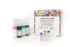
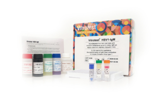
The test kit Vitrotest® HSV1/2-IgM is an enzyme linked immunosorbent assay (ELISA) for the qualitative determination of IgM class antibodies to Herpes Simplex Virus types 1 and 2 (HSV1/2) in human serum or plasma.
Determination of IgM antibodies to HSV1 and HSV2 in the test kit Vitrotest® HSV1/2-IgM is based on a solid phase, «IgM-capture» ELISA in a two-step incubation procedure.
○ ТК008 - 96 tests- Solid phase: breakable microplate ELISA is coated with monoclonal anti-IgM antibodies.
- Conjugate: mixture of recombinant antigens of herpes simplex virus type 1 and type 2 gG1 and gG2 conjugated to horseradish peroxidase.
- Chromogen: ready to use TMB solution.
- Volume of sample for analysis: 10 μl.
- Assay time: 2 hours.
Herpes simplex virus (HSV) is an etiological agent of 2 widespread diseases in humans: orofacial infection, the so called “cold sores”, and genital herpes (GH).
There are two closely related species of HSV, HSV-1 and HSV-2. Both are large enveloped viruses containing double-stranded DNA. HSV-1 is normally (but not always) associated with orofacial infections whereas HSV-2 causes mainly GH (though it could also cause orofacial infections, clinically indistinguishable from those caused by HSV-1). Both viruses could be transmitted from infected mothers to neonates.
HSV infects its host at mucosal or epithelial surfaces and causes primary infection of the epithelium. Then the virus enters sensory neurons and is transported by retrograde flow along axons that connect the point of entry into the body to nuclei of sensory neurons. Viral multiplication occurs in a small number of sensory neurons; the viral genome then remains in a latent state for the life of the host.
HSV frequently reactivates from latency and infects epithelial cells adjacent to neuron where it resides. This process results in lesions with viral shedding or with asymptomatic shedding alone. Reactivation is triggered by another infection, psychological stress, tissue damage, etc.
HSV causes benign vesicular and ulcerative lesions in immunocompetent adults and may cause severe systemic disease in neonates and immunosuppressed hosts. In children >5 years old and immunocompetent adults duration of the first attack is 2-4 weeks. Symptoms of recurrent outbreaks are typically shorter in duration (7-10 days) and even less severe than the first outbreak of genital herpes.
The course of disease is more severe in small children (the so-called neonatal herpes (NH) and immunocompromised persons. Complications of herpes in these patients could remain localized and affect only eyes, mouth and skin; infection could spread to the central nervous system causing meningitis, myelitis, and encephalitis; in the last, disseminated, stage, infection involves multiple organs.
To initiate infection, HSV must contact either mucosal surfaces or abraded skin. Transmission usually occurs by direct oral-oral, oral-genital or genital-genital contact. Orofacial herpes is transmitted mainly by household contact during childhood whereas GH is a disease mainly transmitted by sex. Neonatal HSV infection most commonly results from contact between the newborn and HSV that is present in the birth canal of the mother during delivery.
HSV can be transmitted to child under primary or recurrent herpes, both in the presence or absence of symptoms. However, under recurrent herpes, the risk of transmission to a newborn is low even if open sores are present at the time of birth (the risk is 3/100). On the contrary, the risk is considerably higher if the primary infection is acquired in the third trimester, and especially in the last 6 weeks of pregnancy (the risk increases to around 50%). As a result, seronegative women in the third trimester of pregnancy are the main group at risk, especially, if their partner is seropositive. Intrauterine infection, contrary to neonatal one, is a relatively rare event.
Infection with HHV-6 usually occurs during the first or second year of life, and accordingly, about 95% of adults have antibodies to HHV-6. At birth, most children are seropositive due to maternal antibodies, the titer of which decreases by 5 months. However, by the end of the first year of life, the percentage of seropositive infants is similar to that among older children and adults. -
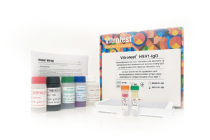
The test kit Vitrotest® HSV1-IgG is an enzyme linked immunosorbent assay (ELISA) for the qualitative and semiquantitative determination of IgG class antibodies to Herpes Simplex Virus type 1 (HSV-1) in human serum or plasma.
Determination of IgG antibodies to HSV-1 in the test kit Vitrotest® HSV1-IgG is based on a solid phase, indirect ELISA in a two-step incubation procedure.
○ ТК026 - 96 tests- Solid phase: breakable microplate ELISA is coated with recombinant antigens of herpes simplex virus type 1.
- Conjugate: a monoclonal antibodies to human IgG conjugated to horseradish peroxidase.
- Chromogen: ready to use TMB solution.
- Volume of sample for analysis: 10 μl.
- Assay time: 1h 15 min.
Herpes simplex virus (HSV) is an etiological agent of 2 widespread diseases in humans: orofacial infection, the so called “cold sores”, and genital herpes (GH).
There are two closely related species of HSV, HSV-1 and HSV-2. Both are large enveloped viruses containing double-stranded DNA. HSV-1 is normally (but not always) associated with orofacial infections whereas HSV-2 causes mainly GH (though it could also cause orofacial infections, clinically indistinguishable from those caused by HSV-1). Both viruses could be transmitted from infected mothers to neonates.
HSV infects its host at mucosal or epithelial surfaces and causes primary infection of the epithelium. Then the virus enters sensory neurons and is transported by retrograde flow along axons that connect the point of entry into the body to nuclei of sensory neurons. Viral multiplication occurs in a small number of sensory neurons; the viral genome then remains in a latent state for the life of the host.
HSV frequently reactivates from latency and infects epithelial cells adjacent to neuron where it resides. This process results in lesions with viral shedding or with asymptomatic shedding alone. Reactivation is triggered by another infection, psychological stress, tissue damage, etc.
HSV causes benign vesicular and ulcerative lesions in immunocompetent adults and may cause severe systemic disease in neonates and immunosuppressed hosts. In children >5 years old and immunocompetent adults duration of the first attack is 2-4 weeks. Symptoms of recurrent outbreaks are typically shorter in duration (7-10 days) and even less severe than the first outbreak of genital herpes.
The course of disease is more severe in small children (the so-called neonatal herpes (NH) and immunocompromised persons. Complications of herpes in these patients could remain localized and affect only eyes, mouth and skin; infection could spread to the central nervous system causing meningitis, myelitis, and encephalitis; in the last, disseminated, stage, infection involves multiple organs.
To initiate infection, HSV must contact either mucosal surfaces or abraded skin. Transmission usually occurs by direct oral-oral, oral-genital or genital-genital contact. Orofacial herpes is transmitted mainly by household contact during childhood whereas GH is a disease mainly transmitted by sex. Neonatal HSV infection most commonly results from contact between the newborn and HSV that is present in the birth canal of the mother during delivery.
HSV can be transmitted to child under primary or recurrent herpes, both in the presence or absence of symptoms. However, under recurrent herpes, the risk of transmission to a newborn is low even if open sores are present at the time of birth (the risk is 3/100). On the contrary, the risk is considerably higher if the primary infection is acquired in the third trimester, and especially in the last 6 weeks of pregnancy (the risk increases to around 50%). As a result, seronegative women in the third trimester of pregnancy are the main group at risk, especially, if their partner is seropositive. Intrauterine infection, contrary to neonatal one, is a relatively rare event.
Infection with HHV-6 usually occurs during the first or second year of life, and accordingly, about 95% of adults have antibodies to HHV-6. At birth, most children are seropositive due to maternal antibodies, the titer of which decreases by 5 months. However, by the end of the first year of life, the percentage of seropositive infants is similar to that among older children and adults. -
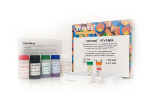
The test kit Vitrotest® HSV2-IgG is an enzyme linked immunosorbent assay (ELISA) for the qualitative and semiquantitative determination of IgG class antibodies to Herpes Simplex Virus type 2 (HSV-2) in human serum or plasma.
Determination of IgG antibodies to HSV-2 in the test kit Vitrotest® HSV2-IgG is based on a solid phase, indirect ELISA in a two-step incubation procedure.
○ ТК011 - 96 tests- Solid phase: breakable microplate ELISA is coated with recombinant antigens of herpes simplex virus type 2.
- Conjugate: a monoclonal antibodies to human IgG conjugated to horseradish peroxidase.
- Chromogen: ready to use TMB solution.
- Volume of sample for analysis: 10 μl.
- Assay time: 1h 15 min.
Herpes simplex virus (HSV) is an etiological agent of 2 widespread diseases in humans: orofacial infection, the so called “cold sores”, and genital herpes (GH).
There are two closely related species of HSV, HSV-1 and HSV-2. Both are large enveloped viruses containing double-stranded DNA. HSV-1 is normally (but not always) associated with orofacial infections whereas HSV-2 causes mainly GH (though it could also cause orofacial infections, clinically indistinguishable from those caused by HSV-1). Both viruses could be transmitted from infected mothers to neonates.
HSV infects its host at mucosal or epithelial surfaces and causes primary infection of the epithelium. Then the virus enters sensory neurons and is transported by retrograde flow along axons that connect the point of entry into the body to nuclei of sensory neurons. Viral multiplication occurs in a small number of sensory neurons; the viral genome then remains in a latent state for the life of the host.
HSV frequently reactivates from latency and infects epithelial cells adjacent to neuron where it resides. This process results in lesions with viral shedding or with asymptomatic shedding alone. Reactivation is triggered by another infection, psychological stress, tissue damage, etc.
HSV causes benign vesicular and ulcerative lesions in immunocompetent adults and may cause severe systemic disease in neonates and immunosuppressed hosts. In children >5 years old and immunocompetent adults duration of the first attack is 2-4 weeks. Symptoms of recurrent outbreaks are typically shorter in duration (7-10 days) and even less severe than the first outbreak of genital herpes.
The course of disease is more severe in small children (the so-called neonatal herpes (NH) and immunocompromised persons. Complications of herpes in these patients could remain localized and affect only eyes, mouth and skin; infection could spread to the central nervous system causing meningitis, myelitis, and encephalitis; in the last, disseminated, stage, infection involves multiple organs.
To initiate infection, HSV must contact either mucosal surfaces or abraded skin. Transmission usually occurs by direct oral-oral, oral-genital or genital-genital contact. Orofacial herpes is transmitted mainly by household contact during childhood whereas GH is a disease mainly transmitted by sex. Neonatal HSV infection most commonly results from contact between the newborn and HSV that is present in the birth canal of the mother during delivery.
HSV can be transmitted to child under primary or recurrent herpes, both in the presence or absence of symptoms. However, under recurrent herpes, the risk of transmission to a newborn is low even if open sores are present at the time of birth (the risk is 3/100). On the contrary, the risk is considerably higher if the primary infection is acquired in the third trimester, and especially in the last 6 weeks of pregnancy (the risk increases to around 50%). As a result, seronegative women in the third trimester of pregnancy are the main group at risk, especially, if their partner is seropositive. Intrauterine infection, contrary to neonatal one, is a relatively rare event.
Infection with HHV-6 usually occurs during the first or second year of life, and accordingly, about 95% of adults have antibodies to HHV-6. At birth, most children are seropositive due to maternal antibodies, the titer of which decreases by 5 months. However, by the end of the first year of life, the percentage of seropositive infants is similar to that among older children and adults. -
The test kit Vitrotest® HSV1-IgM is an enzyme linked immunosorbent assay (ELISA) for the qualitative determination of IgM class antibodies to Herpes Simplex Virus type 1 (HSV-1) in human serum or plasma.
Determination of IgM antibodies to HSV-1 in the test kit Vitrotest® HSV1-IgM is based on a solid phase, «IgM-capture» ELISA in a two-step incubation procedure.
○ ТК009 - 96 tests- Solid phase: breakable microplate ELISA is coated with monoclonal anti-IgM antibodies.
- Conjugate: recombinant HSV1 antigen gG1 conjugated to horseradish peroxidase.
- Chromogen: ready to use TMB solution.
- Volume of sample for analysis: 10 μl.
- Assay time: 2 hours.
Herpes simplex virus (HSV) is an etiological agent of 2 widespread diseases in humans: orofacial infection, the so called “cold sores”, and genital herpes (GH).
There are two closely related species of HSV, HSV-1 and HSV-2. Both are large enveloped viruses containing double-stranded DNA. HSV-1 is normally (but not always) associated with orofacial infections whereas HSV-2 causes mainly GH (though it could also cause orofacial infections, clinically indistinguishable from those caused by HSV-1). Both viruses could be transmitted from infected mothers to neonates.
HSV infects its host at mucosal or epithelial surfaces and causes primary infection of the epithelium. Then the virus enters sensory neurons and is transported by retrograde flow along axons that connect the point of entry into the body to nuclei of sensory neurons. Viral multiplication occurs in a small number of sensory neurons; the viral genome then remains in a latent state for the life of the host.
HSV frequently reactivates from latency and infects epithelial cells adjacent to neuron where it resides. This process results in lesions with viral shedding or with asymptomatic shedding alone. Reactivation is triggered by another infection, psychological stress, tissue damage, etc.
HSV causes benign vesicular and ulcerative lesions in immunocompetent adults and may cause severe systemic disease in neonates and immunosuppressed hosts. In children >5 years old and immunocompetent adults duration of the first attack is 2-4 weeks. Symptoms of recurrent outbreaks are typically shorter in duration (7-10 days) and even less severe than the first outbreak of genital herpes.
The course of disease is more severe in small children (the so-called neonatal herpes (NH) and immunocompromised persons. Complications of herpes in these patients could remain localized and affect only eyes, mouth and skin; infection could spread to the central nervous system causing meningitis, myelitis, and encephalitis; in the last, disseminated, stage, infection involves multiple organs.
To initiate infection, HSV must contact either mucosal surfaces or abraded skin. Transmission usually occurs by direct oral-oral, oral-genital or genital-genital contact. Orofacial herpes is transmitted mainly by household contact during childhood whereas GH is a disease mainly transmitted by sex. Neonatal HSV infection most commonly results from contact between the newborn and HSV that is present in the birth canal of the mother during delivery.
HSV can be transmitted to child under primary or recurrent herpes, both in the presence or absence of symptoms. However, under recurrent herpes, the risk of transmission to a newborn is low even if open sores are present at the time of birth (the risk is 3/100). On the contrary, the risk is considerably higher if the primary infection is acquired in the third trimester, and especially in the last 6 weeks of pregnancy (the risk increases to around 50%). As a result, seronegative women in the third trimester of pregnancy are the main group at risk, especially, if their partner is seropositive. Intrauterine infection, contrary to neonatal one, is a relatively rare event.
Infection with HHV-6 usually occurs during the first or second year of life, and accordingly, about 95% of adults have antibodies to HHV-6. At birth, most children are seropositive due to maternal antibodies, the titer of which decreases by 5 months. However, by the end of the first year of life, the percentage of seropositive infants is similar to that among older children and adults. -
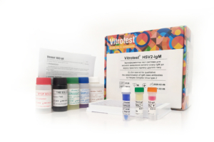
The test kit Vitrotest® HSV2-IgM is an enzyme linked immunosorbent assay (ELISA) for the qualitative determination of IgM class antibodies to Herpes Simplex Virus type 2 (HSV-2) in human serum or plasma.
Determination of IgM antibodies to HSV-2 in the test kit Vitrotest® HSV2-IgM is based on a solid phase, «IgM-capture» ELISA in a two-step incubation procedure.
○ ТК010 - 96 tests- Solid phase: breakable microplate ELISA is coated with monoclonal anti-IgM antibodies.
- Conjugate: recombinant HSV2 antigen gG2 conjugated to horseradish peroxidase.
- Chromogen: ready to use TMB solution.
- Volume of sample for analysis: 10 μl.
- Assay time: 2 hours.
Herpes simplex virus (HSV) is an etiological agent of 2 widespread diseases in humans: orofacial infection, the so called “cold sores”, and genital herpes (GH).
There are two closely related species of HSV, HSV-1 and HSV-2. Both are large enveloped viruses containing double-stranded DNA. HSV-1 is normally (but not always) associated with orofacial infections whereas HSV-2 causes mainly GH (though it could also cause orofacial infections, clinically indistinguishable from those caused by HSV-1). Both viruses could be transmitted from infected mothers to neonates.
HSV infects its host at mucosal or epithelial surfaces and causes primary infection of the epithelium. Then the virus enters sensory neurons and is transported by retrograde flow along axons that connect the point of entry into the body to nuclei of sensory neurons. Viral multiplication occurs in a small number of sensory neurons; the viral genome then remains in a latent state for the life of the host.
HSV frequently reactivates from latency and infects epithelial cells adjacent to neuron where it resides. This process results in lesions with viral shedding or with asymptomatic shedding alone. Reactivation is triggered by another infection, psychological stress, tissue damage, etc.
HSV causes benign vesicular and ulcerative lesions in immunocompetent adults and may cause severe systemic disease in neonates and immunosuppressed hosts. In children >5 years old and immunocompetent adults duration of the first attack is 2-4 weeks. Symptoms of recurrent outbreaks are typically shorter in duration (7-10 days) and even less severe than the first outbreak of genital herpes.
The course of disease is more severe in small children (the so-called neonatal herpes (NH) and immunocompromised persons. Complications of herpes in these patients could remain localized and affect only eyes, mouth and skin; infection could spread to the central nervous system causing meningitis, myelitis, and encephalitis; in the last, disseminated, stage, infection involves multiple organs.
To initiate infection, HSV must contact either mucosal surfaces or abraded skin. Transmission usually occurs by direct oral-oral, oral-genital or genital-genital contact. Orofacial herpes is transmitted mainly by household contact during childhood whereas GH is a disease mainly transmitted by sex. Neonatal HSV infection most commonly results from contact between the newborn and HSV that is present in the birth canal of the mother during delivery.
HSV can be transmitted to child under primary or recurrent herpes, both in the presence or absence of symptoms. However, under recurrent herpes, the risk of transmission to a newborn is low even if open sores are present at the time of birth (the risk is 3/100). On the contrary, the risk is considerably higher if the primary infection is acquired in the third trimester, and especially in the last 6 weeks of pregnancy (the risk increases to around 50%). As a result, seronegative women in the third trimester of pregnancy are the main group at risk, especially, if their partner is seropositive. Intrauterine infection, contrary to neonatal one, is a relatively rare event.
Infection with HHV-6 usually occurs during the first or second year of life, and accordingly, about 95% of adults have antibodies to HHV-6. At birth, most children are seropositive due to maternal antibodies, the titer of which decreases by 5 months. However, by the end of the first year of life, the percentage of seropositive infants is similar to that among older children and adults. -
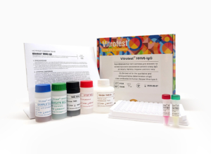
The test kit Vitrotest® HHV6-IgG is an enzyme linked immunosorbent assay (ELISA) for the qualitative and semiquantitative determination of IgG class antibodies to Human Herpes Virus type 6 (HHV-6) in human serum or plasma.
Determination of IgG antibodies to HHV6 in the test kit Vitrotest® HHV6-IgG is based on a solid phase, indirect ELISA in a two-step incubation procedure.
○ ТК090 - 96 tests- Solid phase: breakable microplate ELISA is coated with recombinant antigens of Human Herpes Virus type 6.
- Conjugate: a monoclonal antibodies to human IgG conjugated to horseradish peroxidase.
- Chromogen: ready to use TMB solution.
- Volume of sample for analysis: 10 μl.
- Assay time: 1h 15 min.
Herpes simplex virus (HSV) is an etiological agent of 2 widespread diseases in humans: orofacial infection, the so called “cold sores”, and genital herpes (GH).
There are two closely related species of HSV, HSV-1 and HSV-2. Both are large enveloped viruses containing double-stranded DNA. HSV-1 is normally (but not always) associated with orofacial infections whereas HSV-2 causes mainly GH (though it could also cause orofacial infections, clinically indistinguishable from those caused by HSV-1). Both viruses could be transmitted from infected mothers to neonates.
HSV infects its host at mucosal or epithelial surfaces and causes primary infection of the epithelium. Then the virus enters sensory neurons and is transported by retrograde flow along axons that connect the point of entry into the body to nuclei of sensory neurons. Viral multiplication occurs in a small number of sensory neurons; the viral genome then remains in a latent state for the life of the host.
HSV frequently reactivates from latency and infects epithelial cells adjacent to neuron where it resides. This process results in lesions with viral shedding or with asymptomatic shedding alone. Reactivation is triggered by another infection, psychological stress, tissue damage, etc.
HSV causes benign vesicular and ulcerative lesions in immunocompetent adults and may cause severe systemic disease in neonates and immunosuppressed hosts. In children >5 years old and immunocompetent adults duration of the first attack is 2-4 weeks. Symptoms of recurrent outbreaks are typically shorter in duration (7-10 days) and even less severe than the first outbreak of genital herpes.
The course of disease is more severe in small children (the so-called neonatal herpes (NH) and immunocompromised persons. Complications of herpes in these patients could remain localized and affect only eyes, mouth and skin; infection could spread to the central nervous system causing meningitis, myelitis, and encephalitis; in the last, disseminated, stage, infection involves multiple organs.
To initiate infection, HSV must contact either mucosal surfaces or abraded skin. Transmission usually occurs by direct oral-oral, oral-genital or genital-genital contact. Orofacial herpes is transmitted mainly by household contact during childhood whereas GH is a disease mainly transmitted by sex. Neonatal HSV infection most commonly results from contact between the newborn and HSV that is present in the birth canal of the mother during delivery.
HSV can be transmitted to child under primary or recurrent herpes, both in the presence or absence of symptoms. However, under recurrent herpes, the risk of transmission to a newborn is low even if open sores are present at the time of birth (the risk is 3/100). On the contrary, the risk is considerably higher if the primary infection is acquired in the third trimester, and especially in the last 6 weeks of pregnancy (the risk increases to around 50%). As a result, seronegative women in the third trimester of pregnancy are the main group at risk, especially, if their partner is seropositive. Intrauterine infection, contrary to neonatal one, is a relatively rare event.
Infection with HHV-6 usually occurs during the first or second year of life, and accordingly, about 95% of adults have antibodies to HHV-6. At birth, most children are seropositive due to maternal antibodies, the titer of which decreases by 5 months. However, by the end of the first year of life, the percentage of seropositive infants is similar to that among older children and adults. -
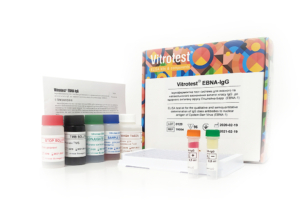
The test kit Vitrotest® EBNA-IgG is an enzyme linked immunosorbent assay (ELISA) for the qualitative and semiquantitative determination of IgG class antibodies to nuclear antigen (EBNA) of Epstein-Barr virus (EBV) in human serum or plasma.
Determination of IgG antibodies to EBV nuclear antigen (EBNA) in the test kit Vitrotest® EBNA-IgG is based on a solid phase, indirect ELISA in a two-step incubation procedure.
○ ТК054 - 96 tests- Solid phase: breakable microplate ELISA is coated with EBV recombinant nuclear antigen.
- Conjugate: a monoclonal antibodies to human IgG conjugated to horseradish peroxidase.
- Chromogen: ready to use TMB solution.
- Volume of sample for analysis: 10 μl.
- Assay time: 1h 15 min.
Epstein-Barr virus (EBV), also known as human herpesvirus 4, is the causative agent of diseases such as infectious mononucleosis (IM), Burkitt’s lymphoma (BL), nasopharyngeal carcinoma.
EBV acquired in early childhood usually does not cause symptoms. Infection in adolescence or early adulthood often results in clinical IM. Typically, the disease presents as pharyngitis, lymphadenopathy, and fever. In most cases, symptoms resolve within 2–4 weeks, but more than 90% of adults develop a latent B-lymphocyte infection. Approximately 1% of immunocompetent individuals may experience severe complications (hepatitis, myocarditis, splenic rupture, neurological complications). In immunosuppressed individuals, primary EBV infection leads to severe disorders (eg, BL).
The clinical symptoms range from mild fever to prolonged debilitating illness. Typically, the disease presents as pharyngitis, lymphadenopathy, and fever. Enlarged spleen and a swollen liver are less common symptoms. Microscopic blood observation reveals lymphocytosis; most lymphocytes have atypical morphology.
EBV spreads most commonly through bodily fluids, especially saliva, sporadically also through blood and semen. Humans are the most infectious during acute IM, but the virus could be shed into saliva even during latency. Transmission occurs mainly via household and sexual routes; however, transmission via blood transfusion and organ transplantation was also demonstrated.
Risk groups for IM are adolescents, senior school students, blood transfusion recipients, as well as people with immunodeficiencies (risk group for complications). Up to 50% of adolescents and young adults in developed countries are seronegative and hence at risk of developing IM.
EBV is a genetically stable virus. EBV is one of the most common human viruses. EBV is found all over the world; virus is able to infect more than 95% of all individuals within the first four decades of life. The current incidence of IM in USA is estimated at approximately 125 000 reported cases/year. EBV DNA is surrounded by a nucleocapsid; protein tegument is situated between the nucleocapsid and envelope; outer envelope has glycoprotein spikes typical of all herpesviruses. The viral genome encodes around 90 proteins. The most of these proteins (80) are expressed only during EBV lytic cycle.
On the basis of their time of expression, EBV genes are designated early or late. Early genes encode mostly non-structural proteins, the most important among them is viral DNA polymerase. Late genes encode mostly structural viral proteins which are required for successful packaging of the viral DNA and formation of the virion. The viral capsids have several polypeptide components ranging in size from 18 to 155 kDa, with a 155-kDa polypeptide being the major component. The membrane antigen (MA) complex is found on the virion envelope and consists of at least three major EBV glycoproteins: gp350/220 (the most abundant protein), gp110, and gp85.
The presence of EBV in epithelial and B cells provokes an intense immune response consisting of antibodies to a large variety of virally encoded antigens. During the acute phase of IM, the humoral response is directed toward viral antigens of the lytic cycle, notably membrane antigen complex, several early antigens (p 54 and p138), and VCA. A strong IgM response (the so-called heterophile antibodies) is directed towards a complex glycoprotein structure on the surface of EBV infected cells. As IM patients recover from clinical symptoms, the IgM response reduces significantly while the IgG response in serum plateaus at a reduced level and is maintained throughout the persistent infection.
The main purpose of the diagnostics of IM is to rule out less common but more serious causes of IM-like symptoms, requiring immediate intervention, such as hepatitis or various leukemic malignancies. The mainstay in the diagnostics of IM are still clinical symptoms (classic triad-fever, lymphoadenopathy, and pharyngytis) in combination with anamnestic aspects.
The specificity of the “classic triad” is around 90%, since IM-like symptoms may be caused by a variety of other pathogens and malignancies, such as cytomegalovirus, human herpesvirus 6, adenovirus, rubella virus, mumps virus, human immunodeficiency virus, hepatitis A virus, influenza A and B viruses, and Toxoplasma gondii, lymphoma and leukemia. Therefore, EBV infection should be confirmed with a blood antibody test.
It is well known for many years that acute IM is characterized by the appearance of heterophile antibodies (i.e. antibodies to EBV antigen described above) in the serum. This peculiarity is used in the so-called “monospot tests”, detecting agglutination of sheep erythrocytes in the presence of serum of patients with acute-phase IM. This test is now not recommended for general use by the Centers for Disease Prevention and Control, USA, since it produces both false positive and false negative results.
Only three serological parameters are essential for the detection of EBV-specific antibodies in immunocompetent individuals: VCA IgG (marker of disease, present or past), VCA IgM (marker of recent or present disease), and EBNA-1 IgG (marker of past disease).
The most widely used serological methods are immunofluorescence assay (IFA) and enzyme immunoassay (EIA). IFA has always been used as a reference method. However, some investigators found EIA to be more sensitive than IFA.
On the contrary, in immunosuppressed individuals serological assays are not recommended for many reasons, such as dysfunctions in the production of antibodies. To date, only detection of viral load by PCR is an established marker for immunosuppressed patients. -
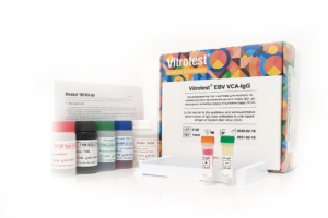
The test kit Vitrotest® EBV VCA-IgG is an enzyme linked immunosorbent assay (ELISA) for the qualitative and semiquantitative determination of IgG class antibodies to viral capsid antigen (VCA) of Epstein-Barr virus (EBV) in human serum or plasma.
Determination of IgG antibodies to EBV capsid antigen (VCA) in the test kit Vitrotest® EBV VCA-IgG is based on a solid phase, indirect ELISA in a two-step incubation procedure.
○ ТК053 - 96 tests- Solid phase: breakable microplate ELISA is coated with EBV recombinant capsid antigen.
- Conjugate: a monoclonal antibodies to human IgG conjugated to horseradish peroxidase.
- Chromogen: ready to use TMB solution.
- Volume of sample for analysis: 10 μl.
- Assay time: 1h 15 min.
Epstein-Barr virus (EBV), also known as human herpesvirus 4, is the causative agent of diseases such as infectious mononucleosis (IM), Burkitt’s lymphoma (BL), nasopharyngeal carcinoma.
EBV acquired in early childhood usually does not cause symptoms. Infection in adolescence or early adulthood often results in clinical IM. Typically, the disease presents as pharyngitis, lymphadenopathy, and fever. In most cases, symptoms resolve within 2–4 weeks, but more than 90% of adults develop a latent B-lymphocyte infection. Approximately 1% of immunocompetent individuals may experience severe complications (hepatitis, myocarditis, splenic rupture, neurological complications). In immunosuppressed individuals, primary EBV infection leads to severe disorders (eg, BL).
The clinical symptoms range from mild fever to prolonged debilitating illness. Typically, the disease presents as pharyngitis, lymphadenopathy, and fever. Enlarged spleen and a swollen liver are less common symptoms. Microscopic blood observation reveals lymphocytosis; most lymphocytes have atypical morphology.
EBV spreads most commonly through bodily fluids, especially saliva, sporadically also through blood and semen. Humans are the most infectious during acute IM, but the virus could be shed into saliva even during latency. Transmission occurs mainly via household and sexual routes; however, transmission via blood transfusion and organ transplantation was also demonstrated.
Risk groups for IM are adolescents, senior school students, blood transfusion recipients, as well as people with immunodeficiencies (risk group for complications). Up to 50% of adolescents and young adults in developed countries are seronegative and hence at risk of developing IM.
EBV is a genetically stable virus. EBV is one of the most common human viruses. EBV is found all over the world; virus is able to infect more than 95% of all individuals within the first four decades of life. The current incidence of IM in USA is estimated at approximately 125 000 reported cases/year. EBV DNA is surrounded by a nucleocapsid; protein tegument is situated between the nucleocapsid and envelope; outer envelope has glycoprotein spikes typical of all herpesviruses. The viral genome encodes around 90 proteins. The most of these proteins (80) are expressed only during EBV lytic cycle.
On the basis of their time of expression, EBV genes are designated early or late. Early genes encode mostly non-structural proteins, the most important among them is viral DNA polymerase. Late genes encode mostly structural viral proteins which are required for successful packaging of the viral DNA and formation of the virion. The viral capsids have several polypeptide components ranging in size from 18 to 155 kDa, with a 155-kDa polypeptide being the major component. The membrane antigen (MA) complex is found on the virion envelope and consists of at least three major EBV glycoproteins: gp350/220 (the most abundant protein), gp110, and gp85.
The presence of EBV in epithelial and B cells provokes an intense immune response consisting of antibodies to a large variety of virally encoded antigens. During the acute phase of IM, the humoral response is directed toward viral antigens of the lytic cycle, notably membrane antigen complex, several early antigens (p 54 and p138), and VCA. A strong IgM response (the so-called heterophile antibodies) is directed towards a complex glycoprotein structure on the surface of EBV infected cells. As IM patients recover from clinical symptoms, the IgM response reduces significantly while the IgG response in serum plateaus at a reduced level and is maintained throughout the persistent infection.
The main purpose of the diagnostics of IM is to rule out less common but more serious causes of IM-like symptoms, requiring immediate intervention, such as hepatitis or various leukemic malignancies. The mainstay in the diagnostics of IM are still clinical symptoms (classic triad-fever, lymphoadenopathy, and pharyngytis) in combination with anamnestic aspects.
The specificity of the “classic triad” is around 90%, since IM-like symptoms may be caused by a variety of other pathogens and malignancies, such as cytomegalovirus, human herpesvirus 6, adenovirus, rubella virus, mumps virus, human immunodeficiency virus, hepatitis A virus, influenza A and B viruses, and Toxoplasma gondii, lymphoma and leukemia. Therefore, EBV infection should be confirmed with a blood antibody test.
It is well known for many years that acute IM is characterized by the appearance of heterophile antibodies (i.e. antibodies to EBV antigen described above) in the serum. This peculiarity is used in the so-called “monospot tests”, detecting agglutination of sheep erythrocytes in the presence of serum of patients with acute-phase IM. This test is now not recommended for general use by the Centers for Disease Prevention and Control, USA, since it produces both false positive and false negative results.
Only three serological parameters are essential for the detection of EBV-specific antibodies in immunocompetent individuals: VCA IgG (marker of disease, present or past), VCA IgM (marker of recent or present disease), and EBNA-1 IgG (marker of past disease).
The most widely used serological methods are immunofluorescence assay (IFA) and enzyme immunoassay (EIA). IFA has always been used as a reference method. However, some investigators found EIA to be more sensitive than IFA.
On the contrary, in immunosuppressed individuals serological assays are not recommended for many reasons, such as dysfunctions in the production of antibodies. To date, only detection of viral load by PCR is an established marker for immunosuppressed patients. -
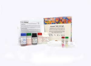
The test kit Vitrotest® EBV VCA-IgА is an enzyme linked immunosorbent assay (ELISA) for the qualitative and semiquantitative determination of IgА class antibodies to viral capsid antigen (VCA) of Epstein-Barr virus (EBV) in human serum or plasma.
Determination of IgА antibodies to EBV capsid antigen (VCA) in the test kit Vitrotest® EBV VCA-IgА is based on a solid phase, indirect ELISA in a two-step incubation procedure.
○ ТК123 - 96 tests- Solid phase: breakable microplate ELISA is coated with EBV recombinant capsid antigen.
- Conjugate: a monoclonal antibodies to human IgА conjugated to horseradish peroxidase.
- Chromogen: ready to use TMB solution.
- Volume of sample for analysis: 10 μl.
- Assay time: 1h 15 min.
Epstein-Barr virus (EBV), also known as human herpesvirus 4, is the causative agent of diseases such as infectious mononucleosis (IM), Burkitt’s lymphoma (BL), nasopharyngeal carcinoma.
EBV acquired in early childhood usually does not cause symptoms. Infection in adolescence or early adulthood often results in clinical IM. Typically, the disease presents as pharyngitis, lymphadenopathy, and fever. In most cases, symptoms resolve within 2–4 weeks, but more than 90% of adults develop a latent B-lymphocyte infection. Approximately 1% of immunocompetent individuals may experience severe complications (hepatitis, myocarditis, splenic rupture, neurological complications). In immunosuppressed individuals, primary EBV infection leads to severe disorders (eg, BL).
The clinical symptoms range from mild fever to prolonged debilitating illness. Typically, the disease presents as pharyngitis, lymphadenopathy, and fever. Enlarged spleen and a swollen liver are less common symptoms. Microscopic blood observation reveals lymphocytosis; most lymphocytes have atypical morphology.
EBV spreads most commonly through bodily fluids, especially saliva, sporadically also through blood and semen. Humans are the most infectious during acute IM, but the virus could be shed into saliva even during latency. Transmission occurs mainly via household and sexual routes; however, transmission via blood transfusion and organ transplantation was also demonstrated.
Risk groups for IM are adolescents, senior school students, blood transfusion recipients, as well as people with immunodeficiencies (risk group for complications). Up to 50% of adolescents and young adults in developed countries are seronegative and hence at risk of developing IM.
EBV is a genetically stable virus. EBV is one of the most common human viruses. EBV is found all over the world; virus is able to infect more than 95% of all individuals within the first four decades of life. The current incidence of IM in USA is estimated at approximately 125 000 reported cases/year. EBV DNA is surrounded by a nucleocapsid; protein tegument is situated between the nucleocapsid and envelope; outer envelope has glycoprotein spikes typical of all herpesviruses. The viral genome encodes around 90 proteins. The most of these proteins (80) are expressed only during EBV lytic cycle.
On the basis of their time of expression, EBV genes are designated early or late. Early genes encode mostly non-structural proteins, the most important among them is viral DNA polymerase. Late genes encode mostly structural viral proteins which are required for successful packaging of the viral DNA and formation of the virion. The viral capsids have several polypeptide components ranging in size from 18 to 155 kDa, with a 155-kDa polypeptide being the major component. The membrane antigen (MA) complex is found on the virion envelope and consists of at least three major EBV glycoproteins: gp350/220 (the most abundant protein), gp110, and gp85.
The presence of EBV in epithelial and B cells provokes an intense immune response consisting of antibodies to a large variety of virally encoded antigens. During the acute phase of IM, the humoral response is directed toward viral antigens of the lytic cycle, notably membrane antigen complex, several early antigens (p 54 and p138), and VCA. A strong IgM response (the so-called heterophile antibodies) is directed towards a complex glycoprotein structure on the surface of EBV infected cells. As IM patients recover from clinical symptoms, the IgM response reduces significantly while the IgG response in serum plateaus at a reduced level and is maintained throughout the persistent infection.
The main purpose of the diagnostics of IM is to rule out less common but more serious causes of IM-like symptoms, requiring immediate intervention, such as hepatitis or various leukemic malignancies. The mainstay in the diagnostics of IM are still clinical symptoms (classic triad-fever, lymphoadenopathy, and pharyngytis) in combination with anamnestic aspects.
The specificity of the “classic triad” is around 90%, since IM-like symptoms may be caused by a variety of other pathogens and malignancies, such as cytomegalovirus, human herpesvirus 6, adenovirus, rubella virus, mumps virus, human immunodeficiency virus, hepatitis A virus, influenza A and B viruses, and Toxoplasma gondii, lymphoma and leukemia. Therefore, EBV infection should be confirmed with a blood antibody test.
It is well known for many years that acute IM is characterized by the appearance of heterophile antibodies (i.e. antibodies to EBV antigen described above) in the serum. This peculiarity is used in the so-called “monospot tests”, detecting agglutination of sheep erythrocytes in the presence of serum of patients with acute-phase IM. This test is now not recommended for general use by the Centers for Disease Prevention and Control, USA, since it produces both false positive and false negative results.
Only three serological parameters are essential for the detection of EBV-specific antibodies in immunocompetent individuals: VCA IgG (marker of disease, present or past), VCA IgM (marker of recent or present disease), and EBNA-1 IgG (marker of past disease).
The most widely used serological methods are immunofluorescence assay (IFA) and enzyme immunoassay (EIA). IFA has always been used as a reference method. However, some investigators found EIA to be more sensitive than IFA.
On the contrary, in immunosuppressed individuals serological assays are not recommended for many reasons, such as dysfunctions in the production of antibodies. To date, only detection of viral load by PCR is an established marker for immunosuppressed patients. -
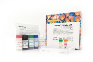
The test kit Vitrotest® EBV VCA-IgM is an enzyme linked immunosorbent assay (ELISA) for the qualitative and semiquantitative determination of IgM class antibodies to viral capsid antigen (VCA) of Epstein-Barr virus (EBV) in human serum or plasma.
Determination of IgM antibodies to EBV VCA in the test kit Vitrotest® EBV VCA-IgM is based on a solid phase «IgM-capture» ELISA in a two-step incubation procedure.
○ ТК052 - 96 tests- Solid phase: breakable microplate ELISA is coated with monoclonal anti-IgM antibodies.
- Conjugate: recombinant Epstein-Barr virus capsid antigen (VCA) conjugated to horseradish peroxidase.
- Chromogen: ready to use TMB solution.
- Volume of sample for analysis: 10 μl.
- Assay time: 1h 15 min.
Epstein-Barr virus (EBV), also known as human herpesvirus 4, is the causative agent of diseases such as infectious mononucleosis (IM), Burkitt’s lymphoma (BL), nasopharyngeal carcinoma.
EBV acquired in early childhood usually does not cause symptoms. Infection in adolescence or early adulthood often results in clinical IM. Typically, the disease presents as pharyngitis, lymphadenopathy, and fever. In most cases, symptoms resolve within 2–4 weeks, but more than 90% of adults develop a latent B-lymphocyte infection. Approximately 1% of immunocompetent individuals may experience severe complications (hepatitis, myocarditis, splenic rupture, neurological complications). In immunosuppressed individuals, primary EBV infection leads to severe disorders (eg, BL).
The clinical symptoms range from mild fever to prolonged debilitating illness. Typically, the disease presents as pharyngitis, lymphadenopathy, and fever. Enlarged spleen and a swollen liver are less common symptoms. Microscopic blood observation reveals lymphocytosis; most lymphocytes have atypical morphology.
EBV spreads most commonly through bodily fluids, especially saliva, sporadically also through blood and semen. Humans are the most infectious during acute IM, but the virus could be shed into saliva even during latency. Transmission occurs mainly via household and sexual routes; however, transmission via blood transfusion and organ transplantation was also demonstrated.
Risk groups for IM are adolescents, senior school students, blood transfusion recipients, as well as people with immunodeficiencies (risk group for complications). Up to 50% of adolescents and young adults in developed countries are seronegative and hence at risk of developing IM.
EBV is a genetically stable virus. EBV is one of the most common human viruses. EBV is found all over the world; virus is able to infect more than 95% of all individuals within the first four decades of life. The current incidence of IM in USA is estimated at approximately 125 000 reported cases/year. EBV DNA is surrounded by a nucleocapsid; protein tegument is situated between the nucleocapsid and envelope; outer envelope has glycoprotein spikes typical of all herpesviruses. The viral genome encodes around 90 proteins. The most of these proteins (80) are expressed only during EBV lytic cycle.
On the basis of their time of expression, EBV genes are designated early or late. Early genes encode mostly non-structural proteins, the most important among them is viral DNA polymerase. Late genes encode mostly structural viral proteins which are required for successful packaging of the viral DNA and formation of the virion. The viral capsids have several polypeptide components ranging in size from 18 to 155 kDa, with a 155-kDa polypeptide being the major component. The membrane antigen (MA) complex is found on the virion envelope and consists of at least three major EBV glycoproteins: gp350/220 (the most abundant protein), gp110, and gp85.
The presence of EBV in epithelial and B cells provokes an intense immune response consisting of antibodies to a large variety of virally encoded antigens. During the acute phase of IM, the humoral response is directed toward viral antigens of the lytic cycle, notably membrane antigen complex, several early antigens (p 54 and p138), and VCA. A strong IgM response (the so-called heterophile antibodies) is directed towards a complex glycoprotein structure on the surface of EBV infected cells. As IM patients recover from clinical symptoms, the IgM response reduces significantly while the IgG response in serum plateaus at a reduced level and is maintained throughout the persistent infection.
The main purpose of the diagnostics of IM is to rule out less common but more serious causes of IM-like symptoms, requiring immediate intervention, such as hepatitis or various leukemic malignancies. The mainstay in the diagnostics of IM are still clinical symptoms (classic triad-fever, lymphoadenopathy, and pharyngytis) in combination with anamnestic aspects.
The specificity of the “classic triad” is around 90%, since IM-like symptoms may be caused by a variety of other pathogens and malignancies, such as cytomegalovirus, human herpesvirus 6, adenovirus, rubella virus, mumps virus, human immunodeficiency virus, hepatitis A virus, influenza A and B viruses, and Toxoplasma gondii, lymphoma and leukemia. Therefore, EBV infection should be confirmed with a blood antibody test.
It is well known for many years that acute IM is characterized by the appearance of heterophile antibodies (i.e. antibodies to EBV antigen described above) in the serum. This peculiarity is used in the so-called “monospot tests”, detecting agglutination of sheep erythrocytes in the presence of serum of patients with acute-phase IM. This test is now not recommended for general use by the Centers for Disease Prevention and Control, USA, since it produces both false positive and false negative results.
Only three serological parameters are essential for the detection of EBV-specific antibodies in immunocompetent individuals: VCA IgG (marker of disease, present or past), VCA IgM (marker of recent or present disease), and EBNA-1 IgG (marker of past disease).
The most widely used serological methods are immunofluorescence assay (IFA) and enzyme immunoassay (EIA). IFA has always been used as a reference method. However, some investigators found EIA to be more sensitive than IFA.
On the contrary, in immunosuppressed individuals serological assays are not recommended for many reasons, such as dysfunctions in the production of antibodies. To date, only detection of viral load by PCR is an established marker for immunosuppressed patients.