-
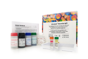
The test kit Vitrotest® Borrelia-IgG is an enzyme linked immunosorbent assay (ELISA) for the qualitative and semiquantitative determination of IgG class antibodies to Borrelia burgdorferi sensu lato in human serum or plasma.
Determination of IgG antibodies to B. burgdorferi in the test kit Vitrotest® Borrelia-IgG is based on a solid phase indirect ELISA in a two-step incubation procedure.
○ ТК084 - 96 tests- Solid phase: breakable microplate ELISA is coated with Borrelia burgdorferi sensu lato antigens.
- Conjugate: a monoclonal antibodies to human IgG conjugated to horseradish peroxidase.
- Chromogen: ready to use TMB solution.
- Volume of sample for analysis: 10 μl.
- Assay time: 1h 15 min.
Lyme disease (tick-borne borreliosis) is the most common tick-borne disease in the Northern hemisphere. The causative agent of borreliosis is spirochetes of the Borrelia burgdorferi group - were first identified in 1982 by the American microbiologist Willie Burgdorfer. Today, the most pathogenic spirochetes belong to the Borrelia burgdorferi sensu lato group, which includes Borrelia burgdorferi sensu stricto, Borrelia afzelii and Borrelia garinii.
Lyme borreliosis is a progressing disease which encompasses several clinical syndromes (erythema migrans (EM), neuroborreliosis, Lyme arthritis, and acrodermatitis chronica atrophicans). The most common clinical manifestation of early localized LD is EM, which appears in approximately 70% of patients 3-30 (average 7) days after bite. EM is a rash which appears at the site of the tick bite and gradually expands to a ring with a central clear zone. If the infection remains unnoticed and untreated in this early localized stage, B. burgdorferi can spread to other tissues and organs. The second, so-called early disseminated stage of the disease causes more severe manifestations that can involve the skin, nervous system, joints, or heart. This stage is mainly characterized by neurological signs (neuroborreliosis) or joint aches (Lyme arthritis). The third stadium is late disseminated LD. It’s typical presentations are acrodermatitis chronica atrophicans and arthritis. Manifestations of LD are similar to such immune-mediated disorders as rheumatoid arthritis, multiple sclerosis and systemic lupus.
Ticks most frequently acquire spirochetes from infected rodents during their larval feeding. Once infected, a tick can transmit infection throughout its life.
Not all ticks of competent species are carriers of an infectious agent. The prevalence of B. burgdorferi among ticks was measured to be as different as 19% and 60%. The probability to be infected is regarded as high if the tick remains attached to skin for more than 24 hours.
People living in or visiting rural areas, particularly campers and hikers, are most at risk. The infection risk appears not only in forests but more and more in parks, which attract more attention from the public. Ixodes ticks are the only natural agents through which humans have been shown to become infected. There is no evidence that LD is transmitted from person-to-person.
B.burgdorferi infection is known to induce strong humoral immune response in the host. IgM titres are typically highest between 3rd and 6th week after disease onset and then decline, however in many patients IgM remains high during later manifestations of illness, reflecting disease activity. IgG titres start increasing 1-2 weeks after onset, but reach highest titers only months later when arthritis is already present.
If the patient is cured, antibody levels in the first weeks after convalescence increase, and only then begin to decrease. If LD reaches an advanced stage, IgM and especially IgG are still detectable after cure for many years in the majority of patients. On the contrary, prompt antibiotic treatment at localized stage often aborts the development of a sustained humoral response, resulting in an absence of IgG and IgM after cure. Hence, reinfection is possible after successfully treated early, but not late LD.
B. burgdorferi genome encodes around 1500 proteins; almost half of them are plasmid encoded. Of these proteins, around 100 are immunogenic. Significant serological cross-reactivity exists between B.burdhoferi and causative agents of human spirochetal infections, particularly relapsing fever and syphilis.
Only several proteins are proven specific antigens and therefore valuable for serodiagnosis. These proteins include FlaB (BB0147), the P66 outer membrane protein (BB0603), OspA and OspB (BBA15 and BBA16), decorinbinding protein B (BBA25), OspC (BBB19), fibronectin-binding protein (BBK32), and VlsE (BBF33). FlaB is a part of flagellar apparatus; the rest are outer membrane proteins.
Currently, there is no “gold standard” in the diagnostics of LD. LD diagnosis is based upon:
a) signs and symptoms;
b) history of exposure to infectious ticks;
c) laboratory diagnosis.
The only manifestation characteristic to LD is EM, whereas clinical features of later stage presentations are not unique to B. burgdorferi infection. Unfortunately, only around 70% of patients develop EM; many patients present to the clinics at disseminated stage. Exposure to ticks may easily left unnoticed as many humans are bitten by ticks at their juvenile stage, called nymphs, which are much smaller in size. Therefore, laboratory support of LD must be sought by isolation of B. burgdorferi in culture, PCR or immunoassays.
Assays available for serology are ELISAs (or EIAs) and indirect fluorescent antibody (IFA) assays. Their performance is best at the late stages of LB. ELISAs are available as first, second or third generation tests. First generation ELISAs use whole cell lysates as antigens; second generation ELISAs use native purified antigens; and third generation ELISAs use recombinant or synthetic antigens. Immunoblots are mainly used as confirmatory tests. Apart from testing for antibody response in serum, antibody response may also be measured in cerebrospinal fluid. -
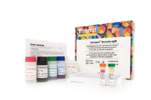
The test kit Vitrotest® Borrelia-IgM is an enzyme linked immunosorbent assay (ELISA) for the qualitative and semiquantitative determination of IgM class antibodies to Borrelia burgdorferi sensu lato in human serum or plasma.
Determination of IgM antibodies to B. burgdorferi in the test kit Vitrotest® Borrelia-IgM is based on a solid phase indirect ELISA in a two-step incubation procedure.
○ ТК085 - 96 tests- Solid phase: breakable microplate ELISA is coated with Borrelia burgdorferi sensu lato antigens.
- Conjugate: a monoclonal antibodies to human IgM conjugated to horseradish peroxidase.
- Chromogen: ready to use TMB solution.
- Volume of sample for analysis: 10 μl.
- Assay time: 1h 15 min.
Lyme disease (tick-borne borreliosis) is the most common tick-borne disease in the Northern hemisphere. The causative agent of borreliosis is spirochetes of the Borrelia burgdorferi group - were first identified in 1982 by the American microbiologist Willie Burgdorfer. Today, the most pathogenic spirochetes belong to the Borrelia burgdorferi sensu lato group, which includes Borrelia burgdorferi sensu stricto, Borrelia afzelii and Borrelia garinii.
Lyme borreliosis is a progressing disease which encompasses several clinical syndromes (erythema migrans (EM), neuroborreliosis, Lyme arthritis, and acrodermatitis chronica atrophicans). The most common clinical manifestation of early localized LD is EM, which appears in approximately 70% of patients 3-30 (average 7) days after bite. EM is a rash which appears at the site of the tick bite and gradually expands to a ring with a central clear zone. If the infection remains unnoticed and untreated in this early localized stage, B. burgdorferi can spread to other tissues and organs. The second, so-called early disseminated stage of the disease causes more severe manifestations that can involve the skin, nervous system, joints, or heart. This stage is mainly characterized by neurological signs (neuroborreliosis) or joint aches (Lyme arthritis). The third stadium is late disseminated LD. It’s typical presentations are acrodermatitis chronica atrophicans and arthritis. Manifestations of LD are similar to such immune-mediated disorders as rheumatoid arthritis, multiple sclerosis and systemic lupus.
Ticks most frequently acquire spirochetes from infected rodents during their larval feeding. Once infected, a tick can transmit infection throughout its life.
Not all ticks of competent species are carriers of an infectious agent. The prevalence of B. burgdorferi among ticks was measured to be as different as 19% and 60%. The probability to be infected is regarded as high if the tick remains attached to skin for more than 24 hours.
People living in or visiting rural areas, particularly campers and hikers, are most at risk. The infection risk appears not only in forests but more and more in parks, which attract more attention from the public. Ixodes ticks are the only natural agents through which humans have been shown to become infected. There is no evidence that LD is transmitted from person-to-person.
B.burgdorferi infection is known to induce strong humoral immune response in the host. IgM titres are typically highest between 3rd and 6th week after disease onset and then decline, however in many patients IgM remains high during later manifestations of illness, reflecting disease activity. IgG titres start increasing 1-2 weeks after onset, but reach highest titers only months later when arthritis is already present.
If the patient is cured, antibody levels in the first weeks after convalescence increase, and only then begin to decrease. If LD reaches an advanced stage, IgM and especially IgG are still detectable after cure for many years in the majority of patients. On the contrary, prompt antibiotic treatment at localized stage often aborts the development of a sustained humoral response, resulting in an absence of IgG and IgM after cure. Hence, reinfection is possible after successfully treated early, but not late LD.
B. burgdorferi genome encodes around 1500 proteins; almost half of them are plasmid encoded. Of these proteins, around 100 are immunogenic. Significant serological cross-reactivity exists between B.burdhoferi and causative agents of human spirochetal infections, particularly relapsing fever and syphilis.
Only several proteins are proven specific antigens and therefore valuable for serodiagnosis. These proteins include FlaB (BB0147), the P66 outer membrane protein (BB0603), OspA and OspB (BBA15 and BBA16), decorinbinding protein B (BBA25), OspC (BBB19), fibronectin-binding protein (BBK32), and VlsE (BBF33). FlaB is a part of flagellar apparatus; the rest are outer membrane proteins.
Currently, there is no “gold standard” in the diagnostics of LD. LD diagnosis is based upon:
a) signs and symptoms;
b) history of exposure to infectious ticks;
c) laboratory diagnosis.
The only manifestation characteristic to LD is EM, whereas clinical features of later stage presentations are not unique to B. burgdorferi infection. Unfortunately, only around 70% of patients develop EM; many patients present to the clinics at disseminated stage. Exposure to ticks may easily left unnoticed as many humans are bitten by ticks at their juvenile stage, called nymphs, which are much smaller in size. Therefore, laboratory support of LD must be sought by isolation of B. burgdorferi in culture, PCR or immunoassays.
Assays available for serology are ELISAs (or EIAs) and indirect fluorescent antibody (IFA) assays. Their performance is best at the late stages of LB. ELISAs are available as first, second or third generation tests. First generation ELISAs use whole cell lysates as antigens; second generation ELISAs use native purified antigens; and third generation ELISAs use recombinant or synthetic antigens. Immunoblots are mainly used as confirmatory tests. Apart from testing for antibody response in serum, antibody response may also be measured in cerebrospinal fluid.