-
The test-kit Vitrotest® Helicobacter screen is an enzyme linked immunosorbent assay (ELISA) for the detection of IgG, IgA and IgM specific antibodies to Helicobacter pylori in human serum or plasma.
Detection of antibodies to H. pylori in the test-kit Vitrotest® Helicobacter screen is based on a solid phase, indirect ELISA in a two-step incubation procedure.
○ ТК045 - 96 tests- Solid phase: breakable microplate ELISA is coated with recombinant antigens of H. pylori.
- Conjugate: a monoclonal antibodies to human IgA, IgG and IgM conjugated to horseradish peroxidase.
- Chromogen: ready to use TMB solution.
- Volume of sample for analysis: 10 μl.
- Assay time: 1h 15 min.
Helicobacter pylori is a widespread microorganism with which the half of the world’s population has been infected. Its prevalence is very high in developing countries and is quite low in the developed world. According to the World Gastroenterology Organization, the rate of infected adults in Eastern Europe and Asia is about 70-80 %.
Studies during recent decades have shown the key role of bacterium Helicobacter pylori in the pathogenesis of stomach and duodenal lesions. H. pylori is detected almost in 100% of adult patients with duodenal ulcer, approximately in 80 % of patients with peptic ulcer, in 92% of patients with gastric cancer and in 92 % of patients with active chronic gastritis. Research has demonstrated that elimination of helicobacter leads to the disappearance of gastritis and significant reduction in the incidence of duodenal ulcer recurrence.
Helicobacteriosis is a chronic infection with long, often asymptomatic course. Its symptoms do not differ from clinical manifestations of gastro-duodenitis since usual constant pain in the epigastrium occurs. H. pylori is often present in patients with no clinical manifestations of disease.
The infection usually starts from non–acid-secreting antral region of the stomach and stimulates the increased release of gastrin. The increased gastrin levels in turn stimulate excess acid secretion from the more proximal acid-secreting fundic mucosa which is relatively free of inflammation. The increased duodenal acid load damages the duodenal mucosa, causing ulceration. If infection progresses, stomach body is damaged, which could finally cause the development of gastric adenocarcinoma. This tumour is preceded by sequential pathological changes of gastric mucosa, from normal mucosa to superficial gastritis, atrophic gastritis, gastric ulcers and intestinal metaplasia.
Main oncogenic factors are both nitrosating bacteria present in the lumen of hypochloric stomach, capable of generating potentially carcinogenic N-nitrosamines and reactive oxygen species, and H. pylori itself. Duodenal or gastric ulcers are reported to develop in 1 to 10% of infected patients, gastric cancer- in 0.1 to 3%; at the same time, the great majority of patients with H. pylori remain asymptomatic carriers.
H. pylori is usually acquired in childhood. The bacteria are most likely spread through fecal-oral or oral-oral routes from person to person; additional transmission routes, such as water, may be important in developing countries. -
The test kit Vitrotest® Helicobacter-IgG is an enzyme linked immunosorbent assay (ELISA) for the quantitative and semiquantitative determination of IgG class antibodies to CagA protein of Helicobacter pylori in human serum or plasma.
Determination of IgG antibodies to CagA protein of H. pylori in the test kit Vitrotest® Helicobacter-IgG is based on a solid phase, indirect ELISA in a two-step incubation procedure.
○ ТК046 - 96 tests- Solid phase: breakable microplate ELISA is coated with recombinant CagA protein of H. pylori.
- Conjugate: a monoclonal antibodies to human IgG conjugated to horseradish peroxidase.
- Chromogen: ready to use TMB solution.
- Volume of sample for analysis: 10 μl.
- Assay time: 1h 15 min.
Helicobacter pylori is a widespread microorganism with which the half of the world’s population has been infected. Its prevalence is very high in developing countries and is quite low in the developed world. According to the World Gastroenterology Organization, the rate of infected adults in Eastern Europe and Asia is about 70-80 %.
Studies during recent decades have shown the key role of bacterium Helicobacter pylori in the pathogenesis of stomach and duodenal lesions. H. pylori is detected almost in 100% of adult patients with duodenal ulcer, approximately in 80 % of patients with peptic ulcer, in 92% of patients with gastric cancer and in 92 % of patients with active chronic gastritis. Research has demonstrated that elimination of helicobacter leads to the disappearance of gastritis and significant reduction in the incidence of duodenal ulcer recurrence.
Helicobacteriosis is a chronic infection with long, often asymptomatic course. Its symptoms do not differ from clinical manifestations of gastro-duodenitis since usual constant pain in the epigastrium occurs. H. pylori is often present in patients with no clinical manifestations of disease.
The infection usually starts from non–acid-secreting antral region of the stomach and stimulates the increased release of gastrin. The increased gastrin levels in turn stimulate excess acid secretion from the more proximal acid-secreting fundic mucosa which is relatively free of inflammation. The increased duodenal acid load damages the duodenal mucosa, causing ulceration. If infection progresses, stomach body is damaged, which could finally cause the development of gastric adenocarcinoma. This tumour is preceded by sequential pathological changes of gastric mucosa, from normal mucosa to superficial gastritis, atrophic gastritis, gastric ulcers and intestinal metaplasia.
Main oncogenic factors are both nitrosating bacteria present in the lumen of hypochloric stomach, capable of generating potentially carcinogenic N-nitrosamines and reactive oxygen species, and H. pylori itself. Duodenal or gastric ulcers are reported to develop in 1 to 10% of infected patients, gastric cancer- in 0.1 to 3%; at the same time, the great majority of patients with H. pylori remain asymptomatic carriers.
H. pylori is usually acquired in childhood. The bacteria are most likely spread through fecal-oral or oral-oral routes from person to person; additional transmission routes, such as water, may be important in developing countries. -
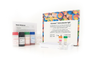
The test kit Vitrotest® Helicobacter-IgА is an enzyme linked immunosorbent assay (ELISA) for the qualitative and semiquantitative determination of IgА class antibodies to CagA protein of Helicobacter pylori in human serum or plasma.
Determination of IgА antibodies to CagA protein of H. pylori in the test kit Vitrotest® Helicobacter-IgА is based on a solid phase, indirect ELISA in a two-step incubation procedure.
○ ТК047 - 96 tests- Solid phase: breakable microplate ELISA is coated with recombinant CagA protein of H. pylori.
- Conjugate: a monoclonal antibodies to human IgА conjugated to horseradish peroxidase.
- Chromogen: ready to use TMB solution.
- Volume of sample for analysis: 10 μl.
- Assay time: 1h 15 min.
Helicobacter pylori is a widespread microorganism with which the half of the world’s population has been infected. Its prevalence is very high in developing countries and is quite low in the developed world. According to the World Gastroenterology Organization, the rate of infected adults in Eastern Europe and Asia is about 70-80 %.
Studies during recent decades have shown the key role of bacterium Helicobacter pylori in the pathogenesis of stomach and duodenal lesions. H. pylori is detected almost in 100% of adult patients with duodenal ulcer, approximately in 80 % of patients with peptic ulcer, in 92% of patients with gastric cancer and in 92 % of patients with active chronic gastritis. Research has demonstrated that elimination of helicobacter leads to the disappearance of gastritis and significant reduction in the incidence of duodenal ulcer recurrence.
Helicobacteriosis is a chronic infection with long, often asymptomatic course. Its symptoms do not differ from clinical manifestations of gastro-duodenitis since usual constant pain in the epigastrium occurs. H. pylori is often present in patients with no clinical manifestations of disease.
The infection usually starts from non–acid-secreting antral region of the stomach and stimulates the increased release of gastrin. The increased gastrin levels in turn stimulate excess acid secretion from the more proximal acid-secreting fundic mucosa which is relatively free of inflammation. The increased duodenal acid load damages the duodenal mucosa, causing ulceration. If infection progresses, stomach body is damaged, which could finally cause the development of gastric adenocarcinoma. This tumour is preceded by sequential pathological changes of gastric mucosa, from normal mucosa to superficial gastritis, atrophic gastritis, gastric ulcers and intestinal metaplasia.
Main oncogenic factors are both nitrosating bacteria present in the lumen of hypochloric stomach, capable of generating potentially carcinogenic N-nitrosamines and reactive oxygen species, and H. pylori itself. Duodenal or gastric ulcers are reported to develop in 1 to 10% of infected patients, gastric cancer- in 0.1 to 3%; at the same time, the great majority of patients with H. pylori remain asymptomatic carriers.
H. pylori is usually acquired in childhood. The bacteria are most likely spread through fecal-oral or oral-oral routes from person to person; additional transmission routes, such as water, may be important in developing countries. -
The test kit Vitrotest® Helicobacter-IgМ is an enzyme linked immunosorbent assay (ELISA) for the qualitative and semiquantitative determination of IgМ class antibodies to CagA protein of Helicobacter pylori in human serum or plasma.
Determination of IgМ antibodies to CagA protein of H. pylori in the test kit Vitrotest® Helicobacter-IgМ is based on a solid phase, indirect ELISA in a two-step incubation procedure.
○ ТК048 - 96 tests- Solid phase: breakable microplate ELISA is coated with recombinant CagA protein of H. pylori.
- Conjugate: a monoclonal antibodies to human IgМ conjugated to horseradish peroxidase.
- Chromogen: ready to use TMB solution.
- Volume of sample for analysis: 10 μl.
- Assay time: 1h 15 min.
Helicobacter pylori is a widespread microorganism with which the half of the world’s population has been infected. Its prevalence is very high in developing countries and is quite low in the developed world. According to the World Gastroenterology Organization, the rate of infected adults in Eastern Europe and Asia is about 70-80 %.
Studies during recent decades have shown the key role of bacterium Helicobacter pylori in the pathogenesis of stomach and duodenal lesions. H. pylori is detected almost in 100% of adult patients with duodenal ulcer, approximately in 80 % of patients with peptic ulcer, in 92% of patients with gastric cancer and in 92 % of patients with active chronic gastritis. Research has demonstrated that elimination of helicobacter leads to the disappearance of gastritis and significant reduction in the incidence of duodenal ulcer recurrence.
Helicobacteriosis is a chronic infection with long, often asymptomatic course. Its symptoms do not differ from clinical manifestations of gastro-duodenitis since usual constant pain in the epigastrium occurs. H. pylori is often present in patients with no clinical manifestations of disease.
The infection usually starts from non–acid-secreting antral region of the stomach and stimulates the increased release of gastrin. The increased gastrin levels in turn stimulate excess acid secretion from the more proximal acid-secreting fundic mucosa which is relatively free of inflammation. The increased duodenal acid load damages the duodenal mucosa, causing ulceration. If infection progresses, stomach body is damaged, which could finally cause the development of gastric adenocarcinoma. This tumour is preceded by sequential pathological changes of gastric mucosa, from normal mucosa to superficial gastritis, atrophic gastritis, gastric ulcers and intestinal metaplasia.
Main oncogenic factors are both nitrosating bacteria present in the lumen of hypochloric stomach, capable of generating potentially carcinogenic N-nitrosamines and reactive oxygen species, and H. pylori itself. Duodenal or gastric ulcers are reported to develop in 1 to 10% of infected patients, gastric cancer- in 0.1 to 3%; at the same time, the great majority of patients with H. pylori remain asymptomatic carriers.
H. pylori is usually acquired in childhood. The bacteria are most likely spread through fecal-oral or oral-oral routes from person to person; additional transmission routes, such as water, may be important in developing countries. -
The test kit Vitrotest® Anti-Lamblia is an enzyme linked immunosorbent assay (ELISA) for the detection of total antibodies to Giardia lamblia (intestinalis) in human serum or plasma.
Determination of total antibodies to Giardia lamblia in the test kit Vitrotest® Anti-Lamblia is based on a solid phase, indirect ELISA in a two-step incubation procedure.
○ ТК030 - 96 tests
○ ТК108 - 192 tests- Solid phase: breakable microplate ELISA is coated Giardia lamblia purified antigens.
- Conjugate: a monoclonal antibodies to human IgG, IgA and IgM conjugated to horseradish peroxidase.
- Chromogen: ready to use TMB solution.
- Volume of sample for analysis 10 μl.
- Assay time: 1h 15 min.
Giardia lamblia (intestinalis) causes giardiasis, a parasitic infestation that can manifest as latent parasitic carriage or in more pronounced forms, such as intestinal dysfunction. Giardiasis has been recorded on all five continents and in most countries worldwide. Infection rates vary from less than 1% to 50%. In many developing countries, where basic sanitary conditions are lacking, infection with Giardia in children by the age of 2 is almost universal. In contrast, in developed countries, the infection rate of G.lamblia is only 3–7%. The disease affects all age groups, but preschool-aged children are the most affected demographic.
The primary route of transmission for G.lamblia is fecal-oral. The parasite has a simple two-stage life cycle. After the host ingests cysts, they release trophozoites in the duodenum, which then attach to the mucosal lining of the small intestine.
Trophozoites exist only on the surface of the mucosa in the upper part of the small intestine. Therefore, Giardia mechanically blocks the mucosa and disrupts membrane digestion and the motor activity of the small intestine. Giardia impairs the absorption of fats, carbohydrates, vitamins C and B12, and leads to secondary bacterial infections. Symptoms of giardiasis can include diarrhea, fatigue, bloating, apathy, weight loss, decreased appetite, pallor, and muscle twitching. From the gastrointestinal tract perspective, giardiasis mainly manifests as enterocolitis with catarrhal symptoms.
Numerous findings indicate the role of the humoral immune response in the elimination of G.lamblia. As shown in an experimental human infection model, IgM antibody levels significantly increased on days 14-21 post-infection and gradually declined following therapy. In contrast, IgG antibody levels remained elevated after successful treatment. The dynamics of IgA levels were similar to those of IgM.
The diagnosis of giardiasis is based on clinical history, symptoms, and the presence of cysts in fecal samples or trophozoites in material obtained from the small intestine during duodenal aspiration or duodenal biopsy. Alternative methods include the detection of G.lamblia antigen in feces and the determination of specific antibodies against Giardia in the patient’s serum. Serological testing is considered a valuable adjunct in the diagnosis of giardiasis. In addition to aiding in diagnosis, it can be useful for assessing the patient’s immune response and for epidemiological studies. -
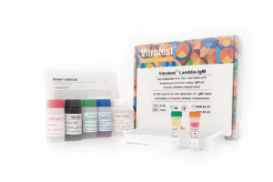
The test kit Vitrotest® Lamblia-IgM is an enzyme linked immunosorbent assay (ELISA) for the detection of IgM class antibodies to Giardia lamblia (intestinalis) in human serum or plasma.
Determination of IgM antibodies to Giardia lamblia in the test kit Vitrotest® Lamblia-IgM is based on a solid phase, indirect ELISA in a two-step incubation procedure.
○ ТК031 - 96 tests- Solid phase: breakable microplate ELISA is coated Giardia lamblia purified antigens.
- Conjugate: a monoclonal antibodies to human IgM conjugated to horseradish peroxidase.
- Chromogen: ready to use TMB solution.
- Volume of sample for analysis 10 μl.
- Assay time: 1h 15 min.
Giardia lamblia (intestinalis) causes giardiasis, a parasitic infestation that can manifest as latent parasitic carriage or in more pronounced forms, such as intestinal dysfunction. Giardiasis has been recorded on all five continents and in most countries worldwide. Infection rates vary from less than 1% to 50%. In many developing countries, where basic sanitary conditions are lacking, infection with Giardia in children by the age of 2 is almost universal. In contrast, in developed countries, the infection rate of G.lamblia is only 3–7%. The disease affects all age groups, but preschool-aged children are the most affected demographic.
The primary route of transmission for G.lamblia is fecal-oral. The parasite has a simple two-stage life cycle. After the host ingests cysts, they release trophozoites in the duodenum, which then attach to the mucosal lining of the small intestine.
Trophozoites exist only on the surface of the mucosa in the upper part of the small intestine. Therefore, Giardia mechanically blocks the mucosa and disrupts membrane digestion and the motor activity of the small intestine. Giardia impairs the absorption of fats, carbohydrates, vitamins C and B12, and leads to secondary bacterial infections. Symptoms of giardiasis can include diarrhea, fatigue, bloating, apathy, weight loss, decreased appetite, pallor, and muscle twitching. From the gastrointestinal tract perspective, giardiasis mainly manifests as enterocolitis with catarrhal symptoms.
Numerous findings indicate the role of the humoral immune response in the elimination of G.lamblia. As shown in an experimental human infection model, IgM antibody levels significantly increased on days 14-21 post-infection and gradually declined following therapy. In contrast, IgG antibody levels remained elevated after successful treatment. The dynamics of IgA levels were similar to those of IgM.
The diagnosis of giardiasis is based on clinical history, symptoms, and the presence of cysts in fecal samples or trophozoites in material obtained from the small intestine during duodenal aspiration or duodenal biopsy. Alternative methods include the detection of G.lamblia antigen in feces and the determination of specific antibodies against Giardia in the patient’s serum. Serological testing is considered a valuable adjunct in the diagnosis of giardiasis. In addition to aiding in diagnosis, it can be useful for assessing the patient’s immune response and for epidemiological studies.