-
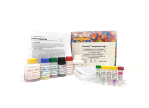
The test kit Vitrotest® Toxoplasma-IgG is an enzyme linked immunosorbent assay (ELISA) for the quantitative determination of IgG class antibodies to Toxoplasma gondii in human serum or plasma.
Determination of IgG antibodies to Toxoplasma gondii in the test kit Vitrotest® Toxoplasma-IgG is based on a solid phase, indirect ELISA in a two-step incubation procedure.
○ ТК001 - 96 tests- Solid phase: breakable microplate ELISA is coated Toxoplasma gondii antigens.
- Conjugate: a monoclonal antibodies to human IgG conjugated to horseradish peroxidase.
- Chromogen: ready to use TMB solution.
- Volume of sample for analysis: 10 μl.
- Assay time: 1h 15 min.
Toxoplasma gondii is a protozoan parasite firstly described in 1909 by Charles Nicolle and Louis Manceaux. T. gondii is an intracellular parasite, which has a complex life cycle consisting of three stages:
a) tachyzoite - this form of the parasite invades and replicates within cells during the acute stage of infection;
b) bradyzoite - present in tissue cysts;
c) sporozoite - is found in environmentally resistant oocysts excreted by the members of the family Felidae (including domestic and feral cats), which are the only definitive hosts of T. gondii.
Incubation period of primary toxoplasmosis is 5–23 days. Central nervous system (CNS), retina and cardiac/skeletal muscles are the main and ultimate target organs for tachyzoites. Symptoms, if they arise, in otherwise healthy people include: fever, mild flu-like symptoms, enlarged lymph nodes in the head and neck, muscle pain, pneumonia, and disturbances of CNS. Tissue cysts in immunocompetent patients usually remain latent for life. In immunosuppressed individuals (patients with AIDS or lymphoproliferative disorders, organ transplant patients) the course of toxoplasmosis is less favourable, often resulting in meningoencephalitis. In this group of patients toxoplasmosis is mostly a result of reactivation of pre-existing latent T. gondii infections. Another group at risk are pregnant females. Damage to the unborn child is sometimes severe and pregnancy can result in miscarriage, stillbirth, or a child born with signs of toxoplasmosis. The classic triad of signs suggestive of congenital toxoplasmosis (CT) include chorioretinitis, intracranial calcifications, and hydrocephalus. Although most severe cases are diagnosed during the first month of life, severe disease can sometimes only become obvious in the second or third month of life. Visual impairment is the most common long-term sequelae, and can greatly impact the quality of life of congenitally infected children.
Women infected with T. gondii before conception, with rare exceptions, do not transmit the infection to their fetuses. Women infected with T. gondii less than 6 months before conception or after it can transmit the infection across the placenta to their fetuses. The overall risk of transmission of CT from mother acutely ill with toxoplasmosis to fetus could be as high as 50-60% in untreated cases. The frequency of vertical transmission increases with the gestational age. In contrast, more severe clinical signs in the infected infant are more commonly observed in offspring of women whose infection was acquired early in gestation.
Toxoplasma can be transmitted to humans via three principal routes:
a) foodborne-by ingestion of raw or inadequately cooked infected meat;
b)catborne- due to ingestion of oocysts that cats pass in their feces through exposure to cat litter or soil;
c) vertical-an infected pregnant woman passing the infection to her fetus.
The infection leaves a long-lasting immunity except for cases of infection with more virulent strain. It is estimated that more than a third of the world’s population has been infected with the parasite, but infection is unevenly distributed across countries, being mostly in the range of 10-80% .The incidence of CT per 10,000 live births also varies significantly across countries. In the USA, it is estimated in one case per 10,000 live births. In France, it was reported in 2.9 per 10,000 live births. In certain countries, such as Brazil, rates as high as nine per 10,000 live births have been reported, while in other areas, such as the UK, CT was estimated in 0.33-1 per 10,000.
A well-orchestrated cellular, humoral and innate immune response must be triggered upon parasite invasion, in order to prevent the uncontrolled proliferation of tachyzoites. IgM are considered as the earliest immunoglobulines in acute infection, since they are produced during the first week after infection, and become undetectable after several months. IgA antibodies show kinetics similar to IgM. IgG can usually be detected 2–3 weeks after IgM, then decreases to reach a plateau in 2–3 months, leading to persistent detectability.
Acute toxoplasmosis in immunocompetent patients is rarely diagnosed by directly detecting the parasite in body fluids, tissue, or secretions; the clinical symptoms are also not specific enough. Therefore, the most common methods of diagnosis are based on antibody detection. A number of serological methods are used to detect antibodies to T. gondii (dye test, indirect immunofluorescence assays, agglutination assays, such as the immunosorbent agglutination assay (ISAGA), immunoenzyme assays). IgG and IgM ELISA are the methods most often used in non-specialized laboratories, whereas many other serological methods should be used by trained personnel of reference laboratories.
In immunosuppressed patients, antibody production can be affected. Therefore, serological tests may not be as efficient as in immunocompetent people. Hence, direct evidence of the parasites must be sought by PCR or parasite isolation.
Serological methods are mostly used in following clinical situations:
1) In pregnant women-to determine serological status before conception/in the first trimester of pregnancy.
2) In pregnant seronegative women-to carry out systematic screening for possible CT during pregnancy.
3) In newborns and infants-to diagnose possible CT (IgG at 12 months is a ‘gold standard’ for ultimate and definite CT diagnosis).
4) In organ donors and recipients-to determine serological status before transplantation.
In the case of CT the early diagnosis and treatment has proved to be efficient. In countries where serological screening and prenatal treatment is systematically offered to pregnant women, such as France, the majority of the cases of congenital T. gondii infection do not have overt clinical disease during gestation or the neonatal period. In Austria, nearly all women who become pregnant are serologically screened early in pregnancy and, if found to be negative initially, are tested again during the second and third trimesters. Women with T. gondii infections are treated as soon as infection is detected. These measures helped reduce the incidence of CT from 50-70 cases per 10,000 births before the program to 1 per 10,000 births thereafter.
It needs to be emphasized that a positive IgM antibody test result does not necessarily mean a recently acquired infection. IgM antibodies may persist for 1 year following acute infection, and most positive IgM antibody test results ( 60%) are obtained in pregnant women with past infection. Therefore, IgM results should not be used by alone; the greatest value of a positive IgM antibody test result is that it raises the question of a recently acquired infection, thereby necessitating confirmatory testing, for example, IgG avidity test.
High-avidity IgG antibodies develop at least 12–16 weeks after infection. Therefore, the presence of high-avidity antibodies in the serum taken during the first trimester indicates that infection was acquired before pregnancy, and the risk for fetus is negligible. Low-avidity IgG results indicate the necessity for further tests, since the peculiarity of T. gondii infection is the persistence of low-avidity antibodies in some individuals for many months-year or more after the primary infection. -
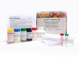
The test kit Vitrotest® Toxoplasma-IgM is an enzyme linked immunosorbent assay (ELISA) for the qualitative and semiquantitative determination of IgM class antibodies to Toxoplasma gondii in human serum or plasma.
Determination of IgM antibodies to Toxoplasma gondii in the test kit Vitrotest® Toxoplasma-IgM is based on a solid phase, indirect ELISA in a two-step incubation procedure.
○ ТК002 - 96 tests- Solid phase: breakable microplate ELISA is coated Toxoplasma gondii antigens.
- Conjugate: a monoclonal antibodies to human IgM conjugated to horseradish peroxidase.
- Chromogen: ready to use TMB solution.
- Volume of sample for analysis: 10 μl.
- Assay time: 1h 15 min.
Toxoplasma gondii is a protozoan parasite firstly described in 1909 by Charles Nicolle and Louis Manceaux. T. gondii is an intracellular parasite, which has a complex life cycle consisting of three stages:
a) tachyzoite - this form of the parasite invades and replicates within cells during the acute stage of infection;
b) bradyzoite - present in tissue cysts;
c) sporozoite - is found in environmentally resistant oocysts excreted by the members of the family Felidae (including domestic and feral cats), which are the only definitive hosts of T. gondii.
Incubation period of primary toxoplasmosis is 5–23 days. Central nervous system (CNS), retina and cardiac/skeletal muscles are the main and ultimate target organs for tachyzoites. Symptoms, if they arise, in otherwise healthy people include: fever, mild flu-like symptoms, enlarged lymph nodes in the head and neck, muscle pain, pneumonia, and disturbances of CNS. Tissue cysts in immunocompetent patients usually remain latent for life. In immunosuppressed individuals (patients with AIDS or lymphoproliferative disorders, organ transplant patients) the course of toxoplasmosis is less favourable, often resulting in meningoencephalitis. In this group of patients toxoplasmosis is mostly a result of reactivation of pre-existing latent T. gondii infections. Another group at risk are pregnant females. Damage to the unborn child is sometimes severe and pregnancy can result in miscarriage, stillbirth, or a child born with signs of toxoplasmosis. The classic triad of signs suggestive of congenital toxoplasmosis (CT) include chorioretinitis, intracranial calcifications, and hydrocephalus. Although most severe cases are diagnosed during the first month of life, severe disease can sometimes only become obvious in the second or third month of life. Visual impairment is the most common long-term sequelae, and can greatly impact the quality of life of congenitally infected children.
Women infected with T. gondii before conception, with rare exceptions, do not transmit the infection to their fetuses. Women infected with T. gondii less than 6 months before conception or after it can transmit the infection across the placenta to their fetuses. The overall risk of transmission of CT from mother acutely ill with toxoplasmosis to fetus could be as high as 50-60% in untreated cases. The frequency of vertical transmission increases with the gestational age. In contrast, more severe clinical signs in the infected infant are more commonly observed in offspring of women whose infection was acquired early in gestation.
Toxoplasma can be transmitted to humans via three principal routes:
a) foodborne-by ingestion of raw or inadequately cooked infected meat;
b)catborne- due to ingestion of oocysts that cats pass in their feces through exposure to cat litter or soil;
c) vertical-an infected pregnant woman passing the infection to her fetus.
The infection leaves a long-lasting immunity except for cases of infection with more virulent strain. It is estimated that more than a third of the world’s population has been infected with the parasite, but infection is unevenly distributed across countries, being mostly in the range of 10-80% .The incidence of CT per 10,000 live births also varies significantly across countries. In the USA, it is estimated in one case per 10,000 live births. In France, it was reported in 2.9 per 10,000 live births. In certain countries, such as Brazil, rates as high as nine per 10,000 live births have been reported, while in other areas, such as the UK, CT was estimated in 0.33-1 per 10,000.
A well-orchestrated cellular, humoral and innate immune response must be triggered upon parasite invasion, in order to prevent the uncontrolled proliferation of tachyzoites. IgM are considered as the earliest immunoglobulines in acute infection, since they are produced during the first week after infection, and become undetectable after several months. IgA antibodies show kinetics similar to IgM. IgG can usually be detected 2–3 weeks after IgM, then decreases to reach a plateau in 2–3 months, leading to persistent detectability.
Acute toxoplasmosis in immunocompetent patients is rarely diagnosed by directly detecting the parasite in body fluids, tissue, or secretions; the clinical symptoms are also not specific enough. Therefore, the most common methods of diagnosis are based on antibody detection. A number of serological methods are used to detect antibodies to T. gondii (dye test, indirect immunofluorescence assays, agglutination assays, such as the immunosorbent agglutination assay (ISAGA), immunoenzyme assays). IgG and IgM ELISA are the methods most often used in non-specialized laboratories, whereas many other serological methods should be used by trained personnel of reference laboratories.
In immunosuppressed patients, antibody production can be affected. Therefore, serological tests may not be as efficient as in immunocompetent people. Hence, direct evidence of the parasites must be sought by PCR or parasite isolation.
Serological methods are mostly used in following clinical situations:
1) In pregnant women-to determine serological status before conception/in the first trimester of pregnancy.
2) In pregnant seronegative women-to carry out systematic screening for possible CT during pregnancy.
3) In newborns and infants-to diagnose possible CT (IgG at 12 months is a ‘gold standard’ for ultimate and definite CT diagnosis).
4) In organ donors and recipients-to determine serological status before transplantation.
In the case of CT the early diagnosis and treatment has proved to be efficient. In countries where serological screening and prenatal treatment is systematically offered to pregnant women, such as France, the majority of the cases of congenital T. gondii infection do not have overt clinical disease during gestation or the neonatal period. In Austria, nearly all women who become pregnant are serologically screened early in pregnancy and, if found to be negative initially, are tested again during the second and third trimesters. Women with T. gondii infections are treated as soon as infection is detected. These measures helped reduce the incidence of CT from 50-70 cases per 10,000 births before the program to 1 per 10,000 births thereafter.
It needs to be emphasized that a positive IgM antibody test result does not necessarily mean a recently acquired infection. IgM antibodies may persist for 1 year following acute infection, and most positive IgM antibody test results ( 60%) are obtained in pregnant women with past infection. Therefore, IgM results should not be used by alone; the greatest value of a positive IgM antibody test result is that it raises the question of a recently acquired infection, thereby necessitating confirmatory testing, for example, IgG avidity test.
High-avidity IgG antibodies develop at least 12–16 weeks after infection. Therefore, the presence of high-avidity antibodies in the serum taken during the first trimester indicates that infection was acquired before pregnancy, and the risk for fetus is negligible. Low-avidity IgG results indicate the necessity for further tests, since the peculiarity of T. gondii infection is the persistence of low-avidity antibodies in some individuals for many months-year or more after the primary infection. -
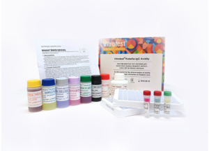
The test kit Vitrotest® Toxoplasma-IgG Avidity is an enzyme linked immunosorbent assay (ELISA) for the determination of avidity index of IgG class antibodies to Toxoplasma gondii in human serum or plasma.
Determination of avidity index of IgG antibodies to Toxoplasma gondii in the test kit Vitrotest® Toxoplasma-IgG Avidity is based on a solid phase, indirect ELISA.
○ ТК064 - 48 tests- Solid phase: breakable microplate ELISA is coated Toxoplasma gondii antigens.
- Conjugate: a monoclonal antibodies to human IgG conjugated to horseradish peroxidase.
- Chromogen: ready to use TMB solution.
- Volume of sample for analysis: 10 μl.
- Assay time: 1h 30 min.
Toxoplasma gondii is a protozoan parasite firstly described in 1909 by Charles Nicolle and Louis Manceaux. T. gondii is an intracellular parasite, which has a complex life cycle consisting of three stages:
a) tachyzoite - this form of the parasite invades and replicates within cells during the acute stage of infection;
b) bradyzoite - present in tissue cysts;
c) sporozoite - is found in environmentally resistant oocysts excreted by the members of the family Felidae (including domestic and feral cats), which are the only definitive hosts of T. gondii.
Incubation period of primary toxoplasmosis is 5–23 days. Central nervous system (CNS), retina and cardiac/skeletal muscles are the main and ultimate target organs for tachyzoites. Symptoms, if they arise, in otherwise healthy people include: fever, mild flu-like symptoms, enlarged lymph nodes in the head and neck, muscle pain, pneumonia, and disturbances of CNS. Tissue cysts in immunocompetent patients usually remain latent for life. In immunosuppressed individuals (patients with AIDS or lymphoproliferative disorders, organ transplant patients) the course of toxoplasmosis is less favourable, often resulting in meningoencephalitis. In this group of patients toxoplasmosis is mostly a result of reactivation of pre-existing latent T. gondii infections. Another group at risk are pregnant females. Damage to the unborn child is sometimes severe and pregnancy can result in miscarriage, stillbirth, or a child born with signs of toxoplasmosis. The classic triad of signs suggestive of congenital toxoplasmosis (CT) include chorioretinitis, intracranial calcifications, and hydrocephalus. Although most severe cases are diagnosed during the first month of life, severe disease can sometimes only become obvious in the second or third month of life. Visual impairment is the most common long-term sequelae, and can greatly impact the quality of life of congenitally infected children.
Women infected with T. gondii before conception, with rare exceptions, do not transmit the infection to their fetuses. Women infected with T. gondii less than 6 months before conception or after it can transmit the infection across the placenta to their fetuses. The overall risk of transmission of CT from mother acutely ill with toxoplasmosis to fetus could be as high as 50-60% in untreated cases. The frequency of vertical transmission increases with the gestational age. In contrast, more severe clinical signs in the infected infant are more commonly observed in offspring of women whose infection was acquired early in gestation.
Toxoplasma can be transmitted to humans via three principal routes:
a) foodborne-by ingestion of raw or inadequately cooked infected meat;
b)catborne- due to ingestion of oocysts that cats pass in their feces through exposure to cat litter or soil;
c) vertical-an infected pregnant woman passing the infection to her fetus.
The infection leaves a long-lasting immunity except for cases of infection with more virulent strain. It is estimated that more than a third of the world’s population has been infected with the parasite, but infection is unevenly distributed across countries, being mostly in the range of 10-80% .The incidence of CT per 10,000 live births also varies significantly across countries. In the USA, it is estimated in one case per 10,000 live births. In France, it was reported in 2.9 per 10,000 live births. In certain countries, such as Brazil, rates as high as nine per 10,000 live births have been reported, while in other areas, such as the UK, CT was estimated in 0.33-1 per 10,000.
A well-orchestrated cellular, humoral and innate immune response must be triggered upon parasite invasion, in order to prevent the uncontrolled proliferation of tachyzoites. IgM are considered as the earliest immunoglobulines in acute infection, since they are produced during the first week after infection, and become undetectable after several months. IgA antibodies show kinetics similar to IgM. IgG can usually be detected 2–3 weeks after IgM, then decreases to reach a plateau in 2–3 months, leading to persistent detectability.
Acute toxoplasmosis in immunocompetent patients is rarely diagnosed by directly detecting the parasite in body fluids, tissue, or secretions; the clinical symptoms are also not specific enough. Therefore, the most common methods of diagnosis are based on antibody detection. A number of serological methods are used to detect antibodies to T. gondii (dye test, indirect immunofluorescence assays, agglutination assays, such as the immunosorbent agglutination assay (ISAGA), immunoenzyme assays). IgG and IgM ELISA are the methods most often used in non-specialized laboratories, whereas many other serological methods should be used by trained personnel of reference laboratories.
In immunosuppressed patients, antibody production can be affected. Therefore, serological tests may not be as efficient as in immunocompetent people. Hence, direct evidence of the parasites must be sought by PCR or parasite isolation.
Serological methods are mostly used in following clinical situations:
1) In pregnant women-to determine serological status before conception/in the first trimester of pregnancy.
2) In pregnant seronegative women-to carry out systematic screening for possible CT during pregnancy.
3) In newborns and infants-to diagnose possible CT (IgG at 12 months is a ‘gold standard’ for ultimate and definite CT diagnosis).
4) In organ donors and recipients-to determine serological status before transplantation.
In the case of CT the early diagnosis and treatment has proved to be efficient. In countries where serological screening and prenatal treatment is systematically offered to pregnant women, such as France, the majority of the cases of congenital T. gondii infection do not have overt clinical disease during gestation or the neonatal period. In Austria, nearly all women who become pregnant are serologically screened early in pregnancy and, if found to be negative initially, are tested again during the second and third trimesters. Women with T. gondii infections are treated as soon as infection is detected. These measures helped reduce the incidence of CT from 50-70 cases per 10,000 births before the program to 1 per 10,000 births thereafter.
It needs to be emphasized that a positive IgM antibody test result does not necessarily mean a recently acquired infection. IgM antibodies may persist for 1 year following acute infection, and most positive IgM antibody test results ( 60%) are obtained in pregnant women with past infection. Therefore, IgM results should not be used by alone; the greatest value of a positive IgM antibody test result is that it raises the question of a recently acquired infection, thereby necessitating confirmatory testing, for example, IgG avidity test.
High-avidity IgG antibodies develop at least 12–16 weeks after infection. Therefore, the presence of high-avidity antibodies in the serum taken during the first trimester indicates that infection was acquired before pregnancy, and the risk for fetus is negligible. Low-avidity IgG results indicate the necessity for further tests, since the peculiarity of T. gondii infection is the persistence of low-avidity antibodies in some individuals for many months-year or more after the primary infection. -
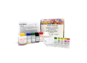
The test kit Vitrotest® Rubella-IgG is an enzyme linked immunosorbent assay (ELISA) for the quantitative determination of IgG class antibodies to Rubella virus in human serum or plasma.
Determination of IgG antibodies to Rubella virus in the test kit Vitrotest® Rubella-IgG is based on a solid phase, indirect ELISA in a two-step incubation procedure.
○ ТК003 - 96 tests- Solid phase: breakable microplate ELISA is coated with Rubella virus antigens.
- Conjugate: a monoclonal antibodies to human IgG conjugated to horseradish peroxidase.
- Chromogen: ready to use TMB solution.
- Volume of sample for analysis: 10 μl.
- Assay time: 1h 15 min.
Rubella is an acute viral infectious disease with significant teratogenic effects. The causative agent of rubella is an RNA virus Rubivirus genus Togaviridae family and was first isolated in 1962. Rubella is an air-bourne disease, takes few days to pass without a special treatment in most patients and does not cause health problems later. The main danger occurs during the first contact of a pregnant woman with the Rubella virus during the first trimester of pregnancy when the foetus is the most vulnerable to rubella. If the virus is being passed on from mother to foetus, it can be cause of a misbirth, a stillbirth or congenital rubella syndrome (CRS) - all of which comprise a group of serious deficiencies that can cause a developmental delay, mental retardation, deafness, cataracts, microcephaly, liver problems and a newborn heart disease. Because of the strong teratogenic effect of the Rubella virus a large-scale vaccination that provides permanent immunity to infection is conducted in most countries.
Extensive vaccination against rubella over the past decade has led to the near eradication of rubella and CRS in many developed countries and some developing countries. Before the introduction of vaccination, up to 4 children with CRS were born per 1,000 live births worldwide. Even now, more than 100,000 children are born with CRS annually in developing countries. In Ukraine, the incidence of rubella is about 4 cases per 100,000 population per year.
Many studies have shown that viral protection is mostly induced by neutralizing antibodies. The rubella specific IgM antibodies are usually detected within 4 days after the onset of rash and for 4–8 weeks thereafter, but in some cases these antibodies can persist for over a year. IgG antibodies appear during the acute phase (7 to 30 days postonset) and persist at varying levels for life. During viral infection, antibodies specific to three structural proteins of the rubella virus develop; the protective immune response is predominantly directed toward the glycoproteins, mainly against the glycoprotein E1.
Since the symptoms of rubella are often not specific, and many cases of rubella are asymptomatic, the diagnosis of rubella is rarely based upon clinical symptoms. The presence of rubella virus in nasal, throat, urine, blood, and cerebrospinal fluid specimens from persons with suspected rubella should be proven by virus isolation or alternatively viral nucleic acid should be detected by polymerase chain reaction. Immunological tests are by far the most popular in the diagnostics of rubella.
Serum assays are used for 3 main purposes:
1) for determining seroconversion after RV vaccination by the level of IgG;
2) for evaluation of the immunity to virus by the level of IgG, determining the need to vaccinate women at reproductive age;
3) to diagnose possible infection in pregnant women by the level of IgM and by the avidity of IgG.
Detection of rubella-specific IgM alone cannot be considered absolute proof of a recent primary infection for several reasons. IgM response after primary infection may be prolonged, lasting up to several years. Furthermore, sometimes rubella IgM is detectable in the case of mild secondary infection occuring despite a vaccination. This secondary infection is regarded as safe for the fetus. False-positive IgM results may also be artifacts due to various reasons.
This issue of doubtful IgM results is especially important when investigating suspected rubella in pregnant women because of the risk of CRS, so additional diagnostic tests should be used in such situations. Аvidity of IgG, which in most cases begins to increase 3 months following rubella infection, is another independent parameter allowing to differentiate between recent and past infection. For example, in one study low avidity specific IgG was detected in 91% of sera taken at 3–4 months after exposure to rubella virus; at 5–7 months after exposure only 21% of sera remained low avidity.
The IgG avidity assay is gaining popularity as a diagnostic method for the assessment of the time of infection. According to the recommendations of the Center for Disease Control and Prevention, USA, if IgM of the first probe is positive, avidity of IgG of the second probe, collected in 5-10 days, should be also measured. If the IgM and IgG of this second probe are positive, but the IgG avidity is high, this may indicate either a false-positive IgM result or a benign secondary infection. -
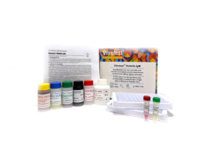
The test kit Vitrotest® Rubella-IgM is an enzyme linked immunosorbent assay (ELISA) for the qualitative and semiquantitative determination of IgM class antibodies to Rubella virus in human serum or plasma.
Determination of IgM antibodies to Rubella virus in the test kit Vitrotest® Rubella-IgM is based on a solid phase, indirect ELISA in a two-step incubation procedure.
○ ТК004 - 96 tests- Solid phase: breakable microplate ELISA is coated with Rubella virus antigens.
- Conjugate: a monoclonal antibodies to human IgM conjugated to horseradish peroxidase.
- Chromogen: ready to use TMB solution.
- Volume of sample for analysis: 10 μl.
- Assay time: 1h 15 min.
Rubella is an acute viral infectious disease with significant teratogenic effects. The causative agent of rubella is an RNA virus Rubivirus genus Togaviridae family and was first isolated in 1962. Rubella is an air-bourne disease, takes few days to pass without a special treatment in most patients and does not cause health problems later. The main danger occurs during the first contact of a pregnant woman with the Rubella virus during the first trimester of pregnancy when the foetus is the most vulnerable to rubella. If the virus is being passed on from mother to foetus, it can be cause of a misbirth, a stillbirth or congenital rubella syndrome (CRS) - all of which comprise a group of serious deficiencies that can cause a developmental delay, mental retardation, deafness, cataracts, microcephaly, liver problems and a newborn heart disease. Because of the strong teratogenic effect of the Rubella virus a large-scale vaccination that provides permanent immunity to infection is conducted in most countries.
Extensive vaccination against rubella over the past decade has led to the near eradication of rubella and CRS in many developed countries and some developing countries. Before the introduction of vaccination, up to 4 children with CRS were born per 1,000 live births worldwide. Even now, more than 100,000 children are born with CRS annually in developing countries. In Ukraine, the incidence of rubella is about 4 cases per 100,000 population per year.
Many studies have shown that viral protection is mostly induced by neutralizing antibodies. The rubella specific IgM antibodies are usually detected within 4 days after the onset of rash and for 4–8 weeks thereafter, but in some cases these antibodies can persist for over a year. IgG antibodies appear during the acute phase (7 to 30 days postonset) and persist at varying levels for life. During viral infection, antibodies specific to three structural proteins of the rubella virus develop; the protective immune response is predominantly directed toward the glycoproteins, mainly against the glycoprotein E1.
Since the symptoms of rubella are often not specific, and many cases of rubella are asymptomatic, the diagnosis of rubella is rarely based upon clinical symptoms. The presence of rubella virus in nasal, throat, urine, blood, and cerebrospinal fluid specimens from persons with suspected rubella should be proven by virus isolation or alternatively viral nucleic acid should be detected by polymerase chain reaction. Immunological tests are by far the most popular in the diagnostics of rubella.
Serum assays are used for 3 main purposes:
1) for determining seroconversion after RV vaccination by the level of IgG;
2) for evaluation of the immunity to virus by the level of IgG, determining the need to vaccinate women at reproductive age;
3) to diagnose possible infection in pregnant women by the level of IgM and by the avidity of IgG.
Detection of rubella-specific IgM alone cannot be considered absolute proof of a recent primary infection for several reasons. IgM response after primary infection may be prolonged, lasting up to several years. Furthermore, sometimes rubella IgM is detectable in the case of mild secondary infection occuring despite a vaccination. This secondary infection is regarded as safe for the fetus. False-positive IgM results may also be artifacts due to various reasons.
This issue of doubtful IgM results is especially important when investigating suspected rubella in pregnant women because of the risk of CRS, so additional diagnostic tests should be used in such situations. Аvidity of IgG, which in most cases begins to increase 3 months following rubella infection, is another independent parameter allowing to differentiate between recent and past infection. For example, in one study low avidity specific IgG was detected in 91% of sera taken at 3–4 months after exposure to rubella virus; at 5–7 months after exposure only 21% of sera remained low avidity.
The IgG avidity assay is gaining popularity as a diagnostic method for the assessment of the time of infection. According to the recommendations of the Center for Disease Control and Prevention, USA, if IgM of the first probe is positive, avidity of IgG of the second probe, collected in 5-10 days, should be also measured. If the IgM and IgG of this second probe are positive, but the IgG avidity is high, this may indicate either a false-positive IgM result or a benign secondary infection. -
The test kit Vitrotest® Rubella-IgG Avidity is an enzyme linked immunosorbent assay (ELISA) for the determination of avidity index of IgG class antibodies to Rubella virus in human serum or plasma.
Determination of avidity index of IgG antibodies to Rubella virus in the test kit Vitrotest® Rubella-IgG Avidity is based on a solid phase, indirect ELISA.
○ ТК065 - 48 tests- Solid phase: breakable microplate ELISA is coated with Rubella virus antigens.
- Conjugate: a monoclonal antibodies to human IgG conjugated to horseradish peroxidase.
- Chromogen: ready to use TMB solution.
- Volume of sample for analysis: 10 μl.
- Assay time: 1h 25 min.
Rubella is an acute viral infectious disease with significant teratogenic effects. The causative agent of rubella is an RNA virus Rubivirus genus Togaviridae family and was first isolated in 1962. Rubella is an air-bourne disease, takes few days to pass without a special treatment in most patients and does not cause health problems later. The main danger occurs during the first contact of a pregnant woman with the Rubella virus during the first trimester of pregnancy when the foetus is the most vulnerable to rubella. If the virus is being passed on from mother to foetus, it can be cause of a misbirth, a stillbirth or congenital rubella syndrome (CRS) - all of which comprise a group of serious deficiencies that can cause a developmental delay, mental retardation, deafness, cataracts, microcephaly, liver problems and a newborn heart disease. Because of the strong teratogenic effect of the Rubella virus a large-scale vaccination that provides permanent immunity to infection is conducted in most countries.
Extensive vaccination against rubella over the past decade has led to the near eradication of rubella and CRS in many developed countries and some developing countries. Before the introduction of vaccination, up to 4 children with CRS were born per 1,000 live births worldwide. Even now, more than 100,000 children are born with CRS annually in developing countries. In Ukraine, the incidence of rubella is about 4 cases per 100,000 population per year.
Many studies have shown that viral protection is mostly induced by neutralizing antibodies. The rubella specific IgM antibodies are usually detected within 4 days after the onset of rash and for 4–8 weeks thereafter, but in some cases these antibodies can persist for over a year. IgG antibodies appear during the acute phase (7 to 30 days postonset) and persist at varying levels for life. During viral infection, antibodies specific to three structural proteins of the rubella virus develop; the protective immune response is predominantly directed toward the glycoproteins, mainly against the glycoprotein E1.
Since the symptoms of rubella are often not specific, and many cases of rubella are asymptomatic, the diagnosis of rubella is rarely based upon clinical symptoms. The presence of rubella virus in nasal, throat, urine, blood, and cerebrospinal fluid specimens from persons with suspected rubella should be proven by virus isolation or alternatively viral nucleic acid should be detected by polymerase chain reaction. Immunological tests are by far the most popular in the diagnostics of rubella.
Serum assays are used for 3 main purposes:
1) for determining seroconversion after RV vaccination by the level of IgG;
2) for evaluation of the immunity to virus by the level of IgG, determining the need to vaccinate women at reproductive age;
3) to diagnose possible infection in pregnant women by the level of IgM and by the avidity of IgG.
Detection of rubella-specific IgM alone cannot be considered absolute proof of a recent primary infection for several reasons. IgM response after primary infection may be prolonged, lasting up to several years. Furthermore, sometimes rubella IgM is detectable in the case of mild secondary infection occuring despite a vaccination. This secondary infection is regarded as safe for the fetus. False-positive IgM results may also be artifacts due to various reasons.
This issue of doubtful IgM results is especially important when investigating suspected rubella in pregnant women because of the risk of CRS, so additional diagnostic tests should be used in such situations. Аvidity of IgG, which in most cases begins to increase 3 months following rubella infection, is another independent parameter allowing to differentiate between recent and past infection. For example, in one study low avidity specific IgG was detected in 91% of sera taken at 3–4 months after exposure to rubella virus; at 5–7 months after exposure only 21% of sera remained low avidity.
The IgG avidity assay is gaining popularity as a diagnostic method for the assessment of the time of infection. According to the recommendations of the Center for Disease Control and Prevention, USA, if IgM of the first probe is positive, avidity of IgG of the second probe, collected in 5-10 days, should be also measured. If the IgM and IgG of this second probe are positive, but the IgG avidity is high, this may indicate either a false-positive IgM result or a benign secondary infection. -
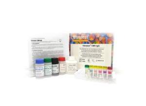
The test kit Vitrotest® CMV-IgG is an enzyme linked immunosorbent assay (ELISA) for the quantitative and semiquantitative determination of IgG class antibodies to Human Cytomegalovirus in human serum or plasma.
Determination of IgG antibodies to human CMV in the test kit Vitrotest® CMV-IgG is based on a solid phase, indirect ELISA in a two-step incubation procedure.
○ ТК005 - 96 tests- Solid phase: breakable microplate ELISA is coated with CMV antigens.
- Conjugate: a monoclonal antibodies to human IgG conjugated to horseradish peroxidase.
- Chromogen: ready to use TMB solution.
- Volume of sample for analysis: 10 μl.
- Assay time: 1h 15 min.
Human cytomegalovirus (CMV, herpesvirus 5) is a member of the herpesvirus family. This group includes herpes simplex virus types 1 and 2, varicella-zoster virus, and Epstein-Barr virus. CMV is a large enveloped virus; its double-stranded DNA genome codes for over 200 proteins. Viral proteins are regulatory and structural. Among structural proteins, glycoproteins and phosphoproteins are described.
CMV, similarly to other herpes viruses, shares a characteristic ability to remain dormant within the body for life. After initial infection, which may cause few symptoms, CMV becomes latent, residing in cells without causing detectable damage or clinical illness. Severe impairment of the body’s immune system by medication or disease may allow the virus to reactivate from the latent or dormant state and become symptomatic.
CMV infects most humans without harm. However infection with CMV causes serious, life-threatening disease in two circumstances: in immunosuppressed adults and in congenital infections of developing fetuses. In immunocompromised patients (organ transplant recipients, patients with lymphoid cancers, and HIV-infected patients) CMV is a major cause of disease and death. The common manifestations of disease in those patients are pneumonia, retinitis, and gastrointestinal diseases.
Congenital CMV (CCMV) infection is mostly (86% of all cases) asymptomatic. Symptoms, if they develop, include jaundice, pneumonia, a rash, an enlarged liver and spleen, low birth weight, seizures, small head. Mortality of children under symptomatic CCMV is around 20%. Around a half of symptomatic infants and around 12% of asymptomatic ones later develop physical or mental problems. These can include hearing loss, visual impairment or blindness, learning difficulties, epilepsy. -
The test kit Vitrotest® CMV-IgM is an enzyme linked immunosorbent assay (ELISA) for the qualitative and semiquantitative determination of IgM class antibodies to Human Cytomegalovirus in human serum or plasma.
Determination of IgM antibodies to human CMV in the test kit Vitrotest® CMV-IgM is based on a solid phase, indirect ELISA in a two-step incubation procedure.
○ ТК006 - 96 tests- Solid phase: breakable microplate ELISA is coated with recombinant CMV antigen.
- Conjugate: a monoclonal antibodies to human IgM conjugated to horseradish peroxidase.
- Chromogen: ready to use TMB solution.
- Volume of sample for analysis: 10 μl.
- Assay time: 1h 15 min.
Human cytomegalovirus (CMV, herpesvirus 5) is a member of the herpesvirus family. This group includes herpes simplex virus types 1 and 2, varicella-zoster virus, and Epstein-Barr virus. CMV is a large enveloped virus; its double-stranded DNA genome codes for over 200 proteins. Viral proteins are regulatory and structural. Among structural proteins, glycoproteins and phosphoproteins are described.
CMV, similarly to other herpes viruses, shares a characteristic ability to remain dormant within the body for life. After initial infection, which may cause few symptoms, CMV becomes latent, residing in cells without causing detectable damage or clinical illness. Severe impairment of the body’s immune system by medication or disease may allow the virus to reactivate from the latent or dormant state and become symptomatic.
CMV infects most humans without harm. However infection with CMV causes serious, life-threatening disease in two circumstances: in immunosuppressed adults and in congenital infections of developing fetuses. In immunocompromised patients (organ transplant recipients, patients with lymphoid cancers, and HIV-infected patients) CMV is a major cause of disease and death. The common manifestations of disease in those patients are pneumonia, retinitis, and gastrointestinal diseases.
Congenital CMV (CCMV) infection is mostly (86% of all cases) asymptomatic. Symptoms, if they develop, include jaundice, pneumonia, a rash, an enlarged liver and spleen, low birth weight, seizures, small head. Mortality of children under symptomatic CCMV is around 20%. Around a half of symptomatic infants and around 12% of asymptomatic ones later develop physical or mental problems. These can include hearing loss, visual impairment or blindness, learning difficulties, epilepsy. -
The test kit Vitrotest® CMV-IgG Avidity is an enzyme linked immunosorbent assay (ELISA) for the determination of avidity index of IgG class antibodies to Human Cytomegalovirus in human serum or plasma.
Determination of avidity index of IgG antibodies to Human Cytomegalovirus in the test kit Vitrotest® CMV-IgG Avidity is based on a solid phase, indirect ELISA.
○ ТК092 - 48 tests- Solid phase: breakable microplate ELISA is coated with CMV antigens.
- Conjugate: a monoclonal antibodies to human IgG conjugated to horseradish peroxidase.
- Chromogen: ready to use TMB solution.
- Volume of sample for analysis: 10 μl.
- Assay time: 1h 30 min.
Human cytomegalovirus (CMV, herpesvirus 5) is a member of the herpesvirus family. This group includes herpes simplex virus types 1 and 2, varicella-zoster virus, and Epstein-Barr virus. CMV is a large enveloped virus; its double-stranded DNA genome codes for over 200 proteins. Viral proteins are regulatory and structural. Among structural proteins, glycoproteins and phosphoproteins are described.
CMV, similarly to other herpes viruses, shares a characteristic ability to remain dormant within the body for life. After initial infection, which may cause few symptoms, CMV becomes latent, residing in cells without causing detectable damage or clinical illness. Severe impairment of the body’s immune system by medication or disease may allow the virus to reactivate from the latent or dormant state and become symptomatic.
CMV infects most humans without harm. However infection with CMV causes serious, life-threatening disease in two circumstances: in immunosuppressed adults and in congenital infections of developing fetuses. In immunocompromised patients (organ transplant recipients, patients with lymphoid cancers, and HIV-infected patients) CMV is a major cause of disease and death. The common manifestations of disease in those patients are pneumonia, retinitis, and gastrointestinal diseases.
Congenital CMV (CCMV) infection is mostly (86% of all cases) asymptomatic. Symptoms, if they develop, include jaundice, pneumonia, a rash, an enlarged liver and spleen, low birth weight, seizures, small head. Mortality of children under symptomatic CCMV is around 20%. Around a half of symptomatic infants and around 12% of asymptomatic ones later develop physical or mental problems. These can include hearing loss, visual impairment or blindness, learning difficulties, epilepsy.