-
The test kit Vitrotest® Anti-Ascaris is an enzyme linked immunosorbent assay (ELISA) for the detection of IgG class antibodies to Ascaris lumbricoides in human serum or plasma.
Determination of IgG antibodies to Ascaris lumbricoides in the test kit Vitrotest® Anti-Ascaris is based on a solid phase, indirect ELISA in a two-step incubation procedure.
○ ТК051 - 96 tests
○ ТК110 - 192 tests- Solid phase: breakable microplate ELISA is coated Ascaris lumbricoides antigens.
- Conjugate: a monoclonal antibodies to human IgG conjugated to horseradish peroxidase.
- Chromogen: ready to use TMB solution.
- Volume of sample for analysis 10 μl.
- Assay time: 1h 15 min.
Ascariasis is the most prevalent helminth infection on earth. Around one-fifth of human population is infected with Ascaris lumbricoides; the majority of infected live in rural or deprived urban settings in developing countries. In these endemic regions, disease prevalence is around 90% whereas in developed countries, the infection is rare. A. lumbricoides is the largest nematode (15-35 cm) parasitizing in the lumen of human small intestine.
Fertile eggs of ascaris become infective after an embryo moults twice within an egg (18 days to several weeks depending on the environmental conditions). After infective eggs are swallowed, the larvae hatch, invade the intestinal mucosa, and are carried via bloodstream to the lungs. The larvae undergo two moults in the lungs (10 to 14 days), penetrate the alveolar walls, ascend the bronchial tree to the throat, and are swallowed. Upon reaching the small intestine, they develop into adult worms which can live 1-2 years.
Daily ascaris egg production is around 200,000 eggs, which are shed in the feces. An infection can occur if a person swallows the microscopic eggs in contaminated food or water, or the eggs are transferred from hands to mouth after touching contaminated soil. Eggs can remain viable in the soil for up to 15 years.
The manifestations of ascariasis can be divided into acute and chronic. Patients experience acute lung inflammation, difficulty in breathing and fever as a result of larval migration through the pulmonary tissue (acute ascariasis). Abdominal distension and pain, nausea and diarrhoea are characteristic symptoms of adult worm invasion (chronic ascariasis). In small percentage of patients, entangled adult worms could lead to mechanical intestinal obstruction. The majority of invasions with A.lumbricoides are asymptomatic, and patients usually seek medical advice because they have seen a worm in their faeces. -
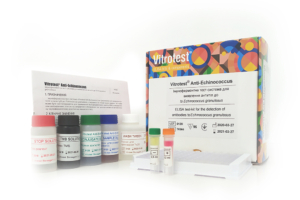
The test kit Vitrotest® Anti-Echinococcus is an enzyme linked immunosorbent assay (ELISA) for the detection of IgG class antibodies to Echinococcus granulosus in human serum or plasma.
Determination of IgG antibodies to Echinococcus granulosus in the test kit Vitrotest® Anti-Echinococcus is based on a solid phase, indirect ELISA in a two-step incubation procedure.
○ ТК066 - 96 tests- Solid phase: breakable microplate ELISA is coated Echinococcus granulosus antigens.
- Conjugate: a monoclonal antibodies to human IgG conjugated to horseradish peroxidase.
- Chromogen: ready to use TMB solution.
- Volume of sample for analysis: 10 μl.
- Assay time: 1h 15 min.
Echinococcosis is a chronic disease of humans and animals caused by parasitizing the larvae of the helminth Echinococcus. The causative agent of this helminthiasis is most often the larva of Echinococcus granulosus. Echinococcosis is quite common all over the world, especially in southern countries, where livestock breeding, mainly sheep breeding, is widespread.
Echinococcus eggs enter the human body through dirty hands after contacting dogs (less often - cats). Also, infection is not excluded when eating unwashed vegetables, berries, fruits, water that are contaminated with helminth eggs.
In the digestive canal of the intermediate host, the egg of the echinococcus is freed from the membrane, and the embryo (oncosphere) deepens into the mucous membrane of the small intestine, entering the internal organs, where, in most cases, they linger and develop into echinococcal cysts. More often, echinococcus affects the liver (in 44-85 % of cases) and lungs (10 % of cases).
The pathological effect of echinococcus is due to the sensitization of the body by the metabolic products of the parasite and mechanical damage to the affected organs and tissues. The sizes of cysts are from 1-5 cm in diameter to large blisters, which can contain several liters of fluid. The mechanical effect of such a cyst leads to dysfunction of the affected organ, its hypertrophy.
To diagnose echinococcosis, cysts visualization methods are used: X-ray and ultrasound studies, computed and magnetic resonance imaging. Puncture biopsy of a cyst is considered dangerous due to the possibility of spreading parasites into adjacent tissues.
The detection of antibodies specific to the antigens of echinococcus in the blood is a reliable indicator of parasite invasion. The level of the immune response largely depends on the organ localization of the cyst and its morphology. Low antibody levels are observed at the onset of cyst formation or at a late inoperable stage of the disease.
Today, methods of indirect hemagglutination and fluorescence, enzyme immunoassay are used to detect specific antibodies to Echinococcus granulosus. These methods are characterized by a sensitivity of 60-90 %, therefore, the best information content is achieved using a combination of serological methods.
Serological methods are also quite informative for monitoring the patient’s postoperative state - a gradual decrease in the level of specific antibodies 4-6 months after surgical removal of the cyst indicates a successful result of the surgical intervention. With relapses of cyst formation, specific antibodies are kept at a high level for years. -
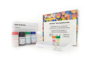
The test kit Vitrotest® Anti-Opisthorchis is an enzyme linked immunosorbent assay (ELISA) for the detection of IgG class antibodies to Opisthorchis felineus in human serum or plasma.
Determination of IgG antibodies to Opisthorchis felineus in the test kit Vitrotest® Anti-Opisthorchis is based on a solid phase, indirect ELISA in a two-step incubation procedure.
○ ТК057 - 96 tests- Solid phase: breakable microplate ELISA is coated Opisthorchis felineus antigens.
- Conjugate: a monoclonal antibodies to human IgG conjugated to horseradish peroxidase.
- Chromogen: ready to use TMB solution.
- Volume of sample for analysis: 10 μl.
- Assay time: 1h 15 min.
Opisthorchiasis - helminthiasis, affecting mainly the hepatobiliary system and the pancreas of humans, cats, dogs, etc. The disease is marked by a long course (in human body the parasite exists for 10-20 years), proceeds with frequent exacerbations, contributes to the occurrence of primary cancer of the liver and pancreas.
The causative agents of opisthorchiasis are two species of trematodes of the Opisthorchidae family - Opisthorchis felineus (Siberian fluke), common in Western Siberia, Kazakhstan and some areas of the Dnieper region; and Opisthorchis viverrini (squirrel fluke), found in countries with tropical climates (mainly in Thailand).
These small trematodes have a flat body 4-13 mm long and 1-3.5 mm wide. The oral sucker is located at the anterior end of the body, and the abdominal sucker is located at the border of the first and second quarter of the body.
Infection occurs when eating raw fish (thawed, frozen), slightly salted and insufficiently calcined carp fish containing helminth larvae - metacercariae. In the stomach, the upper parts of the small intestine, metacercariae are freed from the membrane and through 3-5 h reach the gallbladder, liver, pancreatic ducts, where after 2 weeks turn into sexually mature forms, capable of further releasing eggs.
At the early stage of invasion, a pronounced allergization of the body is observed. Mature opisthorchis injure mucous membranes of the pancreatic and bile ducts, create barriers to the outflow of bile, contribute to the development of cystic enlargements and neoplasms of the liver and carry out toxic and neuro-reflex effect.
Diagnosis of opisthorchiasis according to the clinical manifestations of the disease is difficult due to the absence of symptoms and syndromes characteristic only for this disease. Therefore, it is necessary to carry out a thorough clinical, laboratory and X-ray (ultrasound inclusive) examination.
In the laboratory analysis, it is possible to detect the invasion of opisthorchis one month after the infection when helminths begin to lay eggs (ovoscopic examination of feces and duodenal juice of the patient). More significant complications arise in recognizing early phases of opisthorchiasis. However, more and more studies are being carried out to detect specific antibodies to Opisthorchis felineus by enzyme immunoassay. In the chronic stage invasion is diagnosed by detecting helminth eggs in bile and stools, as well as by ELISA. -
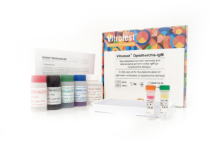
The test kit Vitrotest® Opisthorchis-IgM is an enzyme linked immunosorbent assay (ELISA) for the detection of IgM class antibodies to Opisthorchis felineus in human serum or plasma.
Determination of IgM antibodies to Opisthorchis felineus in the test kit Vitrotest® Opisthorchis-IgM is based on a solid phase, indirect ELISA in a two-step incubation procedure.
○ ТК012 - 96 tests- Solid phase: breakable microplate ELISA is coated Opisthorchis felineus antigens.
- Conjugate: a monoclonal antibodies to human IgM conjugated to horseradish peroxidase.
- Chromogen: ready to use TMB solution.
- Volume of sample for analysis: 10 μl.
- Assay time: 1h 15 min.
Opisthorchiasis - helminthiasis, affecting mainly the hepatobiliary system and the pancreas of humans, cats, dogs, etc. The disease is marked by a long course (in human body the parasite exists for 10-20 years), proceeds with frequent exacerbations, contributes to the occurrence of primary cancer of the liver and pancreas.
The causative agents of opisthorchiasis are two species of trematodes of the Opisthorchidae family - Opisthorchis felineus (Siberian fluke), common in Western Siberia, Kazakhstan and some areas of the Dnieper region; and Opisthorchis viverrini (squirrel fluke), found in countries with tropical climates (mainly in Thailand).
These small trematodes have a flat body 4-13 mm long and 1-3.5 mm wide. The oral sucker is located at the anterior end of the body, and the abdominal sucker is located at the border of the first and second quarter of the body.
Infection occurs when eating raw fish (thawed, frozen), slightly salted and insufficiently calcined carp fish containing helminth larvae - metacercariae. In the stomach, the upper parts of the small intestine, metacercariae are freed from the membrane and through 3-5 h reach the gallbladder, liver, pancreatic ducts, where after 2 weeks turn into sexually mature forms, capable of further releasing eggs.
At the early stage of invasion, a pronounced allergization of the body is observed. Mature opisthorchis injure mucous membranes of the pancreatic and bile ducts, create barriers to the outflow of bile, contribute to the development of cystic enlargements and neoplasms of the liver and carry out toxic and neuro-reflex effect.
Diagnosis of opisthorchiasis according to the clinical manifestations of the disease is difficult due to the absence of symptoms and syndromes characteristic only for this disease. Therefore, it is necessary to carry out a thorough clinical, laboratory and X-ray (ultrasound inclusive) examination.
In the laboratory analysis, it is possible to detect the invasion of opisthorchis one month after the infection when helminths begin to lay eggs (ovoscopic examination of feces and duodenal juice of the patient). More significant complications arise in recognizing early phases of opisthorchiasis. However, more and more studies are being carried out to detect specific antibodies to Opisthorchis felineus by enzyme immunoassay. In the chronic stage invasion is diagnosed by detecting helminth eggs in bile and stools, as well as by ELISA. -
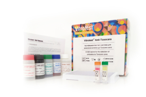
The test kit Vitrotest® Anti-Toxocara is an enzyme linked immunosorbent assay (ELISA) for the detection of IgG class antibodies to Toxocara canis in human serum or plasma.
Determination of IgG antibodies to Toxocara canis in the test kit Vitrotest® Anti-Toxocara is based on a solid phase, indirect ELISA in a two-step incubation procedure.
○ ТК058 - 96 tests
○ ТК112 - 192 tests- Solid phase: breakable microplate ELISA is coated Toxocara canis larvae antigens.
- Conjugate: a monoclonal antibodies to human IgG conjugated to horseradish peroxidase.
- Chromogen: ready to use TMB solution.
- Volume of sample for analysis: 10 μl.
- Assay time: 1h 15 min.
Toxocariasis is an widespread parasitic disease of humans caused by infection with the second stage larvae of two main species of parasitic nematodes, Toxocara canis and Toxocara cati. The role of toxocaras in human disease was disclosed in the 1950th by Wilder and Beaver et al. who first identified toxocara larvae in ocular and visceral tissues respectively.
Adult worms of T. canis and T.cati live within the lumen of the small intestine of dogs and cats respectively which serve as definitive hosts for these helminths. Unembryonated eggs produced by adult worms are shed in the feces of the definitive hosts. Eggs embryonate in the environment, and could be ingested by definitive hosts as well as accidental, paratenic hosts. After ingestion by the definitive hosts, microscopic (300 µm long and 20 µm in diameter) larvae hatch in the intestine, penetrate the intestinal wall, reach lungs via bloodstream, penetrate alveolar walls, ascend the bronchial tree to the throat where they are swallowed, and mature into adult worms. In humans and other paratenic hosts, the larvae are unable to undergo the full development cycle described above; instead, they are carried by the circulation to a wide variety of organs and tissues (liver, heart, lungs, brain, muscle, eyes), and cause severe local reactions that are the basis of toxocariasis.
The degree of host damage, and the concomitant signs and symptoms, varies with regard to which tissue has been invaded; the liver, lungs, and CNS (including the eyes) appear to be most sensitive. In the eye, migrating larvae can damage the retina, inducing granulomatous reactions leading to impaired sight or even loss of sight. The number of migrating larvae and the age of the host are two additional factors defining the presence and severity of symptoms.
Lifespan of the larvae could be as long as several years, and clinical disease could present in any time during this period, or even later, representing pathological immune response to dying or dead larvae.
For most people, an infection with these helminths causes no symptoms. Possible symptoms symptoms are nonspecific and include fever, fatigue, anorexia, or lymphadenopathy. Pulmonary symptoms and abdominal symptoms are similar to those under many other diseases; they are present when larvae migrate to the lungs or abdominal organs, respectively. Neurologic findings are diverse and also nonspecific. -
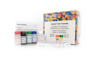
The test kit Vitrotest® Anti-Trichinella is an enzyme linked immunosorbent assay (ELISA) for the detection of IgG class antibodies to Trichinella spiralis in human serum or plasma.
Determination of IgG antibodies to Trichinella spiralis in the test kit Vitrotest® Anti-Trichinella is based on a solid phase, indirect ELISA in a two-step incubation procedure.
○ ТК067 - 96 tests- Solid phase: breakable microplate ELISA is coated Trichinella spiralis larvae antigens.
- Conjugate: a monoclonal antibodies to human IgG conjugated to horseradish peroxidase.
- Chromogen: ready to use TMB solution.
- Volume of sample for analysis: 10 μl.
- Assay time: 1h 15 min.
Trichinosis is a helminthiasis caused by a nematode Trichinella spiralis. Trichinellas are small, thread-like worms covered with a striped cuticle.
Transmission occurs by ingestion of meat containing encapsulated trichinella larvae. During digestion under the action of the gastric juice larvae release from capsules penetrate the submucosal layer of the small intestine, adhere to the mucosa and begin to proliferate. Soon after fertilization of females, the males die and the females start producing larvae, which enter the blood and lymphatic vessels through tissue mucosa, spread throughout the body and settle in striated muscles. Thereafter this capsule is impregnated with calcium salts leading to calcification. The larvae remain viable for many years.
The incubation period of human trichinosis lasts 10-25 days. Trichinosis is characterized by fever, myalgia, facial swelling, skin rash, blood eosinophilia, and in severe cases – by damage to internal organs and central nervous system.
The diagnosis of trichinosis is based on clinical signs, epidemiological history, serological tests (complement fixation tests, the reaction of indirect hemagglutination) and ELISA. The latter method is recommended by OIE for serological diagnosis of trichinosis.
The most specific and successful method to confirm infestation is the detection of IgG antibodies to trichinella antigens in the blood. These antibodies could be determined from 2-3 to 4-6 weeks after the eating of contaminated meat. Specific IgE class antibodies are also present in the blood during the acute stage of the disease, however, they are rarely detected due to the short period of their circulation in the bloodstream. During the early stage of invasion, specific antibodies might be still undetectable. Therefore, another sample should be taken in 1-2 weeks to confirm or reject suspected trichinosis. Seroconversion usually occurs 2-5 weeks after infection depending on the infectious dose. Assessment of antibody dynamics is a very informative marker of therapy effectiveness. In cases of ineffective or untimely therapy, specific antibodies are detected for up to 20 years if trichinosis is effectively treated in the first two weeks after infection, antibodies disappear within a year. -
The test kit Vitrotest® Anti-Strongyloides is an enzyme linked immunosorbent assay (ELISA) for the detection of IgG class antibodies to Strongyloides stercoralis in human serum or plasma.
Determination of IgG antibodies to Strongyloides stercoralis in the test kit Vitrotest® Anti-Strongyloides is based on a solid phase, indirect ELISA in a two-step incubation procedure.
- ТК146 - 96 tests
- Solid phase: breakable microplate ELISA is coated Strongyloides stercoralis antigens.
- Conjugate: a monoclonal antibodies to human IgG conjugated to horseradish peroxidase.
- Chromogen: ready to use TMB solution.
- Volume of sample for analysis: 10 μl.
- Assay time: 40 min.
Strongyloidiasis is a chronic parasitic infection of humans caused by Strongyloides stercoralis. This helminth is predominantly found in tropical and subtropical regions but can also be present in temperate climates. It is estimated that approximately 300 to 600 million people worldwide are infected.
Primary infection occurs when S. stercoralis larvae penetrate human skin through direct contact with contaminated soil. The larvae migrate via the bloodstream and lymphatic system to the respiratory tract, where they are then coughed up and swallowed, reaching the intestines. In the intestines, the parasites develop into adult worms, lay eggs, and produce larvae that can be excreted in the feces. A unique feature of S. stercoralis is its ability to cause autoinfection, as larvae may re-enter the intestines or perianal skin without leaving the host, leading to persistent infection.
In immunocompetent individuals, uncomplicated strongyloidiasis may be asymptomatic or present with mild cutaneous and gastrointestinal symptoms. Often, the only sign of infection is unexplained peripheral eosinophilia. However, in cases of heavy infestation or in immunosuppressed patients, the disease can cause severe manifestations such as abdominal pain, watery diarrhea, constipation, weight loss, vomiting, or small bowel obstruction. Hyperinfection syndrome is the most severe manifestation of the disease, with high mortality rates.
Parasitological methods (microscopy, culture) are traditionally used for diagnosis. The sensitivity and specifi city of these tests, however, are not high. The standard stool examination has a sensitivity of only 21% (5). Serological methods, especially enzyme-linked immunosorbent assay (ELISA), are highly sensitive and convenient for detecting antibodies to S. stercoralis larvae. ELISA is widely used for screening, diagnosing strongyloidiasis, and monitoring treatment effi cacy, as antibody titers signifi cantly decline within the fi rst 6 months following successful eradication therapy.