
-
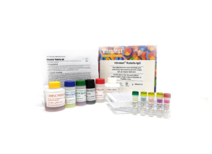
The test kit Vitrotest® Rubella-IgG is an enzyme linked immunosorbent assay (ELISA) for the quantitative determination of IgG class antibodies to Rubella virus in human serum or plasma.
Determination of IgG antibodies to Rubella virus in the test kit Vitrotest® Rubella-IgG is based on a solid phase, indirect ELISA in a two-step incubation procedure.
○ ТК003 - 96 tests- Solid phase: breakable microplate ELISA is coated with Rubella virus antigens.
- Conjugate: a monoclonal antibodies to human IgG conjugated to horseradish peroxidase.
- Chromogen: ready to use TMB solution.
- Volume of sample for analysis: 10 μl.
- Assay time: 1h 15 min.
- Analytical sensitivity: 0,4 IU/ml.
- Linearity range: 4-180 IU/ml.
Rubella is an acute viral infectious disease with significant teratogenic effects. The causative agent of rubella is an RNA virus Rubivirus genus Togaviridae family and was first isolated in 1962. Rubella is an air-bourne disease, takes few days to pass without a special treatment in most patients and does not cause health problems later. The main danger occurs during the first contact of a pregnant woman with the Rubella virus during the first trimester of pregnancy when the foetus is the most vulnerable to rubella. If the virus is being passed on from mother to foetus, it can be cause of a misbirth, a stillbirth or congenital rubella syndrome (CRS) - all of which comprise a group of serious deficiencies that can cause a developmental delay, mental retardation, deafness, cataracts, microcephaly, liver problems and a newborn heart disease. Because of the strong teratogenic effect of the Rubella virus a large-scale vaccination that provides permanent immunity to infection is conducted in most countries.
Extensive vaccination against rubella over the past decade has led to the near eradication of rubella and CRS in many developed countries and some developing countries. Before the introduction of vaccination, up to 4 children with CRS were born per 1,000 live births worldwide. Even now, more than 100,000 children are born with CRS annually in developing countries. In Ukraine, the incidence of rubella is about 4 cases per 100,000 population per year.
Many studies have shown that viral protection is mostly induced by neutralizing antibodies. The rubella specific IgM antibodies are usually detected within 4 days after the onset of rash and for 4–8 weeks thereafter, but in some cases these antibodies can persist for over a year. IgG antibodies appear during the acute phase (7 to 30 days postonset) and persist at varying levels for life. During viral infection, antibodies specific to three structural proteins of the rubella virus develop; the protective immune response is predominantly directed toward the glycoproteins, mainly against the glycoprotein E1.
Since the symptoms of rubella are often not specific, and many cases of rubella are asymptomatic, the diagnosis of rubella is rarely based upon clinical symptoms. The presence of rubella virus in nasal, throat, urine, blood, and cerebrospinal fluid specimens from persons with suspected rubella should be proven by virus isolation or alternatively viral nucleic acid should be detected by polymerase chain reaction. Immunological tests are by far the most popular in the diagnostics of rubella.
Serum assays are used for 3 main purposes:
1) for determining seroconversion after RV vaccination by the level of IgG;
2) for evaluation of the immunity to virus by the level of IgG, determining the need to vaccinate women at reproductive age;
3) to diagnose possible infection in pregnant women by the level of IgM and by the avidity of IgG.
Detection of rubella-specific IgM alone cannot be considered absolute proof of a recent primary infection for several reasons. IgM response after primary infection may be prolonged, lasting up to several years. Furthermore, sometimes rubella IgM is detectable in the case of mild secondary infection occuring despite a vaccination. This secondary infection is regarded as safe for the fetus. False-positive IgM results may also be artifacts due to various reasons.
This issue of doubtful IgM results is especially important when investigating suspected rubella in pregnant women because of the risk of CRS, so additional diagnostic tests should be used in such situations. Аvidity of IgG, which in most cases begins to increase 3 months following rubella infection, is another independent parameter allowing to differentiate between recent and past infection. For example, in one study low avidity specific IgG was detected in 91% of sera taken at 3–4 months after exposure to rubella virus; at 5–7 months after exposure only 21% of sera remained low avidity.
The IgG avidity assay is gaining popularity as a diagnostic method for the assessment of the time of infection. According to the recommendations of the Center for Disease Control and Prevention, USA, if IgM of the first probe is positive, avidity of IgG of the second probe, collected in 5-10 days, should be also measured. If the IgM and IgG of this second probe are positive, but the IgG avidity is high, this may indicate either a false-positive IgM result or a benign secondary infection. -
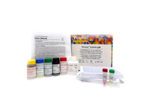
The test kit Vitrotest® Rubella-IgM is an enzyme linked immunosorbent assay (ELISA) for the qualitative and semiquantitative determination of IgM class antibodies to Rubella virus in human serum or plasma.
Determination of IgM antibodies to Rubella virus in the test kit Vitrotest® Rubella-IgM is based on a solid phase, indirect ELISA in a two-step incubation procedure.
○ ТК004 - 96 tests- Solid phase: breakable microplate ELISA is coated with Rubella virus antigens.
- Conjugate: a monoclonal antibodies to human IgM conjugated to horseradish peroxidase.
- Chromogen: ready to use TMB solution.
- Volume of sample for analysis: 10 μl.
- Assay time: 1h 15 min.
Rubella is an acute viral infectious disease with significant teratogenic effects. The causative agent of rubella is an RNA virus Rubivirus genus Togaviridae family and was first isolated in 1962. Rubella is an air-bourne disease, takes few days to pass without a special treatment in most patients and does not cause health problems later. The main danger occurs during the first contact of a pregnant woman with the Rubella virus during the first trimester of pregnancy when the foetus is the most vulnerable to rubella. If the virus is being passed on from mother to foetus, it can be cause of a misbirth, a stillbirth or congenital rubella syndrome (CRS) - all of which comprise a group of serious deficiencies that can cause a developmental delay, mental retardation, deafness, cataracts, microcephaly, liver problems and a newborn heart disease. Because of the strong teratogenic effect of the Rubella virus a large-scale vaccination that provides permanent immunity to infection is conducted in most countries.
Extensive vaccination against rubella over the past decade has led to the near eradication of rubella and CRS in many developed countries and some developing countries. Before the introduction of vaccination, up to 4 children with CRS were born per 1,000 live births worldwide. Even now, more than 100,000 children are born with CRS annually in developing countries. In Ukraine, the incidence of rubella is about 4 cases per 100,000 population per year.
Many studies have shown that viral protection is mostly induced by neutralizing antibodies. The rubella specific IgM antibodies are usually detected within 4 days after the onset of rash and for 4–8 weeks thereafter, but in some cases these antibodies can persist for over a year. IgG antibodies appear during the acute phase (7 to 30 days postonset) and persist at varying levels for life. During viral infection, antibodies specific to three structural proteins of the rubella virus develop; the protective immune response is predominantly directed toward the glycoproteins, mainly against the glycoprotein E1.
Since the symptoms of rubella are often not specific, and many cases of rubella are asymptomatic, the diagnosis of rubella is rarely based upon clinical symptoms. The presence of rubella virus in nasal, throat, urine, blood, and cerebrospinal fluid specimens from persons with suspected rubella should be proven by virus isolation or alternatively viral nucleic acid should be detected by polymerase chain reaction. Immunological tests are by far the most popular in the diagnostics of rubella.
Serum assays are used for 3 main purposes:
1) for determining seroconversion after RV vaccination by the level of IgG;
2) for evaluation of the immunity to virus by the level of IgG, determining the need to vaccinate women at reproductive age;
3) to diagnose possible infection in pregnant women by the level of IgM and by the avidity of IgG.
Detection of rubella-specific IgM alone cannot be considered absolute proof of a recent primary infection for several reasons. IgM response after primary infection may be prolonged, lasting up to several years. Furthermore, sometimes rubella IgM is detectable in the case of mild secondary infection occuring despite a vaccination. This secondary infection is regarded as safe for the fetus. False-positive IgM results may also be artifacts due to various reasons.
This issue of doubtful IgM results is especially important when investigating suspected rubella in pregnant women because of the risk of CRS, so additional diagnostic tests should be used in such situations. Аvidity of IgG, which in most cases begins to increase 3 months following rubella infection, is another independent parameter allowing to differentiate between recent and past infection. For example, in one study low avidity specific IgG was detected in 91% of sera taken at 3–4 months after exposure to rubella virus; at 5–7 months after exposure only 21% of sera remained low avidity.
The IgG avidity assay is gaining popularity as a diagnostic method for the assessment of the time of infection. According to the recommendations of the Center for Disease Control and Prevention, USA, if IgM of the first probe is positive, avidity of IgG of the second probe, collected in 5-10 days, should be also measured. If the IgM and IgG of this second probe are positive, but the IgG avidity is high, this may indicate either a false-positive IgM result or a benign secondary infection. -
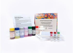
The test kit Vitrotest® Rubella-IgG Avidity is an enzyme linked immunosorbent assay (ELISA) for the determination of avidity index of IgG class antibodies to Rubella virus in human serum or plasma.
Determination of avidity index of IgG antibodies to Rubella virus in the test kit Vitrotest® Rubella-IgG Avidity is based on a solid phase, indirect ELISA.
○ ТК065 - 48 tests- Solid phase: breakable microplate ELISA is coated with Rubella virus antigens.
- Conjugate: a monoclonal antibodies to human IgG conjugated to horseradish peroxidase.
- Chromogen: ready to use TMB solution.
- Volume of sample for analysis: 2×10 μl.
- Assay time: 1h 25 min.
Rubella is an acute viral infectious disease with significant teratogenic effects. The causative agent of rubella is an RNA virus Rubivirus genus Togaviridae family and was first isolated in 1962. Rubella is an air-bourne disease, takes few days to pass without a special treatment in most patients and does not cause health problems later. The main danger occurs during the first contact of a pregnant woman with the Rubella virus during the first trimester of pregnancy when the foetus is the most vulnerable to rubella. If the virus is being passed on from mother to foetus, it can be cause of a misbirth, a stillbirth or congenital rubella syndrome (CRS) - all of which comprise a group of serious deficiencies that can cause a developmental delay, mental retardation, deafness, cataracts, microcephaly, liver problems and a newborn heart disease. Because of the strong teratogenic effect of the Rubella virus a large-scale vaccination that provides permanent immunity to infection is conducted in most countries.
Extensive vaccination against rubella over the past decade has led to the near eradication of rubella and CRS in many developed countries and some developing countries. Before the introduction of vaccination, up to 4 children with CRS were born per 1,000 live births worldwide. Even now, more than 100,000 children are born with CRS annually in developing countries. In Ukraine, the incidence of rubella is about 4 cases per 100,000 population per year.
Many studies have shown that viral protection is mostly induced by neutralizing antibodies. The rubella specific IgM antibodies are usually detected within 4 days after the onset of rash and for 4–8 weeks thereafter, but in some cases these antibodies can persist for over a year. IgG antibodies appear during the acute phase (7 to 30 days postonset) and persist at varying levels for life. During viral infection, antibodies specific to three structural proteins of the rubella virus develop; the protective immune response is predominantly directed toward the glycoproteins, mainly against the glycoprotein E1.
Since the symptoms of rubella are often not specific, and many cases of rubella are asymptomatic, the diagnosis of rubella is rarely based upon clinical symptoms. The presence of rubella virus in nasal, throat, urine, blood, and cerebrospinal fluid specimens from persons with suspected rubella should be proven by virus isolation or alternatively viral nucleic acid should be detected by polymerase chain reaction. Immunological tests are by far the most popular in the diagnostics of rubella.
Serum assays are used for 3 main purposes:
1) for determining seroconversion after RV vaccination by the level of IgG;
2) for evaluation of the immunity to virus by the level of IgG, determining the need to vaccinate women at reproductive age;
3) to diagnose possible infection in pregnant women by the level of IgM and by the avidity of IgG.
Detection of rubella-specific IgM alone cannot be considered absolute proof of a recent primary infection for several reasons. IgM response after primary infection may be prolonged, lasting up to several years. Furthermore, sometimes rubella IgM is detectable in the case of mild secondary infection occuring despite a vaccination. This secondary infection is regarded as safe for the fetus. False-positive IgM results may also be artifacts due to various reasons.
This issue of doubtful IgM results is especially important when investigating suspected rubella in pregnant women because of the risk of CRS, so additional diagnostic tests should be used in such situations. Аvidity of IgG, which in most cases begins to increase 3 months following rubella infection, is another independent parameter allowing to differentiate between recent and past infection. For example, in one study low avidity specific IgG was detected in 91% of sera taken at 3–4 months after exposure to rubella virus; at 5–7 months after exposure only 21% of sera remained low avidity.
The IgG avidity assay is gaining popularity as a diagnostic method for the assessment of the time of infection. According to the recommendations of the Center for Disease Control and Prevention, USA, if IgM of the first probe is positive, avidity of IgG of the second probe, collected in 5-10 days, should be also measured. If the IgM and IgG of this second probe are positive, but the IgG avidity is high, this may indicate either a false-positive IgM result or a benign secondary infection. -
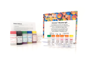
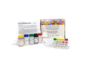
The test kit Vitrotest® Measles-IgG is an enzyme linked immunosorbent assay (ELISA) for the quantitative and semiquantitative determination of IgG class antibodies to measles virus in human serum or plasma.
Determination of IgG antibodies to measles virus in the test kit Vitrotest® Measles-IgG is based on a solid phase, indirect ELISA in a two-step incubation procedure.
○ ТК080 - 96 tests- Solid phase: breakable microplate ELISA is coated with purified antigens of measles virus.
- Conjugate: a monoclonal antibodies to human IgG conjugated to horseradish peroxidase.
- Chromogen: ready to use TMB solution.
- Volume of sample for analysis: 10 μl.
- Assay time: 1h 15 min.
- Analytical sensitivity: 12 mIU/ml.
- Linearity range: 70-3000 mIU/ml.
Measles is a dangerous, rapidly spreading, highly contagious viral disease that can lead to severe complications and death. The causative agent of measles (eng. Measles virus) is an RNA-containing virus of the Paramyxoviruses family.
The measles virus is usually transmitted through direct contact, as well as through the air, infects the mucous membrane of a person, and then spreads throughout the body. The first sign of measles is usually a significant fever, runny nose, cough, red eyes and watery eyes, and small white spots on the inside of the cheeks. After a few days, a rash appears, first on the face and upper neck. After about three days, the rash spreads all over the body, including the arms and legs, lasts for 5 to 6 days, and then disappears.
Most measles deaths are due to complications associated with the disease. Complications are more common in children under the age of five or in adults over 30 years old.
Introduced in 1963, measures to vaccinate against measles have significantly reduced mortality from this disease.
Screening of pregnant women, young people and patients at risk to determine the level of protective immunity is extremely important during periods of epidemic outbreaks. For this purpose, the most common quantitative determination of IgG antibodies specific to the measles virus is by enzyme immunoassay. -
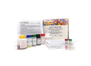
The test kit Vitrotest® Measles-IgM is an enzyme linked immunosorbent assay (ELISA) for the qualitative and semiquantitative determination of IgM class antibodies to measles virus in human serum or plasma.
Determination of IgM antibodies to measles virus in the test kit Vitrotest® Measles-IgM is based on a solid phase, indirect ELISA in a two-step incubation procedure.
○ ТК079 - 96 tests- Solid phase: breakable microplate ELISA is coated with purified antigens of measles virus.
- Conjugate: a monoclonal antibodies to human IgM conjugated to horseradish peroxidase.
- Chromogen: ready to use TMB solution.
- Volume of sample for analysis: 10 μl.
- Assay time: 1h 15 min.
Measles is a dangerous, rapidly spreading, highly contagious viral disease that can lead to severe complications and death. The causative agent of measles (eng. Measles virus) is an RNA-containing virus of the Paramyxoviruses family.
The measles virus is usually transmitted through direct contact, as well as through the air, infects the mucous membrane of a person, and then spreads throughout the body. The first sign of measles is usually a significant fever, runny nose, cough, red eyes and watery eyes, and small white spots on the inside of the cheeks. After a few days, a rash appears, first on the face and upper neck. After about three days, the rash spreads all over the body, including the arms and legs, lasts for 5 to 6 days, and then disappears.
Most measles deaths are due to complications associated with the disease. Complications are more common in children under the age of five or in adults over 30 years old.
Introduced in 1963, measures to vaccinate against measles have significantly reduced mortality from this disease.
Screening of pregnant women, young people and patients at risk to determine the level of protective immunity is extremely important during periods of epidemic outbreaks. For this purpose, the most common quantitative determination of IgG antibodies specific to the measles virus is by enzyme immunoassay. -
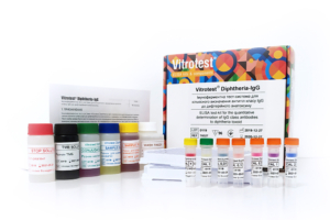
The test kit Vitrotest® Diphtheria-IgG is an enzyme linked immunosorbent assay (ELISA) for the quantitative determination of IgG class antibodies to diphtheria toxoid in human serum or plasma.
Determination of IgG antibodies to diphtheria toxoid in the test kit Vitrotest® Diphtheria-IgG is based on a solid phase, indirect ELISA.
○ ТК027 - 96 tests- Solid phase: breakable microplate ELISA is coated with diphtheria toxoid.
- Conjugate: a monoclonal antibodies to human IgG conjugated to horseradish peroxidase.
- Chromogen: ready to use TMB solution.
- Volume of sample for analysis: 10 μl.
- Assay time: 1h 45 min.
- Analytical sensitivity: 0,0025 IU/ml.
- Linearity range: 0,004 - 1,005 IU/ml.
Diphtheria is an infectious disease that primarily affects children. The causative agent of diphtheria Corynebacterium diphtheriae enters the human body, primarily infecting the throat and upper respiratory tract, and produces a toxin that can affect other organs. The disease is characterized by an acute onset, with the main symptoms being a sore throat, fever, and swollen glands in the neck. In severe cases, the bacteria produce a toxin that causes a thick gray or white film to form on the back of the throat.
The most important virulent factor of C. diphtheriae is the diphtheria toxin (exotoxin), which is secreted by the bacteria, enters the bloodstream, and damages the myocardium, kidneys, and central nervous system. Damage to the heart muscle can lead to arrhythmias, and nerve inflammation can cause paralysis.
Vaccination against diphtheria is a highly effective means of preventing the disease. According to WHO recommendations, all children worldwide should be vaccinated against diphtheria. The primary vaccination with diphtheria toxoid, consisting of three doses of the vaccine, should be administered to infants at the ages of 2, 4, and 6 months (with at least 4-week intervals between vaccinations). This should be followed by three booster immunizations with combined vaccines at the ages of 1-2, 4-7, and 9-15 years. At any age, those who are unvaccinated or incompletely vaccinated against diphtheria should receive the necessary doses to complete the vaccination series.
After the 3-dose primary series of the toxoid-containing vaccine, 94-100% of children show diphtheria antitoxin levels > 0.01 IU/ml, but booster doses of the vaccine are necessary to ensure long-term protection. Quantitative enzyme-linked immunosorbent assay (ELISA) for the determination of IgG antibodies to diphtheria toxoid is widely used to assess the level of protective immunity. -
The test kit Vitrotest® Bordetella pertussis Toxin IgG is an enzyme linked immunosorbent assay (ELISA) for the quantitative determination of IgG class antibodies to Bordetella pertussis toxin in human serum or plasma.
Determination of IgG antibodies to Bordetella pertussis toxin in the test kit Vitrotest® Bordetella pertussis Toxin IgG is based on a solid phase, indirect ELISA in a two-step incubation procedure.
○ ТК125 - 96 tests- Solid phase: breakable microplate ELISA is coated with purified Bordetella pertussis toxin.
- Conjugate: a monoclonal antibodies to human IgG conjugated to horseradish peroxidase.
- Chromogen: ready to use TMB solution.
- Volume of sample for analysis: 10 μl.
- Assay time: 1h 15 min.
- Analytical sensitivity: 3,22 IU/ml.
- Linearity range: 3,5 - 181 IU/ml.
Whooping cough is a dangerous respiratory infection caused by the bacterium Bordetella pertussis. Characteristic symptoms of the disease include paroxysmal spasmodic coughing, a high-pitched "whoop" sound when inhaling, post-cough vomiting, and more. Whooping cough is most severe in infants and young children, often leading to apnea (breathing pauses) and, in some cases, death.
Whooping cough is a globally widespread disease with a cyclic pattern, with peaks in incidence every 3-5 years. Due to the severity of its clinical manifestations, many countries have implemented vaccination programs against whooping cough. As a result of widespread vaccination campaigns conducted in the 1950s and 1960s in developed countries, there was a sharp decrease in both incidence (by more than 90%) and mortality from whooping cough.
However, despite high vaccination coverage, whooping cough remains a public health issue worldwide, with approximately 140,000 cases reported annually.
In Ukraine, according to the National Immunization Schedule, children should be vaccinated against whooping cough at the ages of 2, 4, 6, and 18 months. For vaccinating children against whooping cough during the first year of life, both acellular (aP) and whole-cell (wP) pertussis vaccines can be used. The immunity formed as a result of the full course of vaccination against whooping cough lasts for 5-7 years.
Laboratory methods are used to diagnose whooping cough, with the most common being polymerase chain reaction (PCR) (to detect the pathogen during the first 2-3 weeks of the illness) and enzyme-linked immunosorbent assay (ELISA) (to detect specific antibodies). The presence of IgM antibodies specific to Bordetella pertussis indicates an acute infection or recent vaccination. The detection of IgG antibodies to the pertussis toxin (PT) in the serum of an unvaccinated individual allows for the diagnosis of a past or current infection and provides information about specific immunity after vaccination. -
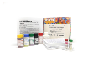
The test kit Vitrotest® Bordetella pertussis IgМ is an enzyme linked immunosorbent assay (ELISA) for the qualitative and semiquantitative determination of IgM class antibodies to Bordetella pertussis in human serum or plasma.
Determination of IgM antibodies to Bordetella pertussis in the test kit Vitrotest® Bordetella pertussis IgM is based on a solid phase, indirect ELISA in a two-step incubation procedure.
○ ТК126 - 96 tests- Solid phase: breakable microplate ELISA is coated with Bordetella pertussis antigens.
- Conjugate: a monoclonal antibodies to human IgM conjugated to horseradish peroxidase.
- Chromogen: ready to use TMB solution.
- Volume of sample for analysis: 10 μl.
- Assay time: 1h 15 min.
Whooping cough is a dangerous respiratory infection caused by the bacterium Bordetella pertussis. Characteristic symptoms of the disease include paroxysmal spasmodic coughing, a high-pitched "whoop" sound when inhaling, post-cough vomiting, and more. Whooping cough is most severe in infants and young children, often leading to apnea (breathing pauses) and, in some cases, death.
Whooping cough is a globally widespread disease with a cyclic pattern, with peaks in incidence every 3-5 years. Due to the severity of its clinical manifestations, many countries have implemented vaccination programs against whooping cough. As a result of widespread vaccination campaigns conducted in the 1950s and 1960s in developed countries, there was a sharp decrease in both incidence (by more than 90%) and mortality from whooping cough.
However, despite high vaccination coverage, whooping cough remains a public health issue worldwide, with approximately 140,000 cases reported annually.
In Ukraine, according to the National Immunization Schedule, children should be vaccinated against whooping cough at the ages of 2, 4, 6, and 18 months. For vaccinating children against whooping cough during the first year of life, both acellular (aP) and whole-cell (wP) pertussis vaccines can be used. The immunity formed as a result of the full course of vaccination against whooping cough lasts for 5-7 years.
Laboratory methods are used to diagnose whooping cough, with the most common being polymerase chain reaction (PCR) (to detect the pathogen during the first 2-3 weeks of the illness) and enzyme-linked immunosorbent assay (ELISA) (to detect specific antibodies). The presence of IgM antibodies specific to Bordetella pertussis indicates an acute infection or recent vaccination. The detection of IgG antibodies to the pertussis toxin (PT) in the serum of an unvaccinated individual allows for the diagnosis of a past or current infection and provides information about specific immunity after vaccination.
-
The test kit Vitrotest® Rubella-IgG is an enzyme linked immunosorbent assay (ELISA) for the quantitative determination of IgG class antibodies to Rubella virus in human serum or plasma.
Determination of IgG antibodies to Rubella virus in the test kit Vitrotest® Rubella-IgG is based on a solid phase, indirect ELISA in a two-step incubation procedure.
○ ТК003 - 96 tests- Solid phase: breakable microplate ELISA is coated with Rubella virus antigens.
- Conjugate: a monoclonal antibodies to human IgG conjugated to horseradish peroxidase.
- Chromogen: ready to use TMB solution.
- Volume of sample for analysis: 10 μl.
- Assay time: 1h 15 min.
- Analytical sensitivity: 0,4 IU/ml.
- Linearity range: 4-180 IU/ml.
Rubella is an acute viral infectious disease with significant teratogenic effects. The causative agent of rubella is an RNA virus Rubivirus genus Togaviridae family and was first isolated in 1962. Rubella is an air-bourne disease, takes few days to pass without a special treatment in most patients and does not cause health problems later. The main danger occurs during the first contact of a pregnant woman with the Rubella virus during the first trimester of pregnancy when the foetus is the most vulnerable to rubella. If the virus is being passed on from mother to foetus, it can be cause of a misbirth, a stillbirth or congenital rubella syndrome (CRS) - all of which comprise a group of serious deficiencies that can cause a developmental delay, mental retardation, deafness, cataracts, microcephaly, liver problems and a newborn heart disease. Because of the strong teratogenic effect of the Rubella virus a large-scale vaccination that provides permanent immunity to infection is conducted in most countries.
Extensive vaccination against rubella over the past decade has led to the near eradication of rubella and CRS in many developed countries and some developing countries. Before the introduction of vaccination, up to 4 children with CRS were born per 1,000 live births worldwide. Even now, more than 100,000 children are born with CRS annually in developing countries. In Ukraine, the incidence of rubella is about 4 cases per 100,000 population per year.
Many studies have shown that viral protection is mostly induced by neutralizing antibodies. The rubella specific IgM antibodies are usually detected within 4 days after the onset of rash and for 4–8 weeks thereafter, but in some cases these antibodies can persist for over a year. IgG antibodies appear during the acute phase (7 to 30 days postonset) and persist at varying levels for life. During viral infection, antibodies specific to three structural proteins of the rubella virus develop; the protective immune response is predominantly directed toward the glycoproteins, mainly against the glycoprotein E1.
Since the symptoms of rubella are often not specific, and many cases of rubella are asymptomatic, the diagnosis of rubella is rarely based upon clinical symptoms. The presence of rubella virus in nasal, throat, urine, blood, and cerebrospinal fluid specimens from persons with suspected rubella should be proven by virus isolation or alternatively viral nucleic acid should be detected by polymerase chain reaction. Immunological tests are by far the most popular in the diagnostics of rubella.
Serum assays are used for 3 main purposes:
1) for determining seroconversion after RV vaccination by the level of IgG;
2) for evaluation of the immunity to virus by the level of IgG, determining the need to vaccinate women at reproductive age;
3) to diagnose possible infection in pregnant women by the level of IgM and by the avidity of IgG.
Detection of rubella-specific IgM alone cannot be considered absolute proof of a recent primary infection for several reasons. IgM response after primary infection may be prolonged, lasting up to several years. Furthermore, sometimes rubella IgM is detectable in the case of mild secondary infection occuring despite a vaccination. This secondary infection is regarded as safe for the fetus. False-positive IgM results may also be artifacts due to various reasons.
This issue of doubtful IgM results is especially important when investigating suspected rubella in pregnant women because of the risk of CRS, so additional diagnostic tests should be used in such situations. Аvidity of IgG, which in most cases begins to increase 3 months following rubella infection, is another independent parameter allowing to differentiate between recent and past infection. For example, in one study low avidity specific IgG was detected in 91% of sera taken at 3–4 months after exposure to rubella virus; at 5–7 months after exposure only 21% of sera remained low avidity.
The IgG avidity assay is gaining popularity as a diagnostic method for the assessment of the time of infection. According to the recommendations of the Center for Disease Control and Prevention, USA, if IgM of the first probe is positive, avidity of IgG of the second probe, collected in 5-10 days, should be also measured. If the IgM and IgG of this second probe are positive, but the IgG avidity is high, this may indicate either a false-positive IgM result or a benign secondary infection. -
The test kit Vitrotest® Rubella-IgM is an enzyme linked immunosorbent assay (ELISA) for the qualitative and semiquantitative determination of IgM class antibodies to Rubella virus in human serum or plasma.
Determination of IgM antibodies to Rubella virus in the test kit Vitrotest® Rubella-IgM is based on a solid phase, indirect ELISA in a two-step incubation procedure.
○ ТК004 - 96 tests- Solid phase: breakable microplate ELISA is coated with Rubella virus antigens.
- Conjugate: a monoclonal antibodies to human IgM conjugated to horseradish peroxidase.
- Chromogen: ready to use TMB solution.
- Volume of sample for analysis: 10 μl.
- Assay time: 1h 15 min.
Rubella is an acute viral infectious disease with significant teratogenic effects. The causative agent of rubella is an RNA virus Rubivirus genus Togaviridae family and was first isolated in 1962. Rubella is an air-bourne disease, takes few days to pass without a special treatment in most patients and does not cause health problems later. The main danger occurs during the first contact of a pregnant woman with the Rubella virus during the first trimester of pregnancy when the foetus is the most vulnerable to rubella. If the virus is being passed on from mother to foetus, it can be cause of a misbirth, a stillbirth or congenital rubella syndrome (CRS) - all of which comprise a group of serious deficiencies that can cause a developmental delay, mental retardation, deafness, cataracts, microcephaly, liver problems and a newborn heart disease. Because of the strong teratogenic effect of the Rubella virus a large-scale vaccination that provides permanent immunity to infection is conducted in most countries.
Extensive vaccination against rubella over the past decade has led to the near eradication of rubella and CRS in many developed countries and some developing countries. Before the introduction of vaccination, up to 4 children with CRS were born per 1,000 live births worldwide. Even now, more than 100,000 children are born with CRS annually in developing countries. In Ukraine, the incidence of rubella is about 4 cases per 100,000 population per year.
Many studies have shown that viral protection is mostly induced by neutralizing antibodies. The rubella specific IgM antibodies are usually detected within 4 days after the onset of rash and for 4–8 weeks thereafter, but in some cases these antibodies can persist for over a year. IgG antibodies appear during the acute phase (7 to 30 days postonset) and persist at varying levels for life. During viral infection, antibodies specific to three structural proteins of the rubella virus develop; the protective immune response is predominantly directed toward the glycoproteins, mainly against the glycoprotein E1.
Since the symptoms of rubella are often not specific, and many cases of rubella are asymptomatic, the diagnosis of rubella is rarely based upon clinical symptoms. The presence of rubella virus in nasal, throat, urine, blood, and cerebrospinal fluid specimens from persons with suspected rubella should be proven by virus isolation or alternatively viral nucleic acid should be detected by polymerase chain reaction. Immunological tests are by far the most popular in the diagnostics of rubella.
Serum assays are used for 3 main purposes:
1) for determining seroconversion after RV vaccination by the level of IgG;
2) for evaluation of the immunity to virus by the level of IgG, determining the need to vaccinate women at reproductive age;
3) to diagnose possible infection in pregnant women by the level of IgM and by the avidity of IgG.
Detection of rubella-specific IgM alone cannot be considered absolute proof of a recent primary infection for several reasons. IgM response after primary infection may be prolonged, lasting up to several years. Furthermore, sometimes rubella IgM is detectable in the case of mild secondary infection occuring despite a vaccination. This secondary infection is regarded as safe for the fetus. False-positive IgM results may also be artifacts due to various reasons.
This issue of doubtful IgM results is especially important when investigating suspected rubella in pregnant women because of the risk of CRS, so additional diagnostic tests should be used in such situations. Аvidity of IgG, which in most cases begins to increase 3 months following rubella infection, is another independent parameter allowing to differentiate between recent and past infection. For example, in one study low avidity specific IgG was detected in 91% of sera taken at 3–4 months after exposure to rubella virus; at 5–7 months after exposure only 21% of sera remained low avidity.
The IgG avidity assay is gaining popularity as a diagnostic method for the assessment of the time of infection. According to the recommendations of the Center for Disease Control and Prevention, USA, if IgM of the first probe is positive, avidity of IgG of the second probe, collected in 5-10 days, should be also measured. If the IgM and IgG of this second probe are positive, but the IgG avidity is high, this may indicate either a false-positive IgM result or a benign secondary infection. -
The test kit Vitrotest® Rubella-IgG Avidity is an enzyme linked immunosorbent assay (ELISA) for the determination of avidity index of IgG class antibodies to Rubella virus in human serum or plasma.
Determination of avidity index of IgG antibodies to Rubella virus in the test kit Vitrotest® Rubella-IgG Avidity is based on a solid phase, indirect ELISA.
○ ТК065 - 48 tests- Solid phase: breakable microplate ELISA is coated with Rubella virus antigens.
- Conjugate: a monoclonal antibodies to human IgG conjugated to horseradish peroxidase.
- Chromogen: ready to use TMB solution.
- Volume of sample for analysis: 2×10 μl.
- Assay time: 1h 25 min.
Rubella is an acute viral infectious disease with significant teratogenic effects. The causative agent of rubella is an RNA virus Rubivirus genus Togaviridae family and was first isolated in 1962. Rubella is an air-bourne disease, takes few days to pass without a special treatment in most patients and does not cause health problems later. The main danger occurs during the first contact of a pregnant woman with the Rubella virus during the first trimester of pregnancy when the foetus is the most vulnerable to rubella. If the virus is being passed on from mother to foetus, it can be cause of a misbirth, a stillbirth or congenital rubella syndrome (CRS) - all of which comprise a group of serious deficiencies that can cause a developmental delay, mental retardation, deafness, cataracts, microcephaly, liver problems and a newborn heart disease. Because of the strong teratogenic effect of the Rubella virus a large-scale vaccination that provides permanent immunity to infection is conducted in most countries.
Extensive vaccination against rubella over the past decade has led to the near eradication of rubella and CRS in many developed countries and some developing countries. Before the introduction of vaccination, up to 4 children with CRS were born per 1,000 live births worldwide. Even now, more than 100,000 children are born with CRS annually in developing countries. In Ukraine, the incidence of rubella is about 4 cases per 100,000 population per year.
Many studies have shown that viral protection is mostly induced by neutralizing antibodies. The rubella specific IgM antibodies are usually detected within 4 days after the onset of rash and for 4–8 weeks thereafter, but in some cases these antibodies can persist for over a year. IgG antibodies appear during the acute phase (7 to 30 days postonset) and persist at varying levels for life. During viral infection, antibodies specific to three structural proteins of the rubella virus develop; the protective immune response is predominantly directed toward the glycoproteins, mainly against the glycoprotein E1.
Since the symptoms of rubella are often not specific, and many cases of rubella are asymptomatic, the diagnosis of rubella is rarely based upon clinical symptoms. The presence of rubella virus in nasal, throat, urine, blood, and cerebrospinal fluid specimens from persons with suspected rubella should be proven by virus isolation or alternatively viral nucleic acid should be detected by polymerase chain reaction. Immunological tests are by far the most popular in the diagnostics of rubella.
Serum assays are used for 3 main purposes:
1) for determining seroconversion after RV vaccination by the level of IgG;
2) for evaluation of the immunity to virus by the level of IgG, determining the need to vaccinate women at reproductive age;
3) to diagnose possible infection in pregnant women by the level of IgM and by the avidity of IgG.
Detection of rubella-specific IgM alone cannot be considered absolute proof of a recent primary infection for several reasons. IgM response after primary infection may be prolonged, lasting up to several years. Furthermore, sometimes rubella IgM is detectable in the case of mild secondary infection occuring despite a vaccination. This secondary infection is regarded as safe for the fetus. False-positive IgM results may also be artifacts due to various reasons.
This issue of doubtful IgM results is especially important when investigating suspected rubella in pregnant women because of the risk of CRS, so additional diagnostic tests should be used in such situations. Аvidity of IgG, which in most cases begins to increase 3 months following rubella infection, is another independent parameter allowing to differentiate between recent and past infection. For example, in one study low avidity specific IgG was detected in 91% of sera taken at 3–4 months after exposure to rubella virus; at 5–7 months after exposure only 21% of sera remained low avidity.
The IgG avidity assay is gaining popularity as a diagnostic method for the assessment of the time of infection. According to the recommendations of the Center for Disease Control and Prevention, USA, if IgM of the first probe is positive, avidity of IgG of the second probe, collected in 5-10 days, should be also measured. If the IgM and IgG of this second probe are positive, but the IgG avidity is high, this may indicate either a false-positive IgM result or a benign secondary infection. -
The test kit Vitrotest® Measles-IgG is an enzyme linked immunosorbent assay (ELISA) for the quantitative and semiquantitative determination of IgG class antibodies to measles virus in human serum or plasma.
Determination of IgG antibodies to measles virus in the test kit Vitrotest® Measles-IgG is based on a solid phase, indirect ELISA in a two-step incubation procedure.
○ ТК080 - 96 tests- Solid phase: breakable microplate ELISA is coated with purified antigens of measles virus.
- Conjugate: a monoclonal antibodies to human IgG conjugated to horseradish peroxidase.
- Chromogen: ready to use TMB solution.
- Volume of sample for analysis: 10 μl.
- Assay time: 1h 15 min.
- Analytical sensitivity: 12 mIU/ml.
- Linearity range: 70-3000 mIU/ml.
Measles is a dangerous, rapidly spreading, highly contagious viral disease that can lead to severe complications and death. The causative agent of measles (eng. Measles virus) is an RNA-containing virus of the Paramyxoviruses family.
The measles virus is usually transmitted through direct contact, as well as through the air, infects the mucous membrane of a person, and then spreads throughout the body. The first sign of measles is usually a significant fever, runny nose, cough, red eyes and watery eyes, and small white spots on the inside of the cheeks. After a few days, a rash appears, first on the face and upper neck. After about three days, the rash spreads all over the body, including the arms and legs, lasts for 5 to 6 days, and then disappears.
Most measles deaths are due to complications associated with the disease. Complications are more common in children under the age of five or in adults over 30 years old.
Introduced in 1963, measures to vaccinate against measles have significantly reduced mortality from this disease.
Screening of pregnant women, young people and patients at risk to determine the level of protective immunity is extremely important during periods of epidemic outbreaks. For this purpose, the most common quantitative determination of IgG antibodies specific to the measles virus is by enzyme immunoassay. -
The test kit Vitrotest® Measles-IgM is an enzyme linked immunosorbent assay (ELISA) for the qualitative and semiquantitative determination of IgM class antibodies to measles virus in human serum or plasma.
Determination of IgM antibodies to measles virus in the test kit Vitrotest® Measles-IgM is based on a solid phase, indirect ELISA in a two-step incubation procedure.
○ ТК079 - 96 tests- Solid phase: breakable microplate ELISA is coated with purified antigens of measles virus.
- Conjugate: a monoclonal antibodies to human IgM conjugated to horseradish peroxidase.
- Chromogen: ready to use TMB solution.
- Volume of sample for analysis: 10 μl.
- Assay time: 1h 15 min.
Measles is a dangerous, rapidly spreading, highly contagious viral disease that can lead to severe complications and death. The causative agent of measles (eng. Measles virus) is an RNA-containing virus of the Paramyxoviruses family.
The measles virus is usually transmitted through direct contact, as well as through the air, infects the mucous membrane of a person, and then spreads throughout the body. The first sign of measles is usually a significant fever, runny nose, cough, red eyes and watery eyes, and small white spots on the inside of the cheeks. After a few days, a rash appears, first on the face and upper neck. After about three days, the rash spreads all over the body, including the arms and legs, lasts for 5 to 6 days, and then disappears.
Most measles deaths are due to complications associated with the disease. Complications are more common in children under the age of five or in adults over 30 years old.
Introduced in 1963, measures to vaccinate against measles have significantly reduced mortality from this disease.
Screening of pregnant women, young people and patients at risk to determine the level of protective immunity is extremely important during periods of epidemic outbreaks. For this purpose, the most common quantitative determination of IgG antibodies specific to the measles virus is by enzyme immunoassay. -
The test kit Vitrotest® Diphtheria-IgG is an enzyme linked immunosorbent assay (ELISA) for the quantitative determination of IgG class antibodies to diphtheria toxoid in human serum or plasma.
Determination of IgG antibodies to diphtheria toxoid in the test kit Vitrotest® Diphtheria-IgG is based on a solid phase, indirect ELISA.
○ ТК027 - 96 tests- Solid phase: breakable microplate ELISA is coated with diphtheria toxoid.
- Conjugate: a monoclonal antibodies to human IgG conjugated to horseradish peroxidase.
- Chromogen: ready to use TMB solution.
- Volume of sample for analysis: 10 μl.
- Assay time: 1h 45 min.
- Analytical sensitivity: 0,0025 IU/ml.
- Linearity range: 0,004 - 1,005 IU/ml.
Diphtheria is an infectious disease that primarily affects children. The causative agent of diphtheria Corynebacterium diphtheriae enters the human body, primarily infecting the throat and upper respiratory tract, and produces a toxin that can affect other organs. The disease is characterized by an acute onset, with the main symptoms being a sore throat, fever, and swollen glands in the neck. In severe cases, the bacteria produce a toxin that causes a thick gray or white film to form on the back of the throat.
The most important virulent factor of C. diphtheriae is the diphtheria toxin (exotoxin), which is secreted by the bacteria, enters the bloodstream, and damages the myocardium, kidneys, and central nervous system. Damage to the heart muscle can lead to arrhythmias, and nerve inflammation can cause paralysis.
Vaccination against diphtheria is a highly effective means of preventing the disease. According to WHO recommendations, all children worldwide should be vaccinated against diphtheria. The primary vaccination with diphtheria toxoid, consisting of three doses of the vaccine, should be administered to infants at the ages of 2, 4, and 6 months (with at least 4-week intervals between vaccinations). This should be followed by three booster immunizations with combined vaccines at the ages of 1-2, 4-7, and 9-15 years. At any age, those who are unvaccinated or incompletely vaccinated against diphtheria should receive the necessary doses to complete the vaccination series.
After the 3-dose primary series of the toxoid-containing vaccine, 94-100% of children show diphtheria antitoxin levels > 0.01 IU/ml, but booster doses of the vaccine are necessary to ensure long-term protection. Quantitative enzyme-linked immunosorbent assay (ELISA) for the determination of IgG antibodies to diphtheria toxoid is widely used to assess the level of protective immunity. -
The test kit Vitrotest® Bordetella pertussis Toxin IgG is an enzyme linked immunosorbent assay (ELISA) for the quantitative determination of IgG class antibodies to Bordetella pertussis toxin in human serum or plasma.
Determination of IgG antibodies to Bordetella pertussis toxin in the test kit Vitrotest® Bordetella pertussis Toxin IgG is based on a solid phase, indirect ELISA in a two-step incubation procedure.
○ ТК125 - 96 tests- Solid phase: breakable microplate ELISA is coated with purified Bordetella pertussis toxin.
- Conjugate: a monoclonal antibodies to human IgG conjugated to horseradish peroxidase.
- Chromogen: ready to use TMB solution.
- Volume of sample for analysis: 10 μl.
- Assay time: 1h 15 min.
- Analytical sensitivity: 3,22 IU/ml.
- Linearity range: 3,5 - 181 IU/ml.
Whooping cough is a dangerous respiratory infection caused by the bacterium Bordetella pertussis. Characteristic symptoms of the disease include paroxysmal spasmodic coughing, a high-pitched "whoop" sound when inhaling, post-cough vomiting, and more. Whooping cough is most severe in infants and young children, often leading to apnea (breathing pauses) and, in some cases, death.
Whooping cough is a globally widespread disease with a cyclic pattern, with peaks in incidence every 3-5 years. Due to the severity of its clinical manifestations, many countries have implemented vaccination programs against whooping cough. As a result of widespread vaccination campaigns conducted in the 1950s and 1960s in developed countries, there was a sharp decrease in both incidence (by more than 90%) and mortality from whooping cough.
However, despite high vaccination coverage, whooping cough remains a public health issue worldwide, with approximately 140,000 cases reported annually.
In Ukraine, according to the National Immunization Schedule, children should be vaccinated against whooping cough at the ages of 2, 4, 6, and 18 months. For vaccinating children against whooping cough during the first year of life, both acellular (aP) and whole-cell (wP) pertussis vaccines can be used. The immunity formed as a result of the full course of vaccination against whooping cough lasts for 5-7 years.
Laboratory methods are used to diagnose whooping cough, with the most common being polymerase chain reaction (PCR) (to detect the pathogen during the first 2-3 weeks of the illness) and enzyme-linked immunosorbent assay (ELISA) (to detect specific antibodies). The presence of IgM antibodies specific to Bordetella pertussis indicates an acute infection or recent vaccination. The detection of IgG antibodies to the pertussis toxin (PT) in the serum of an unvaccinated individual allows for the diagnosis of a past or current infection and provides information about specific immunity after vaccination. -
The test kit Vitrotest® Bordetella pertussis IgМ is an enzyme linked immunosorbent assay (ELISA) for the qualitative and semiquantitative determination of IgM class antibodies to Bordetella pertussis in human serum or plasma.
Determination of IgM antibodies to Bordetella pertussis in the test kit Vitrotest® Bordetella pertussis IgM is based on a solid phase, indirect ELISA in a two-step incubation procedure.
○ ТК126 - 96 tests- Solid phase: breakable microplate ELISA is coated with Bordetella pertussis antigens.
- Conjugate: a monoclonal antibodies to human IgM conjugated to horseradish peroxidase.
- Chromogen: ready to use TMB solution.
- Volume of sample for analysis: 10 μl.
- Assay time: 1h 15 min.
Whooping cough is a dangerous respiratory infection caused by the bacterium Bordetella pertussis. Characteristic symptoms of the disease include paroxysmal spasmodic coughing, a high-pitched "whoop" sound when inhaling, post-cough vomiting, and more. Whooping cough is most severe in infants and young children, often leading to apnea (breathing pauses) and, in some cases, death.
Whooping cough is a globally widespread disease with a cyclic pattern, with peaks in incidence every 3-5 years. Due to the severity of its clinical manifestations, many countries have implemented vaccination programs against whooping cough. As a result of widespread vaccination campaigns conducted in the 1950s and 1960s in developed countries, there was a sharp decrease in both incidence (by more than 90%) and mortality from whooping cough.
However, despite high vaccination coverage, whooping cough remains a public health issue worldwide, with approximately 140,000 cases reported annually.
In Ukraine, according to the National Immunization Schedule, children should be vaccinated against whooping cough at the ages of 2, 4, 6, and 18 months. For vaccinating children against whooping cough during the first year of life, both acellular (aP) and whole-cell (wP) pertussis vaccines can be used. The immunity formed as a result of the full course of vaccination against whooping cough lasts for 5-7 years.
Laboratory methods are used to diagnose whooping cough, with the most common being polymerase chain reaction (PCR) (to detect the pathogen during the first 2-3 weeks of the illness) and enzyme-linked immunosorbent assay (ELISA) (to detect specific antibodies). The presence of IgM antibodies specific to Bordetella pertussis indicates an acute infection or recent vaccination. The detection of IgG antibodies to the pertussis toxin (PT) in the serum of an unvaccinated individual allows for the diagnosis of a past or current infection and provides information about specific immunity after vaccination.