
The Vitrotest ELISA range covers a wide spectrum of immunodiagnostic applications, including HIV infection and viral hepatitis, TORCH infections, parasitic diseases, allergies, tumor markers, hormones, and metabolic disorders. Vitrotest diagnostic kits combine ease of use, reproducibility of results, and simplicity of the testing procedure, supported by standardized incubation times, microplates with detachable wells, clear color development, as well as ready-to-use TMB and сonjugate solutions. TMB, Wash Tween and Stop Solution are interchangeable across Vitrotest kits.
Original synthetic control materials help enhance biosafety. Evaluation of the kits using commercial serum panels and alignment of quantitative methods with international standards help ensure consistent product performance.
-
The test kit Vitrotest Anti-HIV1/2 is an enzyme linked immunosorbent assay (ELISA) for the detection of total antibodies (IgG, IgA, IgM) to HIV1 (group M and O) and HIV2 in human serum or plasma.
Detection of total antibodies to human immunodeficiency virus types 1 and 2 (HIV1/2) in the Vitrotest Anti-HIV1/2 test kit is based on a solid-phase sandwich ELISA in a two-step incubation procedure.
- TK129 - 96 tests
- TK130 - 192 tests
- TK131 - 480 tests
- Solid phase: breakable microplate ELISA is coated with recombinant HIV 1/2 antigens.
- Conjugate: recombinant HIV1/2 antigens conjugated to horseradish peroxidase.
- Chromogen: ready to use TMB solution.
- Volume of sample for analysis: 70 μl.
- Assay time: 60/30/15 min.
-
The test kit Vitrotest HIV Ag/Ab is an enzyme linked immunosorbent assay (ELISA) for the simultaneous detection of HIV p24 antigen and total antibodies (IgG, IgA, IgM) to HIV1 (group M and O) and HIV2 in human serum or plasma.
Simultaneous detection of the HIV1 p24 core antigen and total antibodies to human immunodeficiency virus types 1 and 2 (HIV1/2) in the Vitrotest HIV Ab/Ag test kit is based on a solid-phase sandwich ELISA in a two-step incubation procedure.
○ TK132 - 96 tests
○ TK133 - 192 tests
○ TK134 - 480 tests
- Solid phase: breakable microplate ELISA is coated with monoclonal antibodies specific to p24 HIV1 and recombinant HIV 1/2 antigens.
- Conjugate: solution of streptavidin and recombinant HIV1/2 antigens conjugated to horseradish peroxidase.
- Chromogen: ready to use TMB solution.
- Volume of sample for analysis: 70 μl.
- Assay time: 60/30/15 min.
- Analytical sensitivity: 2 IU/ml.
-
The test kit Vitrotest HIV1 p24 is an enzyme linked immunosorbent assay (ELISA) for quantitative determination of p24 core antigen to HIV1 in human serum or plasma.
Determination of the HIV1 p24 core antigen in the Vitrotest HIV1 p24 test kit is based on a solid-phase sandwich ELISA in a two-step incubation procedure.
- TK138 - 96 tests
- TK139 - 192 tests
- TK140 - 480 tests
- Solid phase: breakable microplate ELISA is coated with monoclonal antibodies specific to p24 HIV1.
- Conjugate: solution of streptavidin conjugated to horseradish peroxidase.
- Chromogen: ready to use TMB solution.
- Volume of sample for analysis: 70 μl.
- Assay time: 60/30/15 min.
-
The test kit Vitrotest HAV-IgM is an enzyme linked immunosorbent assay (ELISA) for the qualitative determination of IgM class antibodies to Hepatitis A Virus in human serum or plasma.
Determination of IgM antibodies to Hepatitis A Virus in the test kit Vitrotest HAV-IgM is based on a solid phase, «IgM-capture» ELISA in a two-step incubation procedure.
- TK069 - 96 tests
- Solid phase: strip ELISA plate pre-coated with monoclonal anti-IgM antibodies.
- Conjugate: monoclonal antibodies to human HAV conjugated with horseradish peroxidase.
- Chromogen: ready to use TMB solution.
- Sample volume: 10 μl.
- Assay time: 30/60/30 min.
-
The test kit Vitrotest HBsAg is an enzyme linked immunosorbent assay (ELISA) for the detection of surface antigen of Hepatitis B Virus (HBsAg) in human serum or plasma.
Detection of the presence of HBsAg in the test kit Vitrotest HBsAg is based on a solid-phase "sandwich" ELISA.
○ ТК016 - 96 tests
○ ТК059 - 192 tests
○ TK127 - 480 tests
- Solid phase: microplate ELISA coated with monoclonal antibodies to HBsAg.
- Conjugate: monoclonal antibodies to HBsAg conjugated with horseradish peroxidase.
- Chromogen: ready to use TMB solution.
- Volume of sample for analysis: 100 µl.
- Assay time: 120/30 min.
- Analitycal sensitivity: 0,05 IU/ml.
-
The Vitrotest HBsAg-Confirmation reagent kit is intended to confirm the presence of the hepatitis B virus surface antigen (HBsAg) in human serum or plasma. The kit is used together with the test kit Vitrotest HBsAg.
Confirmation of the presence of the hepatitis B virus surface antigen (HBsAg) is based on the solid-phase "sandwich" ELISA using the reagent kit Vitrotest HBsAg-Confirmation and the test kit Vitrotest HBsAg.
○ TK017 - 100 tests
-
Сomposition of the set:
- sample diluent;
- neutralizing component (monoclonal antibodies specific for HBsAg).
-
The test kit Vitrotest Anti-HBcore is an enzyme linked immunosorbent assay (ELISA) for the detection of antibodies to core antigen of Hepatitis B Virus (HBcore antigen) in human serum or plasma.
Detection of antibodies to HBcore antigen in the test kit Vitrotest Anti-HBcore is based on a solid phase, indirect ELISA in a two-step incubation procedure.
○ TK018 - 96 tests
○ TK141 - 192 tests
- Solid phase: microplate ELISA is coated with recombinant HBcore antigen.
- Conjugate: a mixture of monoclonal antibodies to human IgG and IgM conjugated with horseradish peroxidase.
- Chromogen: ready to use TMB solution.
- Volume of sample for analysis: 20 μl.
- Assay time: 30/30/30 min.
-
The test kit Vitrotest HBcore-IgG is an enzyme linked immunosorbent assay (ELISA) for the detection of IgG class antibodies to core antigen of Hepatitis B Virus (HBcore antigen) in human serum or plasma.
Detection of IgG class antibodies to HBcore antigen in the test kit Vitrotest HBcore-IgG is based on a solid phase, indirect ELISA in a two-step incubation procedure.
○ TK050 - 96 tests
○ TK142 - 192 tests
- Solid phase: microplate ELISA is coated with recombinant HBcore antigen.
- Conjugate: monoclonal antibodies to human IgG conjugated to horseradish peroxidase.
- Chromogen: ready to use TMB solution.
- Volume of sample for analysis: 20 μl.
- Assay time: 30/30/30 min.
-
The test kit Vitrotest HBcore-IgM is an enzyme linked immunosorbent assay (ELISA) for the detection of IgM class antibodies to the core antigen of the Hepatitis B Virus (HBcore) in human serum or plasma.
Detection of IgM class antibodies to the core antigen of the Hepatitis B Virus in the test kit Vitrotest HBcore-IgM is based on the principle of "IgM-capture" of solid-phase ELISA in two-stage incubation.
○ TK019 - 96 tests
○ TK143 - 192 tests
- Solid phase: microplate ELISA is coated with monoclonal antibodies specific for human immunoglobulin M.
- Conjugate: HBcore recombinant antigen conjugated with horseradish peroxidase.
- Chromogen: ready to use TMB solution.
- Volume of sample for analysis: 10 μl.
- Assay time: 30/30/30 min.
-
The test kit Vitrotest Anti-HCV is an enzyme linked immunosorbent assay (ELISA) for the detection of total antibodies to Hepatitis C Virus (HCV) in human serum or plasma.
Determination of antibodies to HCV in the test kit Vitrotest Anti-HCV is based on a solid phase indirect ELISA in a two-step incubation procedure.
○ TK022 - 96 tests
○ TK060 - 192 tests
○ TK128 - 480 tests
- Solid phase: breakable microplate ELISA is coated with recombinant antigens (core, NS3, NS4 and NS5).
- Conjugate: monoclonal antibodies to human IgG and IgM conjugated to horseradish peroxidase.
- Chromogen: ready to use TMB solution.
- Volume of sample for analysis: 40 μl.
- Assay time: 60/30/30 min.
-
The test kit Vitrotest HCV-IgG is an enzyme linked immunosorbent assay (ELISA) for the detection of IgG class antibodies to Hepatitis C Virus (HCV) in human serum or plasma.
Determination of IgG class antibodies to HCV in the test kit Vitrotest HCV-IgG is based on a solid phase indirect ELISA in a two-step incubation procedure.
○ ТК055 - 96 tests
○ TK144 - 192 tests
- Solid phase: breakable microplate ELISA is coated with recombinant antigens (core, NS3, NS4 and NS5).
- Conjugate: monoclonal antibodies to human IgG conjugated to horseradish peroxidase.
- Chromogen: ready to use TMB solution.
- Volume of sample for analysis: 40 μl.
- Assay time: 60/30/30 min.
-
The test kit Vitrotest HCV-IgМ is an enzyme linked immunosorbent assay (ELISA) for the detection of IgМ class antibodies to Hepatitis C Virus (HCV) in human serum or plasma.
Determination of IgМ class antibodies to HCV in the test kit Vitrotest HCV-IgМ is based on a solid phase indirect ELISA in a two-step incubation procedure.
○ ТК043 - 96 tests
○ TK145 - 192 tests
- Solid phase: breakable microplate ELISA is coated with recombinant antigens (core, NS3 and NS4).
- Conjugate: monoclonal antibodies to human IgМ conjugated to horseradish peroxidase.
- Chromogen: ready to use TMB solution.
- Volume of sample for analysis: 40 μl.
- Assay time: 30/30/30 min.
-
The test kit Vitrotest Anti-HCV Different is an enzyme linked immunosorbent assay (ELISA) for the differential detection of antibodies to antigens of hepatitis C - core, NS3, NS4, NS5 in human serum or plasma.
The differential detection of antibodies to specific antigens of the hepatitis C virus in the test kit Vitrotest Anti-HCV Different is based on a solid phase indirect ELISA in a two-step incubation procedure.
○ TK044 - 24 tests
- Solid phase: breakable microplate ELISA is coated with recombinant HCV antigens in separate wells: core (strips 1, 5, and 9, marked in black), NS3 (strips 2, 6, and 10, marked in green), NS4 (strips 3, 7, and 11, marked in blue), and NS5 (strips 4, 8, and 12, marked in red).
- Conjugate: monoclonal antibodies to human IgG and IgM conjugated to horseradish peroxidase.
- Chromogen: ready to use TMB solution.
- Volume of sample for analysis: 4×40 μl.
- Assay time: 60/30/30 min.
-
The test kit Vitrotest Anti-Treponema is an enzyme linked immunosorbent assay (ELISA) for the detection of IgG and IgM class antibodies to Treponema pallidum in human serum or plasma.
Detection of antibodies to T. pallidum in the test kit Vitrotest Anti-Treponema is based on a solid phase, indirect ELISA in a two-step incubation procedure.
○ ТК023 - 96 tests
○ ТК063 - 192 tests
○ ТК135 - 480 tests
- Solid phase: breakable microplate ELISA is coated with recombinant Treponema pallidum antigens.
- Conjugate: monoclonal antibodies to human IgG and IgM conjugated to horseradish peroxidase.
- Chromogen: ready to use TMB solution.
- Volume of sample for analysis: 20 μl.
- Assay time: 30/30/15 min.
-
The test kit Vitrotest Treponema-IgG is an enzyme linked immunosorbent assay (ELISA) for the detection of IgG class antibodies to Treponema pallidum in human serum or plasma.
Detection of IgG antibodies to T. pallidum in the test kit Vitrotest Treponema-IgG is based on a solid phase, indirect ELISA in a two-step incubation procedure.
○ ТК056 - 96 tests
- Solid phase: breakable microplate ELISA is coated with recombinant Treponema pallidum antigens.
- Conjugate: monoclonal antibodies to human IgG conjugated to horseradish peroxidase.
- Chromogen: ready to use TMB solution.
- Volume of sample for analysis: 20 μl.
- Assay time: 30/30/15 min.
-
The test kit Vitrotest HSV1/2-IgG is an enzyme linked immunosorbent assay (ELISA) for the qualitative and semiquantitative determination of IgG class antibodies to Herpes Simplex Virus types 1 and 2 (HSV1/2) in human serum or plasma.
Determination of IgG antibodies to HSV1/2 in the test kit Vitrotest HSV1/2-IgG is based on a solid phase, indirect ELISA in a two-step incubation procedure.
○ ТК007 - 96 tests
- Solid phase: breakable microplate ELISA is coated with mixture of purified antigens of herpes simplex virus type 1 and type 2.
- Conjugate: a monoclonal antibodies to human IgG conjugated to horseradish peroxidase.
- Chromogen: ready to use TMB solution.
- Volume of sample for analysis: 10 μl.
- Assay time: 30/30/15 min.
-
The test kit Vitrotest HSV1/2-IgM is an enzyme linked immunosorbent assay (ELISA) for the qualitative determination of IgM class antibodies to Herpes Simplex Virus types 1 and 2 (HSV1/2) in human serum or plasma.
Determination of IgM antibodies to HSV1 and HSV2 in the test kit Vitrotest HSV1/2-IgM is based on a solid phase, «IgM-capture» ELISA in a two-step incubation procedure.
○ ТК008 - 96 tests
- Solid phase: breakable microplate ELISA is coated with monoclonal anti-IgM antibodies.
- Conjugate: mixture of recombinant antigens of herpes simplex virus type 1 and type 2 gG1 and gG2 conjugated to horseradish peroxidase.
- Chromogen: ready to use TMB solution.
- Volume of sample for analysis: 10 μl.
- Assay time: 30/60/30 min.
-
The test kit Vitrotest HSV1-IgG is an enzyme linked immunosorbent assay (ELISA) for the qualitative and semiquantitative determination of IgG class antibodies to Herpes Simplex Virus type 1 (HSV-1) in human serum or plasma.
Determination of IgG antibodies to HSV-1 in the test kit Vitrotest HSV1-IgG is based on a solid phase, indirect ELISA in a two-step incubation procedure.
○ ТК026 - 96 tests
- Solid phase: breakable microplate ELISA is coated with recombinant antigens of herpes simplex virus type 1.
- Conjugate: a monoclonal antibodies to human IgG conjugated to horseradish peroxidase.
- Chromogen: ready to use TMB solution.
- Volume of sample for analysis: 10 μl.
- Assay time: 30/30/15 min.
-
The test kit Vitrotest HSV2-IgG is an enzyme linked immunosorbent assay (ELISA) for the qualitative and semiquantitative determination of IgG class antibodies to Herpes Simplex Virus type 2 (HSV-2) in human serum or plasma.
Determination of IgG antibodies to HSV-2 in the test kit Vitrotest HSV2-IgG is based on a solid phase, indirect ELISA in a two-step incubation procedure.
○ ТК011 - 96 tests
- Solid phase: breakable microplate ELISA is coated with recombinant antigens of herpes simplex virus type 2.
- Conjugate: a monoclonal antibodies to human IgG conjugated to horseradish peroxidase.
- Chromogen: ready to use TMB solution.
- Volume of sample for analysis: 10 μl.
- Assay time: 30/30/15 min.
-
The test kit Vitrotest HSV1-IgM is an enzyme linked immunosorbent assay (ELISA) for the qualitative determination of IgM class antibodies to Herpes Simplex Virus type 1 (HSV-1) in human serum or plasma.
Determination of IgM antibodies to HSV-1 in the test kit Vitrotest HSV1-IgM is based on a solid phase, «IgM-capture» ELISA in a two-step incubation procedure.
○ ТК009 - 96 tests
- Solid phase: breakable microplate ELISA is coated with monoclonal anti-IgM antibodies.
- Conjugate: recombinant HSV1 antigen gG1 conjugated to horseradish peroxidase.
- Chromogen: ready to use TMB solution.
- Volume of sample for analysis: 10 μl.
- Assay time: 30/60/30 min.
-
The test kit Vitrotest HSV2-IgM is an enzyme linked immunosorbent assay (ELISA) for the qualitative determination of IgM class antibodies to Herpes Simplex Virus type 2 (HSV-2) in human serum or plasma.
Determination of IgM antibodies to HSV-2 in the test kit Vitrotest HSV2-IgM is based on a solid phase, «IgM-capture» ELISA in a two-step incubation procedure.
○ ТК010 - 96 tests
- Solid phase: breakable microplate ELISA is coated with monoclonal anti-IgM antibodies.
- Conjugate: recombinant HSV2 antigen gG2 conjugated to horseradish peroxidase.
- Chromogen: ready to use TMB solution.
- Volume of sample for analysis: 10 μl.
- Assay time: 30/60/30 min.
-
The test kit Vitrotest® HHV6-IgG is an enzyme linked immunosorbent assay (ELISA) for the qualitative and semiquantitative determination of IgG class antibodies to Human Herpes Virus type 6 (HHV-6) in human serum or plasma.
Determination of IgG antibodies to HHV6 in the test kit Vitrotest® HHV6-IgG is based on a solid phase, indirect ELISA in a two-step incubation procedure.
○ ТК090 - 96 tests- Solid phase: breakable microplate ELISA is coated with recombinant antigens of Human Herpes Virus type 6.
- Conjugate: a monoclonal antibodies to human IgG conjugated to horseradish peroxidase.
- Chromogen: ready to use TMB solution.
- Volume of sample for analysis: 10 μl.
- Assay time: 1h 15 min.
Herpes infections are the cause of the development of many somatic and oncological diseases, occupy a leading place among the causes of stillbirth, premature birth, infant mortality, morbidity of newborns, contributing to early disability of children. This is due to a number of reasons: the prevalence of herpesviruses, the variety of diseases they cause, the existence in the human body in various forms (acute, chronic, latent).
Herpesviruses (Herpesviridae) are a large family of DNA viruses that cause various diseases in humans and other mammals. There are 8 representatives of the herpesvirus family that infect humans. One of them is the human herpesvirus type six (HHV-6). It has been relatively recently added to the list of known human pathogens and is a serious contender for the role of the etiological agent of such diseases as multiple sclerosis, encephalitis, fever in children with convulsive syndrome, infectious mononucleosis, "sudden exanthema". There is evidence that HHV-6 is a cofactor of AIDS, some forms of cervical carcinomas and nasopharyngeal carcinomas.
Infection with HHV-6 usually occurs in the first or second year of life, and accordingly about 95% of adults have antibodies to HHV-6. At birth, most children are seropositive due to maternal antibodies, the titer of which decreases by 5 months. However, by the end of the first year of life, the percentage of seropositive babies is the same as among older children and adults.
HHV-6 is similar to other herpesviruses, but differs from them in biological, immunological properties, spectrum of sensitive cells, antigenic structure, genome composition, number and molecular weight of structural viral proteins. In the diagnosis of HHV-6 infections, immunochemical (IC) and molecular biological (PCR, hybridization) methods are most often used. Among serological methods for determining HHV-6 infection, such as immunofluorescence method, immunoblot, immunoprecipitation, the most common is ELISA. -
The test kit Vitrotest® EBNA-IgG is an enzyme linked immunosorbent assay (ELISA) for the qualitative and semiquantitative determination of IgG class antibodies to nuclear antigen (EBNA) of Epstein-Barr virus (EBV) in human serum or plasma.
Determination of IgG antibodies to EBV nuclear antigen (EBNA) in the test kit Vitrotest® EBNA-IgG is based on a solid phase, indirect ELISA in a two-step incubation procedure.
○ ТК054 - 96 tests- Solid phase: breakable microplate ELISA is coated with EBV recombinant nuclear antigen.
- Conjugate: a monoclonal antibodies to human IgG conjugated to horseradish peroxidase.
- Chromogen: ready to use TMB solution.
- Volume of sample for analysis: 10 μl.
- Assay time: 1h 15 min.
Epstein-Barr virus (EBV), also known as human herpesvirus 4, is the causative agent of diseases such as infectious mononucleosis (IM), Burkitt’s lymphoma (BL), nasopharyngeal carcinoma.
EBV acquired in early childhood usually does not cause symptoms. Infection in adolescence or early adulthood often results in clinical IM. Typically, the disease presents as pharyngitis, lymphadenopathy, and fever. In most cases, symptoms resolve within 2–4 weeks, but more than 90% of adults develop a latent B-lymphocyte infection. Approximately 1% of immunocompetent individuals may experience severe complications (hepatitis, myocarditis, splenic rupture, neurological complications). In immunosuppressed individuals, primary EBV infection leads to severe disorders (eg, BL).
The clinical symptoms range from mild fever to prolonged debilitating illness. Typically, the disease presents as pharyngitis, lymphadenopathy, and fever. Enlarged spleen and a swollen liver are less common symptoms. Microscopic blood observation reveals lymphocytosis; most lymphocytes have atypical morphology.
EBV spreads most commonly through bodily fluids, especially saliva, sporadically also through blood and semen. Humans are the most infectious during acute IM, but the virus could be shed into saliva even during latency. Transmission occurs mainly via household and sexual routes; however, transmission via blood transfusion and organ transplantation was also demonstrated.
Risk groups for IM are adolescents, senior school students, blood transfusion recipients, as well as people with immunodeficiencies (risk group for complications). Up to 50% of adolescents and young adults in developed countries are seronegative and hence at risk of developing IM.
EBV is a genetically stable virus. EBV is one of the most common human viruses. EBV is found all over the world; virus is able to infect more than 95% of all individuals within the first four decades of life. The current incidence of IM in USA is estimated at approximately 125 000 reported cases/year. EBV DNA is surrounded by a nucleocapsid; protein tegument is situated between the nucleocapsid and envelope; outer envelope has glycoprotein spikes typical of all herpesviruses. The viral genome encodes around 90 proteins. The most of these proteins (80) are expressed only during EBV lytic cycle.
On the basis of their time of expression, EBV genes are designated early or late. Early genes encode mostly non-structural proteins, the most important among them is viral DNA polymerase. Late genes encode mostly structural viral proteins which are required for successful packaging of the viral DNA and formation of the virion. The viral capsids have several polypeptide components ranging in size from 18 to 155 kDa, with a 155-kDa polypeptide being the major component. The membrane antigen (MA) complex is found on the virion envelope and consists of at least three major EBV glycoproteins: gp350/220 (the most abundant protein), gp110, and gp85.
The presence of EBV in epithelial and B cells provokes an intense immune response consisting of antibodies to a large variety of virally encoded antigens. During the acute phase of IM, the humoral response is directed toward viral antigens of the lytic cycle, notably membrane antigen complex, several early antigens (p 54 and p138), and VCA. A strong IgM response (the so-called heterophile antibodies) is directed towards a complex glycoprotein structure on the surface of EBV infected cells. As IM patients recover from clinical symptoms, the IgM response reduces significantly while the IgG response in serum plateaus at a reduced level and is maintained throughout the persistent infection.
The main purpose of the diagnostics of IM is to rule out less common but more serious causes of IM-like symptoms, requiring immediate intervention, such as hepatitis or various leukemic malignancies. The mainstay in the diagnostics of IM are still clinical symptoms (classic triad-fever, lymphoadenopathy, and pharyngytis) in combination with anamnestic aspects.
The specificity of the “classic triad” is around 90%, since IM-like symptoms may be caused by a variety of other pathogens and malignancies, such as cytomegalovirus, human herpesvirus 6, adenovirus, rubella virus, mumps virus, human immunodeficiency virus, hepatitis A virus, influenza A and B viruses, and Toxoplasma gondii, lymphoma and leukemia. Therefore, EBV infection should be confirmed with a blood antibody test.
It is well known for many years that acute IM is characterized by the appearance of heterophile antibodies (i.e. antibodies to EBV antigen described above) in the serum. This peculiarity is used in the so-called “monospot tests”, detecting agglutination of sheep erythrocytes in the presence of serum of patients with acute-phase IM. This test is now not recommended for general use by the Centers for Disease Prevention and Control, USA, since it produces both false positive and false negative results.
Only three serological parameters are essential for the detection of EBV-specific antibodies in immunocompetent individuals: VCA IgG (marker of disease, present or past), VCA IgM (marker of recent or present disease), and EBNA-1 IgG (marker of past disease).
The most widely used serological methods are immunofluorescence assay (IFA) and enzyme immunoassay (EIA). IFA has always been used as a reference method. However, some investigators found EIA to be more sensitive than IFA.
On the contrary, in immunosuppressed individuals serological assays are not recommended for many reasons, such as dysfunctions in the production of antibodies. To date, only detection of viral load by PCR is an established marker for immunosuppressed patients. -
The test kit Vitrotest® EBV VCA-IgG is an enzyme linked immunosorbent assay (ELISA) for the qualitative and semiquantitative determination of IgG class antibodies to viral capsid antigen (VCA) of Epstein-Barr virus (EBV) in human serum or plasma.
Determination of IgG antibodies to EBV capsid antigen (VCA) in the test kit Vitrotest® EBV VCA-IgG is based on a solid phase, indirect ELISA in a two-step incubation procedure.
○ ТК053 - 96 tests- Solid phase: breakable microplate ELISA is coated with EBV recombinant capsid antigen.
- Conjugate: a monoclonal antibodies to human IgG conjugated to horseradish peroxidase.
- Chromogen: ready to use TMB solution.
- Volume of sample for analysis: 10 μl.
- Assay time: 1h 15 min.
Epstein-Barr virus (EBV), also known as human herpesvirus 4, is the causative agent of diseases such as infectious mononucleosis (IM), Burkitt’s lymphoma (BL), nasopharyngeal carcinoma.
EBV acquired in early childhood usually does not cause symptoms. Infection in adolescence or early adulthood often results in clinical IM. Typically, the disease presents as pharyngitis, lymphadenopathy, and fever. In most cases, symptoms resolve within 2–4 weeks, but more than 90% of adults develop a latent B-lymphocyte infection. Approximately 1% of immunocompetent individuals may experience severe complications (hepatitis, myocarditis, splenic rupture, neurological complications). In immunosuppressed individuals, primary EBV infection leads to severe disorders (eg, BL).
The clinical symptoms range from mild fever to prolonged debilitating illness. Typically, the disease presents as pharyngitis, lymphadenopathy, and fever. Enlarged spleen and a swollen liver are less common symptoms. Microscopic blood observation reveals lymphocytosis; most lymphocytes have atypical morphology.
EBV spreads most commonly through bodily fluids, especially saliva, sporadically also through blood and semen. Humans are the most infectious during acute IM, but the virus could be shed into saliva even during latency. Transmission occurs mainly via household and sexual routes; however, transmission via blood transfusion and organ transplantation was also demonstrated.
Risk groups for IM are adolescents, senior school students, blood transfusion recipients, as well as people with immunodeficiencies (risk group for complications). Up to 50% of adolescents and young adults in developed countries are seronegative and hence at risk of developing IM.
EBV is a genetically stable virus. EBV is one of the most common human viruses. EBV is found all over the world; virus is able to infect more than 95% of all individuals within the first four decades of life. The current incidence of IM in USA is estimated at approximately 125 000 reported cases/year. EBV DNA is surrounded by a nucleocapsid; protein tegument is situated between the nucleocapsid and envelope; outer envelope has glycoprotein spikes typical of all herpesviruses. The viral genome encodes around 90 proteins. The most of these proteins (80) are expressed only during EBV lytic cycle.
On the basis of their time of expression, EBV genes are designated early or late. Early genes encode mostly non-structural proteins, the most important among them is viral DNA polymerase. Late genes encode mostly structural viral proteins which are required for successful packaging of the viral DNA and formation of the virion. The viral capsids have several polypeptide components ranging in size from 18 to 155 kDa, with a 155-kDa polypeptide being the major component. The membrane antigen (MA) complex is found on the virion envelope and consists of at least three major EBV glycoproteins: gp350/220 (the most abundant protein), gp110, and gp85.
The presence of EBV in epithelial and B cells provokes an intense immune response consisting of antibodies to a large variety of virally encoded antigens. During the acute phase of IM, the humoral response is directed toward viral antigens of the lytic cycle, notably membrane antigen complex, several early antigens (p 54 and p138), and VCA. A strong IgM response (the so-called heterophile antibodies) is directed towards a complex glycoprotein structure on the surface of EBV infected cells. As IM patients recover from clinical symptoms, the IgM response reduces significantly while the IgG response in serum plateaus at a reduced level and is maintained throughout the persistent infection.
The main purpose of the diagnostics of IM is to rule out less common but more serious causes of IM-like symptoms, requiring immediate intervention, such as hepatitis or various leukemic malignancies. The mainstay in the diagnostics of IM are still clinical symptoms (classic triad-fever, lymphoadenopathy, and pharyngytis) in combination with anamnestic aspects.
The specificity of the “classic triad” is around 90%, since IM-like symptoms may be caused by a variety of other pathogens and malignancies, such as cytomegalovirus, human herpesvirus 6, adenovirus, rubella virus, mumps virus, human immunodeficiency virus, hepatitis A virus, influenza A and B viruses, and Toxoplasma gondii, lymphoma and leukemia. Therefore, EBV infection should be confirmed with a blood antibody test.
It is well known for many years that acute IM is characterized by the appearance of heterophile antibodies (i.e. antibodies to EBV antigen described above) in the serum. This peculiarity is used in the so-called “monospot tests”, detecting agglutination of sheep erythrocytes in the presence of serum of patients with acute-phase IM. This test is now not recommended for general use by the Centers for Disease Prevention and Control, USA, since it produces both false positive and false negative results.
Only three serological parameters are essential for the detection of EBV-specific antibodies in immunocompetent individuals: VCA IgG (marker of disease, present or past), VCA IgM (marker of recent or present disease), and EBNA-1 IgG (marker of past disease).
The most widely used serological methods are immunofluorescence assay (IFA) and enzyme immunoassay (EIA). IFA has always been used as a reference method. However, some investigators found EIA to be more sensitive than IFA.
On the contrary, in immunosuppressed individuals serological assays are not recommended for many reasons, such as dysfunctions in the production of antibodies. To date, only detection of viral load by PCR is an established marker for immunosuppressed patients. -
The test kit Vitrotest® EBV VCA-IgА is an enzyme linked immunosorbent assay (ELISA) for the qualitative and semiquantitative determination of IgА class antibodies to viral capsid antigen (VCA) of Epstein-Barr virus (EBV) in human serum or plasma.
Determination of IgА antibodies to EBV capsid antigen (VCA) in the test kit Vitrotest® EBV VCA-IgА is based on a solid phase, indirect ELISA in a two-step incubation procedure.
○ ТК123 - 96 tests- Solid phase: breakable microplate ELISA is coated with EBV recombinant capsid antigen.
- Conjugate: a monoclonal antibodies to human IgА conjugated to horseradish peroxidase.
- Chromogen: ready to use TMB solution.
- Volume of sample for analysis: 10 μl.
- Assay time: 1h 15 min.
Epstein-Barr virus (EBV), also known as human herpesvirus 4, is the causative agent of diseases such as infectious mononucleosis (IM), Burkitt’s lymphoma (BL), nasopharyngeal carcinoma.
EBV acquired in early childhood usually does not cause symptoms. Infection in adolescence or early adulthood often results in clinical IM. Typically, the disease presents as pharyngitis, lymphadenopathy, and fever. In most cases, symptoms resolve within 2–4 weeks, but more than 90% of adults develop a latent B-lymphocyte infection. Approximately 1% of immunocompetent individuals may experience severe complications (hepatitis, myocarditis, splenic rupture, neurological complications). In immunosuppressed individuals, primary EBV infection leads to severe disorders (eg, BL).
The clinical symptoms range from mild fever to prolonged debilitating illness. Typically, the disease presents as pharyngitis, lymphadenopathy, and fever. Enlarged spleen and a swollen liver are less common symptoms. Microscopic blood observation reveals lymphocytosis; most lymphocytes have atypical morphology.
EBV spreads most commonly through bodily fluids, especially saliva, sporadically also through blood and semen. Humans are the most infectious during acute IM, but the virus could be shed into saliva even during latency. Transmission occurs mainly via household and sexual routes; however, transmission via blood transfusion and organ transplantation was also demonstrated.
Risk groups for IM are adolescents, senior school students, blood transfusion recipients, as well as people with immunodeficiencies (risk group for complications). Up to 50% of adolescents and young adults in developed countries are seronegative and hence at risk of developing IM.
EBV is a genetically stable virus. EBV is one of the most common human viruses. EBV is found all over the world; virus is able to infect more than 95% of all individuals within the first four decades of life. The current incidence of IM in USA is estimated at approximately 125 000 reported cases/year. EBV DNA is surrounded by a nucleocapsid; protein tegument is situated between the nucleocapsid and envelope; outer envelope has glycoprotein spikes typical of all herpesviruses. The viral genome encodes around 90 proteins. The most of these proteins (80) are expressed only during EBV lytic cycle.
On the basis of their time of expression, EBV genes are designated early or late. Early genes encode mostly non-structural proteins, the most important among them is viral DNA polymerase. Late genes encode mostly structural viral proteins which are required for successful packaging of the viral DNA and formation of the virion. The viral capsids have several polypeptide components ranging in size from 18 to 155 kDa, with a 155-kDa polypeptide being the major component. The membrane antigen (MA) complex is found on the virion envelope and consists of at least three major EBV glycoproteins: gp350/220 (the most abundant protein), gp110, and gp85.
The presence of EBV in epithelial and B cells provokes an intense immune response consisting of antibodies to a large variety of virally encoded antigens. During the acute phase of IM, the humoral response is directed toward viral antigens of the lytic cycle, notably membrane antigen complex, several early antigens (p 54 and p138), and VCA. A strong IgM response (the so-called heterophile antibodies) is directed towards a complex glycoprotein structure on the surface of EBV infected cells. As IM patients recover from clinical symptoms, the IgM response reduces significantly while the IgG response in serum plateaus at a reduced level and is maintained throughout the persistent infection.
The main purpose of the diagnostics of IM is to rule out less common but more serious causes of IM-like symptoms, requiring immediate intervention, such as hepatitis or various leukemic malignancies. The mainstay in the diagnostics of IM are still clinical symptoms (classic triad-fever, lymphoadenopathy, and pharyngytis) in combination with anamnestic aspects.
The specificity of the “classic triad” is around 90%, since IM-like symptoms may be caused by a variety of other pathogens and malignancies, such as cytomegalovirus, human herpesvirus 6, adenovirus, rubella virus, mumps virus, human immunodeficiency virus, hepatitis A virus, influenza A and B viruses, and Toxoplasma gondii, lymphoma and leukemia. Therefore, EBV infection should be confirmed with a blood antibody test.
It is well known for many years that acute IM is characterized by the appearance of heterophile antibodies (i.e. antibodies to EBV antigen described above) in the serum. This peculiarity is used in the so-called “monospot tests”, detecting agglutination of sheep erythrocytes in the presence of serum of patients with acute-phase IM. This test is now not recommended for general use by the Centers for Disease Prevention and Control, USA, since it produces both false positive and false negative results.
Only three serological parameters are essential for the detection of EBV-specific antibodies in immunocompetent individuals: VCA IgG (marker of disease, present or past), VCA IgM (marker of recent or present disease), and EBNA-1 IgG (marker of past disease).
The most widely used serological methods are immunofluorescence assay (IFA) and enzyme immunoassay (EIA). IFA has always been used as a reference method. However, some investigators found EIA to be more sensitive than IFA.
On the contrary, in immunosuppressed individuals serological assays are not recommended for many reasons, such as dysfunctions in the production of antibodies. To date, only detection of viral load by PCR is an established marker for immunosuppressed patients. -
The test kit Vitrotest EBV VCA-IgM is an enzyme linked immunosorbent assay (ELISA) for the qualitative and semiquantitative determination of IgM class antibodies to viral capsid antigen (VCA) of Epstein-Barr virus (EBV) in human serum or plasma.
Determination of IgM antibodies to EBV VCA in the test kit Vitrotest EBV VCA-IgM is based on a solid phase «IgM-capture» ELISA in a two-step incubation procedure.
○ ТК052 - 96 tests
- Solid phase: breakable microplate ELISA is coated with monoclonal anti-IgM antibodies.
- Conjugate: recombinant Epstein-Barr virus capsid antigen (VCA) conjugated to horseradish peroxidase.
- Chromogen: ready to use TMB solution.
- Volume of sample for analysis: 10 μl.
- Assay time: 30/30/15 min.
Epstein-Barr virus (EBV), also known as human herpesvirus 4, is the causative agent of diseases such as infectious mononucleosis (IM), Burkitt’s lymphoma (BL), nasopharyngeal carcinoma.
EBV acquired in early childhood usually does not cause symptoms. Infection in adolescence or early adulthood often results in clinical IM. Typically, the disease presents as pharyngitis, lymphadenopathy, and fever. In most cases, symptoms resolve within 2–4 weeks, but more than 90% of adults develop a latent B-lymphocyte infection. Approximately 1% of immunocompetent individuals may experience severe complications (hepatitis, myocarditis, splenic rupture, neurological complications). In immunosuppressed individuals, primary EBV infection leads to severe disorders (eg, BL).
The clinical symptoms range from mild fever to prolonged debilitating illness. Typically, the disease presents as pharyngitis, lymphadenopathy, and fever. Enlarged spleen and a swollen liver are less common symptoms. Microscopic blood observation reveals lymphocytosis; most lymphocytes have atypical morphology.
EBV spreads most commonly through bodily fluids, especially saliva, sporadically also through blood and semen. Humans are the most infectious during acute IM, but the virus could be shed into saliva even during latency. Transmission occurs mainly via household and sexual routes; however, transmission via blood transfusion and organ transplantation was also demonstrated.
Risk groups for IM are adolescents, senior school students, blood transfusion recipients, as well as people with immunodeficiencies (risk group for complications). Up to 50% of adolescents and young adults in developed countries are seronegative and hence at risk of developing IM.
EBV is a genetically stable virus. EBV is one of the most common human viruses. EBV is found all over the world; virus is able to infect more than 95% of all individuals within the first four decades of life. The current incidence of IM in USA is estimated at approximately 125 000 reported cases/year. EBV DNA is surrounded by a nucleocapsid; protein tegument is situated between the nucleocapsid and envelope; outer envelope has glycoprotein spikes typical of all herpesviruses. The viral genome encodes around 90 proteins. The most of these proteins (80) are expressed only during EBV lytic cycle.
On the basis of their time of expression, EBV genes are designated early or late. Early genes encode mostly non-structural proteins, the most important among them is viral DNA polymerase. Late genes encode mostly structural viral proteins which are required for successful packaging of the viral DNA and formation of the virion. The viral capsids have several polypeptide components ranging in size from 18 to 155 kDa, with a 155-kDa polypeptide being the major component. The membrane antigen (MA) complex is found on the virion envelope and consists of at least three major EBV glycoproteins: gp350/220 (the most abundant protein), gp110, and gp85.
The presence of EBV in epithelial and B cells provokes an intense immune response consisting of antibodies to a large variety of virally encoded antigens. During the acute phase of IM, the humoral response is directed toward viral antigens of the lytic cycle, notably membrane antigen complex, several early antigens (p 54 and p138), and VCA. A strong IgM response (the so-called heterophile antibodies) is directed towards a complex glycoprotein structure on the surface of EBV infected cells. As IM patients recover from clinical symptoms, the IgM response reduces significantly while the IgG response in serum plateaus at a reduced level and is maintained throughout the persistent infection.
The main purpose of the diagnostics of IM is to rule out less common but more serious causes of IM-like symptoms, requiring immediate intervention, such as hepatitis or various leukemic malignancies. The mainstay in the diagnostics of IM are still clinical symptoms (classic triad-fever, lymphoadenopathy, and pharyngytis) in combination with anamnestic aspects.
The specificity of the “classic triad” is around 90%, since IM-like symptoms may be caused by a variety of other pathogens and malignancies, such as cytomegalovirus, human herpesvirus 6, adenovirus, rubella virus, mumps virus, human immunodeficiency virus, hepatitis A virus, influenza A and B viruses, and Toxoplasma gondii, lymphoma and leukemia. Therefore, EBV infection should be confirmed with a blood antibody test.
It is well known for many years that acute IM is characterized by the appearance of heterophile antibodies (i.e. antibodies to EBV antigen described above) in the serum. This peculiarity is used in the so-called “monospot tests”, detecting agglutination of sheep erythrocytes in the presence of serum of patients with acute-phase IM. This test is now not recommended for general use by the Centers for Disease Prevention and Control, USA, since it produces both false positive and false negative results.
Only three serological parameters are essential for the detection of EBV-specific antibodies in immunocompetent individuals: VCA IgG (marker of disease, present or past), VCA IgM (marker of recent or present disease), and EBNA-1 IgG (marker of past disease).
The most widely used serological methods are immunofluorescence assay (IFA) and enzyme immunoassay (EIA). IFA has always been used as a reference method. However, some investigators found EIA to be more sensitive than IFA.
On the contrary, in immunosuppressed individuals serological assays are not recommended for many reasons, such as dysfunctions in the production of antibodies. To date, only detection of viral load by PCR is an established marker for immunosuppressed patients. -
The test kit Vitrotest Toxoplasma-IgG is an enzyme linked immunosorbent assay (ELISA) for the quantitative determination of IgG class antibodies to Toxoplasma gondii in human serum or plasma.
Determination of IgG antibodies to Toxoplasma gondii in the test kit Vitrotest Toxoplasma-IgG is based on a solid phase, indirect ELISA in a two-step incubation procedure.
○ ТК001 - 96 tests
- Solid phase: breakable microplate ELISA is coated Toxoplasma gondii antigens.
- Conjugate: a monoclonal antibodies to human IgG conjugated to horseradish peroxidase.
- Chromogen: ready to use TMB solution.
- Volume of sample for analysis: 10 μl.
- Assay time: 30/30/15 min.
- Analytical sensitivity: 0,7 IU/ml.
- Linearity range: 10-180 IU/ml.
-
The test kit Vitrotest Toxoplasma-IgM is an enzyme linked immunosorbent assay (ELISA) for the qualitative and semiquantitative determination of IgM class antibodies to Toxoplasma gondii in human serum or plasma.
Determination of IgM antibodies to Toxoplasma gondii in the test kit Vitrotest Toxoplasma-IgM is based on a solid phase, indirect ELISA in a two-step incubation procedure.
○ ТК002 - 96 tests
- Solid phase: breakable microplate ELISA is coated Toxoplasma gondii antigens.
- Conjugate: a monoclonal antibodies to human IgM conjugated to horseradish peroxidase.
- Chromogen: ready to use TMB solution.
- Volume of sample for analysis: 10 μl.
- Assay time: 30/30/15 min.
-
The test kit Vitrotest Toxoplasma-IgG Avidity is an enzyme linked immunosorbent assay (ELISA) for the determination of avidity index of IgG class antibodies to Toxoplasma gondii in human serum or plasma.
Determination of avidity index of IgG antibodies to Toxoplasma gondii in the test kit Vitrotest Toxoplasma-IgG Avidity is based on a solid phase, indirect ELISA.
○ ТК064 - 48 tests
- Solid phase: breakable microplate ELISA is coated Toxoplasma gondii antigens.
- Conjugate: a monoclonal antibodies to human IgG conjugated to horseradish peroxidase.
- Chromogen: ready to use TMB solution.
- Volume of sample for analysis: 2×10 μl.
- Assay time: 30/15/30/15 min.
-
The test kit Vitrotest Rubella-IgG is an enzyme linked immunosorbent assay (ELISA) for the quantitative determination of IgG class antibodies to Rubella virus in human serum or plasma.
Determination of IgG antibodies to Rubella virus in the test kit Vitrotest Rubella-IgG is based on a solid phase, indirect ELISA in a two-step incubation procedure.
○ ТК003 - 96 tests
- Solid phase: breakable microplate ELISA is coated with Rubella virus antigens.
- Conjugate: a monoclonal antibodies to human IgG conjugated to horseradish peroxidase.
- Chromogen: ready to use TMB solution.
- Volume of sample for analysis: 10 μl.
- Assay time: 30/30/15 min.
- Analytical sensitivity: 0,4 IU/ml.
- Linearity range: 4-180 IU/ml.
-
The test kit Vitrotest Rubella-IgM is an enzyme linked immunosorbent assay (ELISA) for the qualitative and semiquantitative determination of IgM class antibodies to Rubella virus in human serum or plasma.
Determination of IgM antibodies to Rubella virus in the test kit Vitrotest Rubella-IgM is based on a solid phase, indirect ELISA in a two-step incubation procedure.
○ ТК004 - 96 tests
- Solid phase: breakable microplate ELISA is coated with Rubella virus antigens.
- Conjugate: a monoclonal antibodies to human IgM conjugated to horseradish peroxidase.
- Chromogen: ready to use TMB solution.
- Volume of sample for analysis: 10 μl.
- Assay time: 30/30/15 min.
-
The test kit Vitrotest Rubella-IgG Avidity is an enzyme linked immunosorbent assay (ELISA) for the determination of avidity index of IgG class antibodies to Rubella virus in human serum or plasma.
Determination of avidity index of IgG antibodies to Rubella virus in the test kit Vitrotest Rubella-IgG Avidity is based on a solid phase, indirect ELISA.
○ ТК065 - 48 tests
- Solid phase: breakable microplate ELISA is coated with Rubella virus antigens.
- Conjugate: a monoclonal antibodies to human IgG conjugated to horseradish peroxidase.
- Chromogen: ready to use TMB solution.
- Volume of sample for analysis: 2×10 μl.
- Assay time: 30/10/30/15 min.
-
The test kit Vitrotest CMV-IgG is an enzyme linked immunosorbent assay (ELISA) for the quantitative and semiquantitative determination of IgG class antibodies to Human Cytomegalovirus in human serum or plasma.
Determination of IgG antibodies to human CMV in the test kit Vitrotest CMV-IgG is based on a solid phase, indirect ELISA in a two-step incubation procedure.
○ ТК005 - 96 tests
- Solid phase: breakable microplate ELISA is coated with CMV antigens.
- Conjugate: a monoclonal antibodies to human IgG conjugated to horseradish peroxidase.
- Chromogen: ready to use TMB solution.
- Volume of sample for analysis: 10 μl.
- Assay time: 30/30/15 min.
- Linearity range: 0,4 - 8 U/ml.
-
The test kit Vitrotest CMV-IgM is an enzyme linked immunosorbent assay (ELISA) for the qualitative and semiquantitative determination of IgM class antibodies to Human Cytomegalovirus in human serum or plasma.
Determination of IgM antibodies to human CMV in the test kit Vitrotest CMV-IgM is based on a solid phase, indirect ELISA in a two-step incubation procedure.
○ ТК006 - 96 tests
- Solid phase: breakable microplate ELISA is coated with recombinant CMV antigen.
- Conjugate: a monoclonal antibodies to human IgM conjugated to horseradish peroxidase.
- Chromogen: ready to use TMB solution.
- Volume of sample for analysis: 10 μl.
- Assay time: 30/30/15 min.
-
The test kit Vitrotest CMV-IgG Avidity is an enzyme linked immunosorbent assay (ELISA) for the determination of avidity index of IgG class antibodies to Human Cytomegalovirus in human serum or plasma.
Determination of avidity index of IgG antibodies to Human Cytomegalovirus in the test kit Vitrotest CMV-IgG Avidity is based on a solid phase, indirect ELISA.
○ ТК092 - 48 tests
- Solid phase: breakable microplate ELISA is coated with CMV antigens.
- Conjugate: a monoclonal antibodies to human IgG conjugated to horseradish peroxidase.
- Chromogen: ready to use TMB solution.
- Volume of sample for analysis: 2×10 μl.
- Assay time: 30/15/30/15 min.
-
The test kit Vitrotest® Measles-IgG is an enzyme linked immunosorbent assay (ELISA) for the quantitative and semiquantitative determination of IgG class antibodies to measles virus in human serum or plasma.
Determination of IgG antibodies to measles virus in the test kit Vitrotest® Measles-IgG is based on a solid phase, indirect ELISA in a two-step incubation procedure.
○ ТК080 - 96 tests- Solid phase: breakable microplate ELISA is coated with purified antigens of measles virus.
- Conjugate: a monoclonal antibodies to human IgG conjugated to horseradish peroxidase.
- Chromogen: ready to use TMB solution.
- Volume of sample for analysis: 10 μl.
- Assay time: 1h 15 min.
- Analytical sensitivity: 12 mIU/ml.
- Linearity range: 70-3000 mIU/ml.
Measles is a dangerous, rapidly spreading, highly contagious viral disease that can lead to severe complications and death. The causative agent of measles (eng. Measles virus) is an RNA-containing virus of the Paramyxoviruses family.
The measles virus is usually transmitted through direct contact, as well as through the air, infects the mucous membrane of a person, and then spreads throughout the body. The first sign of measles is usually a significant fever, runny nose, cough, red eyes and watery eyes, and small white spots on the inside of the cheeks. After a few days, a rash appears, first on the face and upper neck. After about three days, the rash spreads all over the body, including the arms and legs, lasts for 5 to 6 days, and then disappears.
Most measles deaths are due to complications associated with the disease. Complications are more common in children under the age of five or in adults over 30 years old.
Introduced in 1963, measures to vaccinate against measles have significantly reduced mortality from this disease.
Screening of pregnant women, young people and patients at risk to determine the level of protective immunity is extremely important during periods of epidemic outbreaks. For this purpose, the most common quantitative determination of IgG antibodies specific to the measles virus is by enzyme immunoassay. -
The test kit Vitrotest® Measles-IgM is an enzyme linked immunosorbent assay (ELISA) for the qualitative and semiquantitative determination of IgM class antibodies to measles virus in human serum or plasma.
Determination of IgM antibodies to measles virus in the test kit Vitrotest® Measles-IgM is based on a solid phase, indirect ELISA in a two-step incubation procedure.
○ ТК079 - 96 tests- Solid phase: breakable microplate ELISA is coated with purified antigens of measles virus.
- Conjugate: a monoclonal antibodies to human IgM conjugated to horseradish peroxidase.
- Chromogen: ready to use TMB solution.
- Volume of sample for analysis: 10 μl.
- Assay time: 1h 15 min.
Measles is a dangerous, rapidly spreading, highly contagious viral disease that can lead to severe complications and death. The causative agent of measles (eng. Measles virus) is an RNA-containing virus of the Paramyxoviruses family.
The measles virus is usually transmitted through direct contact, as well as through the air, infects the mucous membrane of a person, and then spreads throughout the body. The first sign of measles is usually a significant fever, runny nose, cough, red eyes and watery eyes, and small white spots on the inside of the cheeks. After a few days, a rash appears, first on the face and upper neck. After about three days, the rash spreads all over the body, including the arms and legs, lasts for 5 to 6 days, and then disappears.
Most measles deaths are due to complications associated with the disease. Complications are more common in children under the age of five or in adults over 30 years old.
Introduced in 1963, measures to vaccinate against measles have significantly reduced mortality from this disease.
Screening of pregnant women, young people and patients at risk to determine the level of protective immunity is extremely important during periods of epidemic outbreaks. For this purpose, the most common quantitative determination of IgG antibodies specific to the measles virus is by enzyme immunoassay. -
The test kit Vitrotest® Diphtheria-IgG is an enzyme linked immunosorbent assay (ELISA) for the quantitative determination of IgG class antibodies to diphtheria toxoid in human serum or plasma.
Determination of IgG antibodies to diphtheria toxoid in the test kit Vitrotest® Diphtheria-IgG is based on a solid phase, indirect ELISA.
○ ТК027 - 96 tests- Solid phase: breakable microplate ELISA is coated with diphtheria toxoid.
- Conjugate: a monoclonal antibodies to human IgG conjugated to horseradish peroxidase.
- Chromogen: ready to use TMB solution.
- Volume of sample for analysis: 10 μl.
- Assay time: 1h 45 min.
- Analytical sensitivity: 0,0025 IU/ml.
- Linearity range: 0,004 - 1,005 IU/ml.
Diphtheria is an infectious disease that primarily affects children. The causative agent of diphtheria Corynebacterium diphtheriae enters the human body, primarily infecting the throat and upper respiratory tract, and produces a toxin that can affect other organs. The disease is characterized by an acute onset, with the main symptoms being a sore throat, fever, and swollen glands in the neck. In severe cases, the bacteria produce a toxin that causes a thick gray or white film to form on the back of the throat.
The most important virulent factor of C. diphtheriae is the diphtheria toxin (exotoxin), which is secreted by the bacteria, enters the bloodstream, and damages the myocardium, kidneys, and central nervous system. Damage to the heart muscle can lead to arrhythmias, and nerve inflammation can cause paralysis.
Vaccination against diphtheria is a highly effective means of preventing the disease. According to WHO recommendations, all children worldwide should be vaccinated against diphtheria. The primary vaccination with diphtheria toxoid, consisting of three doses of the vaccine, should be administered to infants at the ages of 2, 4, and 6 months (with at least 4-week intervals between vaccinations). This should be followed by three booster immunizations with combined vaccines at the ages of 1-2, 4-7, and 9-15 years. At any age, those who are unvaccinated or incompletely vaccinated against diphtheria should receive the necessary doses to complete the vaccination series.
After the 3-dose primary series of the toxoid-containing vaccine, 94-100% of children show diphtheria antitoxin levels > 0.01 IU/ml, but booster doses of the vaccine are necessary to ensure long-term protection. Quantitative enzyme-linked immunosorbent assay (ELISA) for the determination of IgG antibodies to diphtheria toxoid is widely used to assess the level of protective immunity. -
The test kit Vitrotest® Bordetella pertussis Toxin IgG is an enzyme linked immunosorbent assay (ELISA) for the quantitative determination of IgG class antibodies to Bordetella pertussis toxin in human serum or plasma.
Determination of IgG antibodies to Bordetella pertussis toxin in the test kit Vitrotest® Bordetella pertussis Toxin IgG is based on a solid phase, indirect ELISA in a two-step incubation procedure.
○ ТК125 - 96 tests- Solid phase: breakable microplate ELISA is coated with purified Bordetella pertussis toxin.
- Conjugate: a monoclonal antibodies to human IgG conjugated to horseradish peroxidase.
- Chromogen: ready to use TMB solution.
- Volume of sample for analysis: 10 μl.
- Assay time: 1h 15 min.
- Analytical sensitivity: 3,22 IU/ml.
- Linearity range: 3,5 - 181 IU/ml.
Whooping cough is a dangerous respiratory infection caused by the bacterium Bordetella pertussis. Characteristic symptoms of the disease include paroxysmal spasmodic coughing, a high-pitched "whoop" sound when inhaling, post-cough vomiting, and more. Whooping cough is most severe in infants and young children, often leading to apnea (breathing pauses) and, in some cases, death.
Whooping cough is a globally widespread disease with a cyclic pattern, with peaks in incidence every 3-5 years. Due to the severity of its clinical manifestations, many countries have implemented vaccination programs against whooping cough. As a result of widespread vaccination campaigns conducted in the 1950s and 1960s in developed countries, there was a sharp decrease in both incidence (by more than 90%) and mortality from whooping cough.
However, despite high vaccination coverage, whooping cough remains a public health issue worldwide, with approximately 140,000 cases reported annually.
In Ukraine, according to the National Immunization Schedule, children should be vaccinated against whooping cough at the ages of 2, 4, 6, and 18 months. For vaccinating children against whooping cough during the first year of life, both acellular (aP) and whole-cell (wP) pertussis vaccines can be used. The immunity formed as a result of the full course of vaccination against whooping cough lasts for 5-7 years.
Laboratory methods are used to diagnose whooping cough, with the most common being polymerase chain reaction (PCR) (to detect the pathogen during the first 2-3 weeks of the illness) and enzyme-linked immunosorbent assay (ELISA) (to detect specific antibodies). The presence of IgM antibodies specific to Bordetella pertussis indicates an acute infection or recent vaccination. The detection of IgG antibodies to the pertussis toxin (PT) in the serum of an unvaccinated individual allows for the diagnosis of a past or current infection and provides information about specific immunity after vaccination. -
The test kit Vitrotest® Bordetella pertussis IgМ is an enzyme linked immunosorbent assay (ELISA) for the qualitative and semiquantitative determination of IgM class antibodies to Bordetella pertussis in human serum or plasma.
Determination of IgM antibodies to Bordetella pertussis in the test kit Vitrotest® Bordetella pertussis IgM is based on a solid phase, indirect ELISA in a two-step incubation procedure.
○ ТК126 - 96 tests- Solid phase: breakable microplate ELISA is coated with Bordetella pertussis antigens.
- Conjugate: a monoclonal antibodies to human IgM conjugated to horseradish peroxidase.
- Chromogen: ready to use TMB solution.
- Volume of sample for analysis: 10 μl.
- Assay time: 1h 15 min.
Whooping cough is a dangerous respiratory infection caused by the bacterium Bordetella pertussis. Characteristic symptoms of the disease include paroxysmal spasmodic coughing, a high-pitched "whoop" sound when inhaling, post-cough vomiting, and more. Whooping cough is most severe in infants and young children, often leading to apnea (breathing pauses) and, in some cases, death.
Whooping cough is a globally widespread disease with a cyclic pattern, with peaks in incidence every 3-5 years. Due to the severity of its clinical manifestations, many countries have implemented vaccination programs against whooping cough. As a result of widespread vaccination campaigns conducted in the 1950s and 1960s in developed countries, there was a sharp decrease in both incidence (by more than 90%) and mortality from whooping cough.
However, despite high vaccination coverage, whooping cough remains a public health issue worldwide, with approximately 140,000 cases reported annually.
In Ukraine, according to the National Immunization Schedule, children should be vaccinated against whooping cough at the ages of 2, 4, 6, and 18 months. For vaccinating children against whooping cough during the first year of life, both acellular (aP) and whole-cell (wP) pertussis vaccines can be used. The immunity formed as a result of the full course of vaccination against whooping cough lasts for 5-7 years.
Laboratory methods are used to diagnose whooping cough, with the most common being polymerase chain reaction (PCR) (to detect the pathogen during the first 2-3 weeks of the illness) and enzyme-linked immunosorbent assay (ELISA) (to detect specific antibodies). The presence of IgM antibodies specific to Bordetella pertussis indicates an acute infection or recent vaccination. The detection of IgG antibodies to the pertussis toxin (PT) in the serum of an unvaccinated individual allows for the diagnosis of a past or current infection and provides information about specific immunity after vaccination. -
The test kit Vitrotest® SARS-CoV-2 IgG is an enzyme linked immunosorbent assay (ELISA) for the qualitative determination of IgG class antibodies to the nucleocapsid antigen of the SARS-CoV-2 coronavirus, as well as the spike protein (S1 and S2 antigen domains) of the SARS-CoV-2 in human serum or plasma.
Determination of IgG class antibodies specific to SARS-CoV-2 in the test kit Vitrotest® SARS-CoV-2 IgG is based on a solid phase indirect ELISA in a two-step incubation procedure.
○ ТК039 - 96 tests- Solid phase: breakable microplate ELISA is coated recombinant antigens SARS-CoV-2.
- Conjugate: a monoclonal antibodies to human IgG conjugated to horseradish peroxidase.
- Chromogen: ready to use TMB solution.
- Volume of sample for analysis: 10 μl.
- Assay time: 1h 15 min.
COVID-19 is an infectious disease caused by a new SARS-CoV-2 coronavirus which had not previously been detected in humans.
The viral infection leads to the development of a respiratory flu-like disease with symptoms such as cough and fever. In more severe cases pneumonia can develop. The average incubation period of the COVID-19 is 6.5 days, but it can range from 3 to 21 days.
SARS-CoV-2 is an RNA-virus with a specific envelope with spikes in the form of a “corona”. The main structural proteins of the virus include envelope protein (E), membrane protein (M), spike (S) glycoprotein, and nucleocapsid (N) protein. S protein on the surface of the SARS-CoV-2 virion mediates the receptor recognition and cell membrane fusion with ACE2 molecules, which are mainly expressed on type II pneumocytes, colon and kidney epithelial cells. It contains three fragments, namely the ectodomain, the transmembrane domain and the short intracellular segment. The ectodomain consists of a receptor-binding subunit S1 containing the RBD domain and a fusion subunit (S2). During viral infection, S1 C-terminal domain binds to the extracellular peptidase (PD) domain of ACE2 to ensure that the virus attaches to the surface of the target cell. The S1 N-terminal domain binds to glycans causing the cleavage of S protein between S1 and S2 fragments by cellular proteases, which, in turn, initiates the fusion of viral and cell membranes by the S2 subunit.
Although most viral proteins are able to induce the production of specific antibodies after SARS-CoV-2 infection, and antibodies to N- and S-protein are widely used in the serological diagnosis of COVID-19, antibodies targeting viral S-protein are more noteworthy because they can block SARS-CoV-2 entry into the host cells. And since most vaccines induce antibodies to the spike protein the determination of IgG specific to this antigen also makes it possible to assess the presence of protective antibodies after the disease or vaccination against COVID-19 -
The test kit Vitrotest® SARS-CoV-2 IgМ is an enzyme linked immunosorbent assay (ELISA) for the qualitative determination of IgМ class antibodies to the nucleocapsid antigen and also spike-protein of coronavirus SARS-CoV-2 in human serum or plasma.
Determination of IgM class antibodies to SARS-CoV-2 in the test kit Vitrotest® SARS-CoV-2 IgМ is based on a solid phase, «IgM-capture» ELISA in a two-step incubation procedure.
○ ТК034 - 96 tests
○ ТК042 - 192 tests- Solid phase: breakable microplate ELISA is coated with monoclonal anti-IgM antibodies.
- Conjugate: recombinant antigens of the SARS-CoV-2 conjugated to horseradish peroxidase.
- Chromogen: ready to use TMB solution.
- Volume of sample for analysis: 10 μl.
- Assay time: 1h 30 min.
COVID-19 is an infectious disease caused by a new SARS-CoV-2 coronavirus which had not previously been detected in humans.
The viral infection leads to the development of a respiratory flu-like disease with symptoms such as cough and fever. In more severe cases pneumonia can develop. The average incubation period of the COVID-19 is 6.5 days, but it can range from 3 to 21 days.
SARS-CoV-2 is an RNA-virus with a specific envelope with spikes in the form of a “corona”. The main structural proteins of the virus include envelope protein (E), membrane protein (M), spike (S) glycoprotein, and nucleocapsid (N) protein. S protein on the surface of the SARS-CoV-2 virion mediates the receptor recognition and cell membrane fusion with ACE2 molecules, which are mainly expressed on type II pneumocytes, colon and kidney epithelial cells. It contains three fragments, namely the ectodomain, the transmembrane domain and the short intracellular segment. The ectodomain consists of a receptor-binding subunit S1 containing the RBD domain and a fusion subunit (S2). During viral infection, S1 C-terminal domain binds to the extracellular peptidase (PD) domain of ACE2 to ensure that the virus attaches to the surface of the target cell. The S1 N-terminal domain binds to glycans causing the cleavage of S protein between S1 and S2 fragments by cellular proteases, which, in turn, initiates the fusion of viral and cell membranes by the S2 subunit.
Although most viral proteins are able to induce the production of specific antibodies after SARS-CoV-2 infection, and antibodies to N- and S-protein are widely used in the serological diagnosis of COVID-19, antibodies targeting viral S-protein are more noteworthy because they can block SARS-CoV-2 entry into the host cells. And since most vaccines induce antibodies to the spike protein the determination of IgG specific to this antigen also makes it possible to assess the presence of protective antibodies after the disease or vaccination against COVID-19 -
The test kit Vitrotest® SARS-CoV-2 IgG QuantiSpikeTM is an enzyme linked immunosorbent assay (ELISA) for the quantitative determination of IgG class antibodies to SARS-CoV-2 Spike protein synthesized in humans due to the disease or vaccination in serum or plasma.
Determination of IgG class antibodies to S-protein of SARS-CoV-2 in the test kit Vitrotest® SARS-CoV-2 IgG QuantiSpikeTM is based on a solid phase, indirect ELISA in a two-step incubation procedure.
○ ТК040 - 96 tests- Solid phase: breakable microplate ELISA is coated recombinant antigens, SARS-CoV-2 S-protein analogues.
- Conjugate: a monoclonal antibodies to human IgG conjugated to horseradish peroxidase.
- Chromogen: ready to use TMB solution.
- Volume of sample for analysis: 10 μl.
- Assay time: 1h 15 min.
- Analytical sensitivity: 3,5 BAU/ml.
- Linearity range: 10-164 BAU/ml.
COVID-19 is an infectious disease caused by a new SARS-CoV-2 coronavirus which had not previously been detected in humans.
The viral infection leads to the development of a respiratory flu-like disease with symptoms such as cough and fever. In more severe cases pneumonia can develop. The average incubation period of the COVID-19 is 6.5 days, but it can range from 3 to 21 days.
SARS-CoV-2 is an RNA-virus with a specific envelope with spikes in the form of a “corona”. The main structural proteins of the virus include envelope protein (E), membrane protein (M), spike (S) glycoprotein, and nucleocapsid (N) protein. S protein on the surface of the SARS-CoV-2 virion mediates the receptor recognition and cell membrane fusion with ACE2 molecules, which are mainly expressed on type II pneumocytes, colon and kidney epithelial cells. It contains three fragments, namely the ectodomain, the transmembrane domain and the short intracellular segment. The ectodomain consists of a receptor-binding subunit S1 containing the RBD domain and a fusion subunit (S2). During viral infection, S1 C-terminal domain binds to the extracellular peptidase (PD) domain of ACE2 to ensure that the virus attaches to the surface of the target cell. The S1 N-terminal domain binds to glycans causing the cleavage of S protein between S1 and S2 fragments by cellular proteases, which, in turn, initiates the fusion of viral and cell membranes by the S2 subunit.
Although most viral proteins are able to induce the production of specific antibodies after SARS-CoV-2 infection, and antibodies to N- and S-protein are widely used in the serological diagnosis of COVID-19, antibodies targeting viral S-protein are more noteworthy because they can block SARS-CoV-2 entry into the host cells. And since most vaccines induce antibodies to the spike protein the determination of IgG specific to this antigen also makes it possible to assess the presence of protective antibodies after the disease or vaccination against COVID-19 -
The test kit Vitrotest® Chlamydia pneumoniae-IgG is an enzyme linked immunosorbent assay (ELISA) for the qualitative and semiquantitative determination of IgG class antibodies to Chlamydia pneumoniae in human serum or plasma. Determination of IgG antibodies to Chlamydia pneumoniae in the test kit Vitrotest® Chlamydia pneumoniae-IgG is based on a solid phase indirect ELISA in a two-step incubation procedure. ○ ТК081 - 96 tests
- Solid phase: breakable microplate ELISA is coated with Chlamydia pneumoniae antigens.
- Conjugate: a monoclonal antibodies to human IgG conjugated to horseradish peroxidase.
- Chromogen: ready to use TMB solution.
- Volume of sample for analysis: 10 μl.
- Assay time: 1h 15 min.
Chlamydia (Chlamydophila) pneumoniae is one of the most common types of intracellular obligate bacteria transmitted by airborne droplets and capable of causing respiratory tract infections. Most respiratory tract infections caused by C. pneumoniae are asymptomatic or have minor clinical manifestations. Common symptoms that appear after a 3-4 week incubation period are usually runny nose, nasal congestion, fatigue, low-grade fever, hoarseness, sore throat, cough, and headache. However, about 30% of C. pneumoniae infections are characterized by more severe respiratory diseases, such as community-acquired pneumonia with atypical manifestations, bronchitis, etc. In addition, C. pneumoniae is involved in the pathogenesis of multiple inflammatory conditions, including chronic obstructive pulmonary disease (COPD), asthma, lung cancer, atherosclerosis, arthritis, neurological disorders (Alzheimer's disease, multiple sclerosis, schizophrenia). According to statistics, about 30-50% of people under the age of 20 and 80% aged 60 to 70 have antibodies to this pathogen. With primary infection with C. pneumoniae, IgM antibodies usually appear in the human blood after 2-3 weeks, IgG antibodies - somewhat later, after 3-5 weeks from the onset of the disease. The simultaneous detection of IgM and IgG antibodies indicates acute or manifest chronic infection. In case of re-infection or exacerbation of the chronic process, IgM antibodies may not be detected or may be detected in low titers, while the level of IgG class antibodies increases rapidly, which has diagnostic value in the study of paired sera taken at intervals of 10-14 days. For the diagnosis of C. pneumoniae-mediated diseases, the most widespread are the culture method, immunohistochemistry, polymerase chain reaction and serological methods. Among the latter, the most informative and widespread is ELISA. -
The test kit Vitrotest Borrelia-IgG is an enzyme linked immunosorbent assay (ELISA) for the qualitative and semiquantitative determination of IgG class antibodies to Borrelia burgdorferi sensu lato in human serum or plasma.
Determination of IgG antibodies to B. burgdorferi in the test kit Vitrotest Borrelia-IgG is based on a solid phase indirect ELISA in a two-step incubation procedure.
○ ТК084 - 96 tests
- Solid phase: breakable microplate ELISA is coated with Borrelia burgdorferi sensu lato antigens.
- Conjugate: a monoclonal antibodies to human IgG conjugated to horseradish peroxidase.
- Chromogen: ready to use TMB solution.
- Volume of sample for analysis: 10 μl.
- Assay time: 30/30/15 min.
-
The test kit Vitrotest Borrelia-IgM is an enzyme linked immunosorbent assay (ELISA) for the qualitative and semiquantitative determination of IgM class antibodies to Borrelia burgdorferi sensu lato in human serum or plasma.
Determination of IgM antibodies to B. burgdorferi in the test kit Vitrotest Borrelia-IgM is based on a solid phase indirect ELISA in a two-step incubation procedure.
○ ТК085 - 96 tests
- Solid phase: breakable microplate ELISA is coated with Borrelia burgdorferi sensu lato antigens.
- Conjugate: a monoclonal antibodies to human IgM conjugated to horseradish peroxidase.
- Chromogen: ready to use TMB solution.
- Volume of sample for analysis: 10 μl.
- Assay time: 30/30/15 min.
-
The test kit Vitrotest® Anti-Ascaris is an enzyme linked immunosorbent assay (ELISA) for the detection of IgG class antibodies to Ascaris lumbricoides in human serum or plasma.
Determination of IgG antibodies to Ascaris lumbricoides in the test kit Vitrotest® Anti-Ascaris is based on a solid phase, indirect ELISA in a two-step incubation procedure.
○ ТК051 - 96 tests
○ ТК110 - 192 tests- Solid phase: breakable microplate ELISA is coated Ascaris lumbricoides antigens.
- Conjugate: a monoclonal antibodies to human IgG conjugated to horseradish peroxidase.
- Chromogen: ready to use TMB solution.
- Volume of sample for analysis 10 μl.
- Assay time: 1h 15 min.
Ascariasis is the most prevalent helminth infection on earth. Around one-fifth of human population is infected with Ascaris lumbricoides; the majority of infected live in rural or deprived urban settings in developing countries. In these endemic regions, disease prevalence is around 90% whereas in developed countries, the infection is rare. A. lumbricoides is the largest nematode (15-35 cm) parasitizing in the lumen of human small intestine.
Fertile eggs of ascaris become infective after an embryo moults twice within an egg (18 days to several weeks depending on the environmental conditions). After infective eggs are swallowed, the larvae hatch, invade the intestinal mucosa, and are carried via bloodstream to the lungs. The larvae undergo two moults in the lungs (10 to 14 days), penetrate the alveolar walls, ascend the bronchial tree to the throat, and are swallowed. Upon reaching the small intestine, they develop into adult worms which can live 1-2 years.
Daily ascaris egg production is around 200,000 eggs, which are shed in the feces. An infection can occur if a person swallows the microscopic eggs in contaminated food or water, or the eggs are transferred from hands to mouth after touching contaminated soil. Eggs can remain viable in the soil for up to 15 years.
The manifestations of ascariasis can be divided into acute and chronic. Patients experience acute lung inflammation, difficulty in breathing and fever as a result of larval migration through the pulmonary tissue (acute ascariasis). Abdominal distension and pain, nausea and diarrhoea are characteristic symptoms of adult worm invasion (chronic ascariasis). In small percentage of patients, entangled adult worms could lead to mechanical intestinal obstruction. The majority of invasions with A.lumbricoides are asymptomatic, and patients usually seek medical advice because they have seen a worm in their faeces. -
The test kit Vitrotest® Anti-Echinococcus is an enzyme linked immunosorbent assay (ELISA) for the detection of IgG class antibodies to Echinococcus granulosus in human serum or plasma.
Determination of IgG antibodies to Echinococcus granulosus in the test kit Vitrotest® Anti-Echinococcus is based on a solid phase, indirect ELISA in a two-step incubation procedure.
○ ТК066 - 96 tests- Solid phase: breakable microplate ELISA is coated Echinococcus granulosus antigens.
- Conjugate: a monoclonal antibodies to human IgG conjugated to horseradish peroxidase.
- Chromogen: ready to use TMB solution.
- Volume of sample for analysis: 10 μl.
- Assay time: 1h 15 min.
Echinococcosis is a chronic disease of humans and animals caused by parasitizing the larvae of the helminth Echinococcus. The causative agent of this helminthiasis is most often the larva of Echinococcus granulosus. Echinococcosis is quite common all over the world, especially in southern countries, where livestock breeding, mainly sheep breeding, is widespread.
Echinococcus eggs enter the human body through dirty hands after contacting dogs (less often - cats). Also, infection is not excluded when eating unwashed vegetables, berries, fruits, water that are contaminated with helminth eggs.
In the digestive canal of the intermediate host, the egg of the echinococcus is freed from the membrane, and the embryo (oncosphere) deepens into the mucous membrane of the small intestine, entering the internal organs, where, in most cases, they linger and develop into echinococcal cysts. More often, echinococcus affects the liver (in 44-85 % of cases) and lungs (10 % of cases).
The pathological effect of echinococcus is due to the sensitization of the body by the metabolic products of the parasite and mechanical damage to the affected organs and tissues. The sizes of cysts are from 1-5 cm in diameter to large blisters, which can contain several liters of fluid. The mechanical effect of such a cyst leads to dysfunction of the affected organ, its hypertrophy.
To diagnose echinococcosis, cysts visualization methods are used: X-ray and ultrasound studies, computed and magnetic resonance imaging. Puncture biopsy of a cyst is considered dangerous due to the possibility of spreading parasites into adjacent tissues.
The detection of antibodies specific to the antigens of echinococcus in the blood is a reliable indicator of parasite invasion. The level of the immune response largely depends on the organ localization of the cyst and its morphology. Low antibody levels are observed at the onset of cyst formation or at a late inoperable stage of the disease.
Today, methods of indirect hemagglutination and fluorescence, enzyme immunoassay are used to detect specific antibodies to Echinococcus granulosus. These methods are characterized by a sensitivity of 60-90 %, therefore, the best information content is achieved using a combination of serological methods.
Serological methods are also quite informative for monitoring the patient’s postoperative state - a gradual decrease in the level of specific antibodies 4-6 months after surgical removal of the cyst indicates a successful result of the surgical intervention. With relapses of cyst formation, specific antibodies are kept at a high level for years. -
The test kit Vitrotest® Anti-Opisthorchis is an enzyme linked immunosorbent assay (ELISA) for the detection of IgG class antibodies to Opisthorchis felineus in human serum or plasma.
Determination of IgG antibodies to Opisthorchis felineus in the test kit Vitrotest® Anti-Opisthorchis is based on a solid phase, indirect ELISA in a two-step incubation procedure.
○ ТК057 - 96 tests- Solid phase: breakable microplate ELISA is coated Opisthorchis felineus antigens.
- Conjugate: a monoclonal antibodies to human IgG conjugated to horseradish peroxidase.
- Chromogen: ready to use TMB solution.
- Volume of sample for analysis: 10 μl.
- Assay time: 1h 15 min.
Opisthorchiasis - helminthiasis, affecting mainly the hepatobiliary system and the pancreas of humans, cats, dogs, etc. The disease is marked by a long course (in human body the parasite exists for 10-20 years), proceeds with frequent exacerbations, contributes to the occurrence of primary cancer of the liver and pancreas.
The causative agents of opisthorchiasis are two species of trematodes of the Opisthorchidae family - Opisthorchis felineus (Siberian fluke), common in Western Siberia, Kazakhstan and some areas of the Dnieper region; and Opisthorchis viverrini (squirrel fluke), found in countries with tropical climates (mainly in Thailand).
These small trematodes have a flat body 4-13 mm long and 1-3.5 mm wide. The oral sucker is located at the anterior end of the body, and the abdominal sucker is located at the border of the first and second quarter of the body.
Infection occurs when eating raw fish (thawed, frozen), slightly salted and insufficiently calcined carp fish containing helminth larvae - metacercariae. In the stomach, the upper parts of the small intestine, metacercariae are freed from the membrane and through 3-5 h reach the gallbladder, liver, pancreatic ducts, where after 2 weeks turn into sexually mature forms, capable of further releasing eggs.
At the early stage of invasion, a pronounced allergization of the body is observed. Mature opisthorchis injure mucous membranes of the pancreatic and bile ducts, create barriers to the outflow of bile, contribute to the development of cystic enlargements and neoplasms of the liver and carry out toxic and neuro-reflex effect.
Diagnosis of opisthorchiasis according to the clinical manifestations of the disease is difficult due to the absence of symptoms and syndromes characteristic only for this disease. Therefore, it is necessary to carry out a thorough clinical, laboratory and X-ray (ultrasound inclusive) examination.
In the laboratory analysis, it is possible to detect the invasion of opisthorchis one month after the infection when helminths begin to lay eggs (ovoscopic examination of feces and duodenal juice of the patient). More significant complications arise in recognizing early phases of opisthorchiasis. However, more and more studies are being carried out to detect specific antibodies to Opisthorchis felineus by enzyme immunoassay. In the chronic stage invasion is diagnosed by detecting helminth eggs in bile and stools, as well as by ELISA. -
The test kit Vitrotest® Opisthorchis-IgM is an enzyme linked immunosorbent assay (ELISA) for the detection of IgM class antibodies to Opisthorchis felineus in human serum or plasma.
Determination of IgM antibodies to Opisthorchis felineus in the test kit Vitrotest® Opisthorchis-IgM is based on a solid phase, indirect ELISA in a two-step incubation procedure.
○ ТК012 - 96 tests- Solid phase: breakable microplate ELISA is coated Opisthorchis felineus antigens.
- Conjugate: a monoclonal antibodies to human IgM conjugated to horseradish peroxidase.
- Chromogen: ready to use TMB solution.
- Volume of sample for analysis: 10 μl.
- Assay time: 1h 15 min.
Opisthorchiasis - helminthiasis, affecting mainly the hepatobiliary system and the pancreas of humans, cats, dogs, etc. The disease is marked by a long course (in human body the parasite exists for 10-20 years), proceeds with frequent exacerbations, contributes to the occurrence of primary cancer of the liver and pancreas.
The causative agents of opisthorchiasis are two species of trematodes of the Opisthorchidae family - Opisthorchis felineus (Siberian fluke), common in Western Siberia, Kazakhstan and some areas of the Dnieper region; and Opisthorchis viverrini (squirrel fluke), found in countries with tropical climates (mainly in Thailand).
These small trematodes have a flat body 4-13 mm long and 1-3.5 mm wide. The oral sucker is located at the anterior end of the body, and the abdominal sucker is located at the border of the first and second quarter of the body.
Infection occurs when eating raw fish (thawed, frozen), slightly salted and insufficiently calcined carp fish containing helminth larvae - metacercariae. In the stomach, the upper parts of the small intestine, metacercariae are freed from the membrane and through 3-5 h reach the gallbladder, liver, pancreatic ducts, where after 2 weeks turn into sexually mature forms, capable of further releasing eggs.
At the early stage of invasion, a pronounced allergization of the body is observed. Mature opisthorchis injure mucous membranes of the pancreatic and bile ducts, create barriers to the outflow of bile, contribute to the development of cystic enlargements and neoplasms of the liver and carry out toxic and neuro-reflex effect.
Diagnosis of opisthorchiasis according to the clinical manifestations of the disease is difficult due to the absence of symptoms and syndromes characteristic only for this disease. Therefore, it is necessary to carry out a thorough clinical, laboratory and X-ray (ultrasound inclusive) examination.
In the laboratory analysis, it is possible to detect the invasion of opisthorchis one month after the infection when helminths begin to lay eggs (ovoscopic examination of feces and duodenal juice of the patient). More significant complications arise in recognizing early phases of opisthorchiasis. However, more and more studies are being carried out to detect specific antibodies to Opisthorchis felineus by enzyme immunoassay. In the chronic stage invasion is diagnosed by detecting helminth eggs in bile and stools, as well as by ELISA.
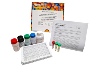
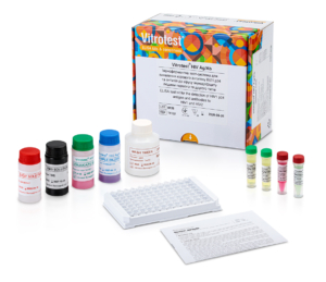

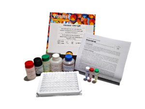
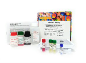
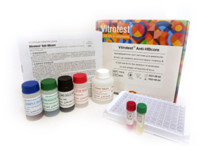
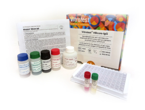
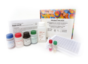
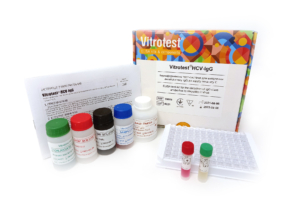
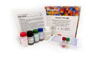
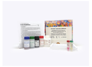
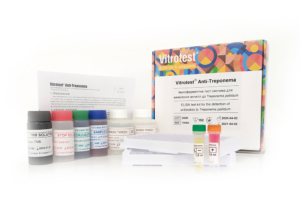
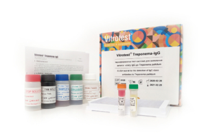
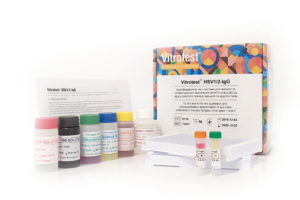
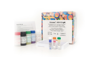
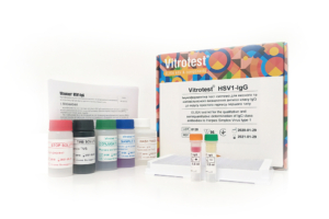
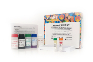
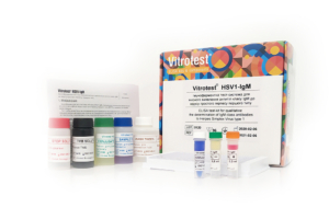
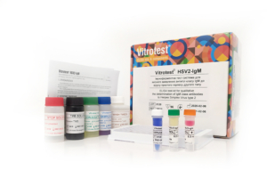
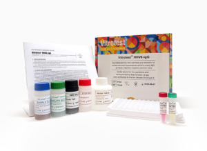
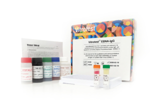
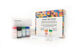
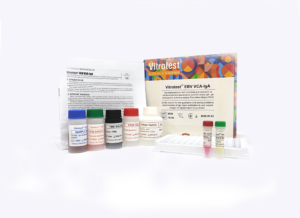
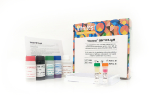
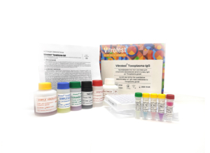
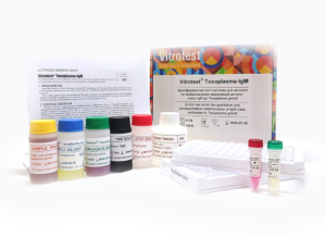
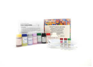
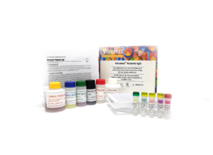
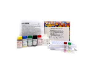
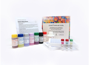
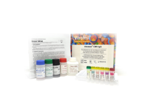
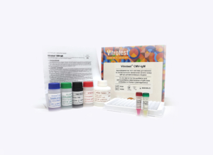
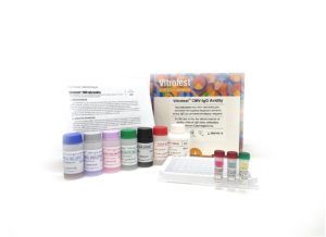
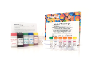
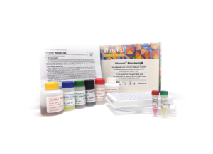
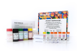
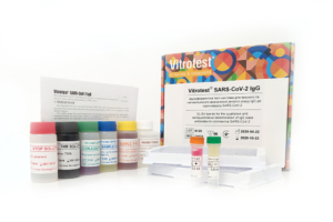
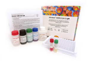
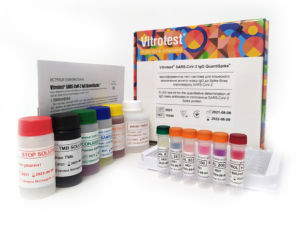
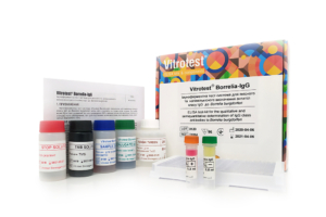
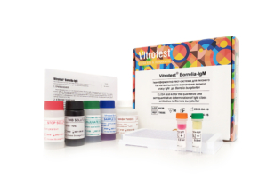
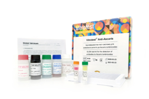
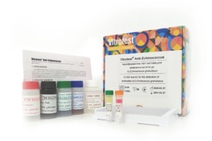
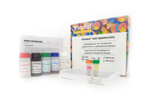
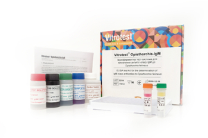
-
The test kit Vitrotest Anti-HIV1/2 is an enzyme linked immunosorbent assay (ELISA) for the detection of total antibodies (IgG, IgA, IgM) to HIV1 (group M and O) and HIV2 in human serum or plasma.
Detection of total antibodies to human immunodeficiency virus types 1 and 2 (HIV1/2) in the Vitrotest Anti-HIV1/2 test kit is based on a solid-phase sandwich ELISA in a two-step incubation procedure.
- TK129 - 96 tests
- TK130 - 192 tests
- TK131 - 480 tests
- Solid phase: breakable microplate ELISA is coated with recombinant HIV 1/2 antigens.
- Conjugate: recombinant HIV1/2 antigens conjugated to horseradish peroxidase.
- Chromogen: ready to use TMB solution.
- Volume of sample for analysis: 70 μl.
- Assay time: 60/30/15 min.
-
The test kit Vitrotest HIV Ag/Ab is an enzyme linked immunosorbent assay (ELISA) for the simultaneous detection of HIV p24 antigen and total antibodies (IgG, IgA, IgM) to HIV1 (group M and O) and HIV2 in human serum or plasma.
Simultaneous detection of the HIV1 p24 core antigen and total antibodies to human immunodeficiency virus types 1 and 2 (HIV1/2) in the Vitrotest HIV Ab/Ag test kit is based on a solid-phase sandwich ELISA in a two-step incubation procedure.
○ TK132 - 96 tests
○ TK133 - 192 tests
○ TK134 - 480 tests
- Solid phase: breakable microplate ELISA is coated with monoclonal antibodies specific to p24 HIV1 and recombinant HIV 1/2 antigens.
- Conjugate: solution of streptavidin and recombinant HIV1/2 antigens conjugated to horseradish peroxidase.
- Chromogen: ready to use TMB solution.
- Volume of sample for analysis: 70 μl.
- Assay time: 60/30/15 min.
- Analytical sensitivity: 2 IU/ml.
-
The test kit Vitrotest HIV1 p24 is an enzyme linked immunosorbent assay (ELISA) for quantitative determination of p24 core antigen to HIV1 in human serum or plasma.
Determination of the HIV1 p24 core antigen in the Vitrotest HIV1 p24 test kit is based on a solid-phase sandwich ELISA in a two-step incubation procedure.
- TK138 - 96 tests
- TK139 - 192 tests
- TK140 - 480 tests
- Solid phase: breakable microplate ELISA is coated with monoclonal antibodies specific to p24 HIV1.
- Conjugate: solution of streptavidin conjugated to horseradish peroxidase.
- Chromogen: ready to use TMB solution.
- Volume of sample for analysis: 70 μl.
- Assay time: 60/30/15 min.
-
The test kit Vitrotest HAV-IgM is an enzyme linked immunosorbent assay (ELISA) for the qualitative determination of IgM class antibodies to Hepatitis A Virus in human serum or plasma.
Determination of IgM antibodies to Hepatitis A Virus in the test kit Vitrotest HAV-IgM is based on a solid phase, «IgM-capture» ELISA in a two-step incubation procedure.
- TK069 - 96 tests
- Solid phase: strip ELISA plate pre-coated with monoclonal anti-IgM antibodies.
- Conjugate: monoclonal antibodies to human HAV conjugated with horseradish peroxidase.
- Chromogen: ready to use TMB solution.
- Sample volume: 10 μl.
- Assay time: 30/60/30 min.
-
The test kit Vitrotest HBsAg is an enzyme linked immunosorbent assay (ELISA) for the detection of surface antigen of Hepatitis B Virus (HBsAg) in human serum or plasma.
Detection of the presence of HBsAg in the test kit Vitrotest HBsAg is based on a solid-phase "sandwich" ELISA.
○ ТК016 - 96 tests
○ ТК059 - 192 tests
○ TK127 - 480 tests
- Solid phase: microplate ELISA coated with monoclonal antibodies to HBsAg.
- Conjugate: monoclonal antibodies to HBsAg conjugated with horseradish peroxidase.
- Chromogen: ready to use TMB solution.
- Volume of sample for analysis: 100 µl.
- Assay time: 120/30 min.
- Analitycal sensitivity: 0,05 IU/ml.
-
The Vitrotest HBsAg-Confirmation reagent kit is intended to confirm the presence of the hepatitis B virus surface antigen (HBsAg) in human serum or plasma. The kit is used together with the test kit Vitrotest HBsAg.
Confirmation of the presence of the hepatitis B virus surface antigen (HBsAg) is based on the solid-phase "sandwich" ELISA using the reagent kit Vitrotest HBsAg-Confirmation and the test kit Vitrotest HBsAg.
○ TK017 - 100 tests
-
Сomposition of the set:
- sample diluent;
- neutralizing component (monoclonal antibodies specific for HBsAg).
-
The test kit Vitrotest Anti-HBcore is an enzyme linked immunosorbent assay (ELISA) for the detection of antibodies to core antigen of Hepatitis B Virus (HBcore antigen) in human serum or plasma.
Detection of antibodies to HBcore antigen in the test kit Vitrotest Anti-HBcore is based on a solid phase, indirect ELISA in a two-step incubation procedure.
○ TK018 - 96 tests
○ TK141 - 192 tests
- Solid phase: microplate ELISA is coated with recombinant HBcore antigen.
- Conjugate: a mixture of monoclonal antibodies to human IgG and IgM conjugated with horseradish peroxidase.
- Chromogen: ready to use TMB solution.
- Volume of sample for analysis: 20 μl.
- Assay time: 30/30/30 min.
-
The test kit Vitrotest HBcore-IgG is an enzyme linked immunosorbent assay (ELISA) for the detection of IgG class antibodies to core antigen of Hepatitis B Virus (HBcore antigen) in human serum or plasma.
Detection of IgG class antibodies to HBcore antigen in the test kit Vitrotest HBcore-IgG is based on a solid phase, indirect ELISA in a two-step incubation procedure.
○ TK050 - 96 tests
○ TK142 - 192 tests
- Solid phase: microplate ELISA is coated with recombinant HBcore antigen.
- Conjugate: monoclonal antibodies to human IgG conjugated to horseradish peroxidase.
- Chromogen: ready to use TMB solution.
- Volume of sample for analysis: 20 μl.
- Assay time: 30/30/30 min.
-
The test kit Vitrotest HBcore-IgM is an enzyme linked immunosorbent assay (ELISA) for the detection of IgM class antibodies to the core antigen of the Hepatitis B Virus (HBcore) in human serum or plasma.
Detection of IgM class antibodies to the core antigen of the Hepatitis B Virus in the test kit Vitrotest HBcore-IgM is based on the principle of "IgM-capture" of solid-phase ELISA in two-stage incubation.
○ TK019 - 96 tests
○ TK143 - 192 tests
- Solid phase: microplate ELISA is coated with monoclonal antibodies specific for human immunoglobulin M.
- Conjugate: HBcore recombinant antigen conjugated with horseradish peroxidase.
- Chromogen: ready to use TMB solution.
- Volume of sample for analysis: 10 μl.
- Assay time: 30/30/30 min.
-
The test kit Vitrotest Anti-HCV is an enzyme linked immunosorbent assay (ELISA) for the detection of total antibodies to Hepatitis C Virus (HCV) in human serum or plasma.
Determination of antibodies to HCV in the test kit Vitrotest Anti-HCV is based on a solid phase indirect ELISA in a two-step incubation procedure.
○ TK022 - 96 tests
○ TK060 - 192 tests
○ TK128 - 480 tests
- Solid phase: breakable microplate ELISA is coated with recombinant antigens (core, NS3, NS4 and NS5).
- Conjugate: monoclonal antibodies to human IgG and IgM conjugated to horseradish peroxidase.
- Chromogen: ready to use TMB solution.
- Volume of sample for analysis: 40 μl.
- Assay time: 60/30/30 min.
-
The test kit Vitrotest HCV-IgG is an enzyme linked immunosorbent assay (ELISA) for the detection of IgG class antibodies to Hepatitis C Virus (HCV) in human serum or plasma.
Determination of IgG class antibodies to HCV in the test kit Vitrotest HCV-IgG is based on a solid phase indirect ELISA in a two-step incubation procedure.
○ ТК055 - 96 tests
○ TK144 - 192 tests
- Solid phase: breakable microplate ELISA is coated with recombinant antigens (core, NS3, NS4 and NS5).
- Conjugate: monoclonal antibodies to human IgG conjugated to horseradish peroxidase.
- Chromogen: ready to use TMB solution.
- Volume of sample for analysis: 40 μl.
- Assay time: 60/30/30 min.
-
The test kit Vitrotest HCV-IgМ is an enzyme linked immunosorbent assay (ELISA) for the detection of IgМ class antibodies to Hepatitis C Virus (HCV) in human serum or plasma.
Determination of IgМ class antibodies to HCV in the test kit Vitrotest HCV-IgМ is based on a solid phase indirect ELISA in a two-step incubation procedure.
○ ТК043 - 96 tests
○ TK145 - 192 tests
- Solid phase: breakable microplate ELISA is coated with recombinant antigens (core, NS3 and NS4).
- Conjugate: monoclonal antibodies to human IgМ conjugated to horseradish peroxidase.
- Chromogen: ready to use TMB solution.
- Volume of sample for analysis: 40 μl.
- Assay time: 30/30/30 min.
-
The test kit Vitrotest Anti-HCV Different is an enzyme linked immunosorbent assay (ELISA) for the differential detection of antibodies to antigens of hepatitis C - core, NS3, NS4, NS5 in human serum or plasma.
The differential detection of antibodies to specific antigens of the hepatitis C virus in the test kit Vitrotest Anti-HCV Different is based on a solid phase indirect ELISA in a two-step incubation procedure.
○ TK044 - 24 tests
- Solid phase: breakable microplate ELISA is coated with recombinant HCV antigens in separate wells: core (strips 1, 5, and 9, marked in black), NS3 (strips 2, 6, and 10, marked in green), NS4 (strips 3, 7, and 11, marked in blue), and NS5 (strips 4, 8, and 12, marked in red).
- Conjugate: monoclonal antibodies to human IgG and IgM conjugated to horseradish peroxidase.
- Chromogen: ready to use TMB solution.
- Volume of sample for analysis: 4×40 μl.
- Assay time: 60/30/30 min.
-
The test kit Vitrotest Anti-Treponema is an enzyme linked immunosorbent assay (ELISA) for the detection of IgG and IgM class antibodies to Treponema pallidum in human serum or plasma.
Detection of antibodies to T. pallidum in the test kit Vitrotest Anti-Treponema is based on a solid phase, indirect ELISA in a two-step incubation procedure.
○ ТК023 - 96 tests
○ ТК063 - 192 tests
○ ТК135 - 480 tests
- Solid phase: breakable microplate ELISA is coated with recombinant Treponema pallidum antigens.
- Conjugate: monoclonal antibodies to human IgG and IgM conjugated to horseradish peroxidase.
- Chromogen: ready to use TMB solution.
- Volume of sample for analysis: 20 μl.
- Assay time: 30/30/15 min.
-
The test kit Vitrotest Treponema-IgG is an enzyme linked immunosorbent assay (ELISA) for the detection of IgG class antibodies to Treponema pallidum in human serum or plasma.
Detection of IgG antibodies to T. pallidum in the test kit Vitrotest Treponema-IgG is based on a solid phase, indirect ELISA in a two-step incubation procedure.
○ ТК056 - 96 tests
- Solid phase: breakable microplate ELISA is coated with recombinant Treponema pallidum antigens.
- Conjugate: monoclonal antibodies to human IgG conjugated to horseradish peroxidase.
- Chromogen: ready to use TMB solution.
- Volume of sample for analysis: 20 μl.
- Assay time: 30/30/15 min.
-
The test kit Vitrotest HSV1/2-IgG is an enzyme linked immunosorbent assay (ELISA) for the qualitative and semiquantitative determination of IgG class antibodies to Herpes Simplex Virus types 1 and 2 (HSV1/2) in human serum or plasma.
Determination of IgG antibodies to HSV1/2 in the test kit Vitrotest HSV1/2-IgG is based on a solid phase, indirect ELISA in a two-step incubation procedure.
○ ТК007 - 96 tests
- Solid phase: breakable microplate ELISA is coated with mixture of purified antigens of herpes simplex virus type 1 and type 2.
- Conjugate: a monoclonal antibodies to human IgG conjugated to horseradish peroxidase.
- Chromogen: ready to use TMB solution.
- Volume of sample for analysis: 10 μl.
- Assay time: 30/30/15 min.
-
The test kit Vitrotest HSV1/2-IgM is an enzyme linked immunosorbent assay (ELISA) for the qualitative determination of IgM class antibodies to Herpes Simplex Virus types 1 and 2 (HSV1/2) in human serum or plasma.
Determination of IgM antibodies to HSV1 and HSV2 in the test kit Vitrotest HSV1/2-IgM is based on a solid phase, «IgM-capture» ELISA in a two-step incubation procedure.
○ ТК008 - 96 tests
- Solid phase: breakable microplate ELISA is coated with monoclonal anti-IgM antibodies.
- Conjugate: mixture of recombinant antigens of herpes simplex virus type 1 and type 2 gG1 and gG2 conjugated to horseradish peroxidase.
- Chromogen: ready to use TMB solution.
- Volume of sample for analysis: 10 μl.
- Assay time: 30/60/30 min.
-
The test kit Vitrotest HSV1-IgG is an enzyme linked immunosorbent assay (ELISA) for the qualitative and semiquantitative determination of IgG class antibodies to Herpes Simplex Virus type 1 (HSV-1) in human serum or plasma.
Determination of IgG antibodies to HSV-1 in the test kit Vitrotest HSV1-IgG is based on a solid phase, indirect ELISA in a two-step incubation procedure.
○ ТК026 - 96 tests
- Solid phase: breakable microplate ELISA is coated with recombinant antigens of herpes simplex virus type 1.
- Conjugate: a monoclonal antibodies to human IgG conjugated to horseradish peroxidase.
- Chromogen: ready to use TMB solution.
- Volume of sample for analysis: 10 μl.
- Assay time: 30/30/15 min.
-
The test kit Vitrotest HSV2-IgG is an enzyme linked immunosorbent assay (ELISA) for the qualitative and semiquantitative determination of IgG class antibodies to Herpes Simplex Virus type 2 (HSV-2) in human serum or plasma.
Determination of IgG antibodies to HSV-2 in the test kit Vitrotest HSV2-IgG is based on a solid phase, indirect ELISA in a two-step incubation procedure.
○ ТК011 - 96 tests
- Solid phase: breakable microplate ELISA is coated with recombinant antigens of herpes simplex virus type 2.
- Conjugate: a monoclonal antibodies to human IgG conjugated to horseradish peroxidase.
- Chromogen: ready to use TMB solution.
- Volume of sample for analysis: 10 μl.
- Assay time: 30/30/15 min.
-
The test kit Vitrotest HSV1-IgM is an enzyme linked immunosorbent assay (ELISA) for the qualitative determination of IgM class antibodies to Herpes Simplex Virus type 1 (HSV-1) in human serum or plasma.
Determination of IgM antibodies to HSV-1 in the test kit Vitrotest HSV1-IgM is based on a solid phase, «IgM-capture» ELISA in a two-step incubation procedure.
○ ТК009 - 96 tests
- Solid phase: breakable microplate ELISA is coated with monoclonal anti-IgM antibodies.
- Conjugate: recombinant HSV1 antigen gG1 conjugated to horseradish peroxidase.
- Chromogen: ready to use TMB solution.
- Volume of sample for analysis: 10 μl.
- Assay time: 30/60/30 min.
-
The test kit Vitrotest HSV2-IgM is an enzyme linked immunosorbent assay (ELISA) for the qualitative determination of IgM class antibodies to Herpes Simplex Virus type 2 (HSV-2) in human serum or plasma.
Determination of IgM antibodies to HSV-2 in the test kit Vitrotest HSV2-IgM is based on a solid phase, «IgM-capture» ELISA in a two-step incubation procedure.
○ ТК010 - 96 tests
- Solid phase: breakable microplate ELISA is coated with monoclonal anti-IgM antibodies.
- Conjugate: recombinant HSV2 antigen gG2 conjugated to horseradish peroxidase.
- Chromogen: ready to use TMB solution.
- Volume of sample for analysis: 10 μl.
- Assay time: 30/60/30 min.
-
The test kit Vitrotest® HHV6-IgG is an enzyme linked immunosorbent assay (ELISA) for the qualitative and semiquantitative determination of IgG class antibodies to Human Herpes Virus type 6 (HHV-6) in human serum or plasma.
Determination of IgG antibodies to HHV6 in the test kit Vitrotest® HHV6-IgG is based on a solid phase, indirect ELISA in a two-step incubation procedure.
○ ТК090 - 96 tests- Solid phase: breakable microplate ELISA is coated with recombinant antigens of Human Herpes Virus type 6.
- Conjugate: a monoclonal antibodies to human IgG conjugated to horseradish peroxidase.
- Chromogen: ready to use TMB solution.
- Volume of sample for analysis: 10 μl.
- Assay time: 1h 15 min.
Herpes infections are the cause of the development of many somatic and oncological diseases, occupy a leading place among the causes of stillbirth, premature birth, infant mortality, morbidity of newborns, contributing to early disability of children. This is due to a number of reasons: the prevalence of herpesviruses, the variety of diseases they cause, the existence in the human body in various forms (acute, chronic, latent).
Herpesviruses (Herpesviridae) are a large family of DNA viruses that cause various diseases in humans and other mammals. There are 8 representatives of the herpesvirus family that infect humans. One of them is the human herpesvirus type six (HHV-6). It has been relatively recently added to the list of known human pathogens and is a serious contender for the role of the etiological agent of such diseases as multiple sclerosis, encephalitis, fever in children with convulsive syndrome, infectious mononucleosis, "sudden exanthema". There is evidence that HHV-6 is a cofactor of AIDS, some forms of cervical carcinomas and nasopharyngeal carcinomas.
Infection with HHV-6 usually occurs in the first or second year of life, and accordingly about 95% of adults have antibodies to HHV-6. At birth, most children are seropositive due to maternal antibodies, the titer of which decreases by 5 months. However, by the end of the first year of life, the percentage of seropositive babies is the same as among older children and adults.
HHV-6 is similar to other herpesviruses, but differs from them in biological, immunological properties, spectrum of sensitive cells, antigenic structure, genome composition, number and molecular weight of structural viral proteins. In the diagnosis of HHV-6 infections, immunochemical (IC) and molecular biological (PCR, hybridization) methods are most often used. Among serological methods for determining HHV-6 infection, such as immunofluorescence method, immunoblot, immunoprecipitation, the most common is ELISA. -
The test kit Vitrotest® EBNA-IgG is an enzyme linked immunosorbent assay (ELISA) for the qualitative and semiquantitative determination of IgG class antibodies to nuclear antigen (EBNA) of Epstein-Barr virus (EBV) in human serum or plasma.
Determination of IgG antibodies to EBV nuclear antigen (EBNA) in the test kit Vitrotest® EBNA-IgG is based on a solid phase, indirect ELISA in a two-step incubation procedure.
○ ТК054 - 96 tests- Solid phase: breakable microplate ELISA is coated with EBV recombinant nuclear antigen.
- Conjugate: a monoclonal antibodies to human IgG conjugated to horseradish peroxidase.
- Chromogen: ready to use TMB solution.
- Volume of sample for analysis: 10 μl.
- Assay time: 1h 15 min.
Epstein-Barr virus (EBV), also known as human herpesvirus 4, is the causative agent of diseases such as infectious mononucleosis (IM), Burkitt’s lymphoma (BL), nasopharyngeal carcinoma.
EBV acquired in early childhood usually does not cause symptoms. Infection in adolescence or early adulthood often results in clinical IM. Typically, the disease presents as pharyngitis, lymphadenopathy, and fever. In most cases, symptoms resolve within 2–4 weeks, but more than 90% of adults develop a latent B-lymphocyte infection. Approximately 1% of immunocompetent individuals may experience severe complications (hepatitis, myocarditis, splenic rupture, neurological complications). In immunosuppressed individuals, primary EBV infection leads to severe disorders (eg, BL).
The clinical symptoms range from mild fever to prolonged debilitating illness. Typically, the disease presents as pharyngitis, lymphadenopathy, and fever. Enlarged spleen and a swollen liver are less common symptoms. Microscopic blood observation reveals lymphocytosis; most lymphocytes have atypical morphology.
EBV spreads most commonly through bodily fluids, especially saliva, sporadically also through blood and semen. Humans are the most infectious during acute IM, but the virus could be shed into saliva even during latency. Transmission occurs mainly via household and sexual routes; however, transmission via blood transfusion and organ transplantation was also demonstrated.
Risk groups for IM are adolescents, senior school students, blood transfusion recipients, as well as people with immunodeficiencies (risk group for complications). Up to 50% of adolescents and young adults in developed countries are seronegative and hence at risk of developing IM.
EBV is a genetically stable virus. EBV is one of the most common human viruses. EBV is found all over the world; virus is able to infect more than 95% of all individuals within the first four decades of life. The current incidence of IM in USA is estimated at approximately 125 000 reported cases/year. EBV DNA is surrounded by a nucleocapsid; protein tegument is situated between the nucleocapsid and envelope; outer envelope has glycoprotein spikes typical of all herpesviruses. The viral genome encodes around 90 proteins. The most of these proteins (80) are expressed only during EBV lytic cycle.
On the basis of their time of expression, EBV genes are designated early or late. Early genes encode mostly non-structural proteins, the most important among them is viral DNA polymerase. Late genes encode mostly structural viral proteins which are required for successful packaging of the viral DNA and formation of the virion. The viral capsids have several polypeptide components ranging in size from 18 to 155 kDa, with a 155-kDa polypeptide being the major component. The membrane antigen (MA) complex is found on the virion envelope and consists of at least three major EBV glycoproteins: gp350/220 (the most abundant protein), gp110, and gp85.
The presence of EBV in epithelial and B cells provokes an intense immune response consisting of antibodies to a large variety of virally encoded antigens. During the acute phase of IM, the humoral response is directed toward viral antigens of the lytic cycle, notably membrane antigen complex, several early antigens (p 54 and p138), and VCA. A strong IgM response (the so-called heterophile antibodies) is directed towards a complex glycoprotein structure on the surface of EBV infected cells. As IM patients recover from clinical symptoms, the IgM response reduces significantly while the IgG response in serum plateaus at a reduced level and is maintained throughout the persistent infection.
The main purpose of the diagnostics of IM is to rule out less common but more serious causes of IM-like symptoms, requiring immediate intervention, such as hepatitis or various leukemic malignancies. The mainstay in the diagnostics of IM are still clinical symptoms (classic triad-fever, lymphoadenopathy, and pharyngytis) in combination with anamnestic aspects.
The specificity of the “classic triad” is around 90%, since IM-like symptoms may be caused by a variety of other pathogens and malignancies, such as cytomegalovirus, human herpesvirus 6, adenovirus, rubella virus, mumps virus, human immunodeficiency virus, hepatitis A virus, influenza A and B viruses, and Toxoplasma gondii, lymphoma and leukemia. Therefore, EBV infection should be confirmed with a blood antibody test.
It is well known for many years that acute IM is characterized by the appearance of heterophile antibodies (i.e. antibodies to EBV antigen described above) in the serum. This peculiarity is used in the so-called “monospot tests”, detecting agglutination of sheep erythrocytes in the presence of serum of patients with acute-phase IM. This test is now not recommended for general use by the Centers for Disease Prevention and Control, USA, since it produces both false positive and false negative results.
Only three serological parameters are essential for the detection of EBV-specific antibodies in immunocompetent individuals: VCA IgG (marker of disease, present or past), VCA IgM (marker of recent or present disease), and EBNA-1 IgG (marker of past disease).
The most widely used serological methods are immunofluorescence assay (IFA) and enzyme immunoassay (EIA). IFA has always been used as a reference method. However, some investigators found EIA to be more sensitive than IFA.
On the contrary, in immunosuppressed individuals serological assays are not recommended for many reasons, such as dysfunctions in the production of antibodies. To date, only detection of viral load by PCR is an established marker for immunosuppressed patients. -
The test kit Vitrotest® EBV VCA-IgG is an enzyme linked immunosorbent assay (ELISA) for the qualitative and semiquantitative determination of IgG class antibodies to viral capsid antigen (VCA) of Epstein-Barr virus (EBV) in human serum or plasma.
Determination of IgG antibodies to EBV capsid antigen (VCA) in the test kit Vitrotest® EBV VCA-IgG is based on a solid phase, indirect ELISA in a two-step incubation procedure.
○ ТК053 - 96 tests- Solid phase: breakable microplate ELISA is coated with EBV recombinant capsid antigen.
- Conjugate: a monoclonal antibodies to human IgG conjugated to horseradish peroxidase.
- Chromogen: ready to use TMB solution.
- Volume of sample for analysis: 10 μl.
- Assay time: 1h 15 min.
Epstein-Barr virus (EBV), also known as human herpesvirus 4, is the causative agent of diseases such as infectious mononucleosis (IM), Burkitt’s lymphoma (BL), nasopharyngeal carcinoma.
EBV acquired in early childhood usually does not cause symptoms. Infection in adolescence or early adulthood often results in clinical IM. Typically, the disease presents as pharyngitis, lymphadenopathy, and fever. In most cases, symptoms resolve within 2–4 weeks, but more than 90% of adults develop a latent B-lymphocyte infection. Approximately 1% of immunocompetent individuals may experience severe complications (hepatitis, myocarditis, splenic rupture, neurological complications). In immunosuppressed individuals, primary EBV infection leads to severe disorders (eg, BL).
The clinical symptoms range from mild fever to prolonged debilitating illness. Typically, the disease presents as pharyngitis, lymphadenopathy, and fever. Enlarged spleen and a swollen liver are less common symptoms. Microscopic blood observation reveals lymphocytosis; most lymphocytes have atypical morphology.
EBV spreads most commonly through bodily fluids, especially saliva, sporadically also through blood and semen. Humans are the most infectious during acute IM, but the virus could be shed into saliva even during latency. Transmission occurs mainly via household and sexual routes; however, transmission via blood transfusion and organ transplantation was also demonstrated.
Risk groups for IM are adolescents, senior school students, blood transfusion recipients, as well as people with immunodeficiencies (risk group for complications). Up to 50% of adolescents and young adults in developed countries are seronegative and hence at risk of developing IM.
EBV is a genetically stable virus. EBV is one of the most common human viruses. EBV is found all over the world; virus is able to infect more than 95% of all individuals within the first four decades of life. The current incidence of IM in USA is estimated at approximately 125 000 reported cases/year. EBV DNA is surrounded by a nucleocapsid; protein tegument is situated between the nucleocapsid and envelope; outer envelope has glycoprotein spikes typical of all herpesviruses. The viral genome encodes around 90 proteins. The most of these proteins (80) are expressed only during EBV lytic cycle.
On the basis of their time of expression, EBV genes are designated early or late. Early genes encode mostly non-structural proteins, the most important among them is viral DNA polymerase. Late genes encode mostly structural viral proteins which are required for successful packaging of the viral DNA and formation of the virion. The viral capsids have several polypeptide components ranging in size from 18 to 155 kDa, with a 155-kDa polypeptide being the major component. The membrane antigen (MA) complex is found on the virion envelope and consists of at least three major EBV glycoproteins: gp350/220 (the most abundant protein), gp110, and gp85.
The presence of EBV in epithelial and B cells provokes an intense immune response consisting of antibodies to a large variety of virally encoded antigens. During the acute phase of IM, the humoral response is directed toward viral antigens of the lytic cycle, notably membrane antigen complex, several early antigens (p 54 and p138), and VCA. A strong IgM response (the so-called heterophile antibodies) is directed towards a complex glycoprotein structure on the surface of EBV infected cells. As IM patients recover from clinical symptoms, the IgM response reduces significantly while the IgG response in serum plateaus at a reduced level and is maintained throughout the persistent infection.
The main purpose of the diagnostics of IM is to rule out less common but more serious causes of IM-like symptoms, requiring immediate intervention, such as hepatitis or various leukemic malignancies. The mainstay in the diagnostics of IM are still clinical symptoms (classic triad-fever, lymphoadenopathy, and pharyngytis) in combination with anamnestic aspects.
The specificity of the “classic triad” is around 90%, since IM-like symptoms may be caused by a variety of other pathogens and malignancies, such as cytomegalovirus, human herpesvirus 6, adenovirus, rubella virus, mumps virus, human immunodeficiency virus, hepatitis A virus, influenza A and B viruses, and Toxoplasma gondii, lymphoma and leukemia. Therefore, EBV infection should be confirmed with a blood antibody test.
It is well known for many years that acute IM is characterized by the appearance of heterophile antibodies (i.e. antibodies to EBV antigen described above) in the serum. This peculiarity is used in the so-called “monospot tests”, detecting agglutination of sheep erythrocytes in the presence of serum of patients with acute-phase IM. This test is now not recommended for general use by the Centers for Disease Prevention and Control, USA, since it produces both false positive and false negative results.
Only three serological parameters are essential for the detection of EBV-specific antibodies in immunocompetent individuals: VCA IgG (marker of disease, present or past), VCA IgM (marker of recent or present disease), and EBNA-1 IgG (marker of past disease).
The most widely used serological methods are immunofluorescence assay (IFA) and enzyme immunoassay (EIA). IFA has always been used as a reference method. However, some investigators found EIA to be more sensitive than IFA.
On the contrary, in immunosuppressed individuals serological assays are not recommended for many reasons, such as dysfunctions in the production of antibodies. To date, only detection of viral load by PCR is an established marker for immunosuppressed patients. -
The test kit Vitrotest® EBV VCA-IgА is an enzyme linked immunosorbent assay (ELISA) for the qualitative and semiquantitative determination of IgА class antibodies to viral capsid antigen (VCA) of Epstein-Barr virus (EBV) in human serum or plasma.
Determination of IgА antibodies to EBV capsid antigen (VCA) in the test kit Vitrotest® EBV VCA-IgА is based on a solid phase, indirect ELISA in a two-step incubation procedure.
○ ТК123 - 96 tests- Solid phase: breakable microplate ELISA is coated with EBV recombinant capsid antigen.
- Conjugate: a monoclonal antibodies to human IgА conjugated to horseradish peroxidase.
- Chromogen: ready to use TMB solution.
- Volume of sample for analysis: 10 μl.
- Assay time: 1h 15 min.
Epstein-Barr virus (EBV), also known as human herpesvirus 4, is the causative agent of diseases such as infectious mononucleosis (IM), Burkitt’s lymphoma (BL), nasopharyngeal carcinoma.
EBV acquired in early childhood usually does not cause symptoms. Infection in adolescence or early adulthood often results in clinical IM. Typically, the disease presents as pharyngitis, lymphadenopathy, and fever. In most cases, symptoms resolve within 2–4 weeks, but more than 90% of adults develop a latent B-lymphocyte infection. Approximately 1% of immunocompetent individuals may experience severe complications (hepatitis, myocarditis, splenic rupture, neurological complications). In immunosuppressed individuals, primary EBV infection leads to severe disorders (eg, BL).
The clinical symptoms range from mild fever to prolonged debilitating illness. Typically, the disease presents as pharyngitis, lymphadenopathy, and fever. Enlarged spleen and a swollen liver are less common symptoms. Microscopic blood observation reveals lymphocytosis; most lymphocytes have atypical morphology.
EBV spreads most commonly through bodily fluids, especially saliva, sporadically also through blood and semen. Humans are the most infectious during acute IM, but the virus could be shed into saliva even during latency. Transmission occurs mainly via household and sexual routes; however, transmission via blood transfusion and organ transplantation was also demonstrated.
Risk groups for IM are adolescents, senior school students, blood transfusion recipients, as well as people with immunodeficiencies (risk group for complications). Up to 50% of adolescents and young adults in developed countries are seronegative and hence at risk of developing IM.
EBV is a genetically stable virus. EBV is one of the most common human viruses. EBV is found all over the world; virus is able to infect more than 95% of all individuals within the first four decades of life. The current incidence of IM in USA is estimated at approximately 125 000 reported cases/year. EBV DNA is surrounded by a nucleocapsid; protein tegument is situated between the nucleocapsid and envelope; outer envelope has glycoprotein spikes typical of all herpesviruses. The viral genome encodes around 90 proteins. The most of these proteins (80) are expressed only during EBV lytic cycle.
On the basis of their time of expression, EBV genes are designated early or late. Early genes encode mostly non-structural proteins, the most important among them is viral DNA polymerase. Late genes encode mostly structural viral proteins which are required for successful packaging of the viral DNA and formation of the virion. The viral capsids have several polypeptide components ranging in size from 18 to 155 kDa, with a 155-kDa polypeptide being the major component. The membrane antigen (MA) complex is found on the virion envelope and consists of at least three major EBV glycoproteins: gp350/220 (the most abundant protein), gp110, and gp85.
The presence of EBV in epithelial and B cells provokes an intense immune response consisting of antibodies to a large variety of virally encoded antigens. During the acute phase of IM, the humoral response is directed toward viral antigens of the lytic cycle, notably membrane antigen complex, several early antigens (p 54 and p138), and VCA. A strong IgM response (the so-called heterophile antibodies) is directed towards a complex glycoprotein structure on the surface of EBV infected cells. As IM patients recover from clinical symptoms, the IgM response reduces significantly while the IgG response in serum plateaus at a reduced level and is maintained throughout the persistent infection.
The main purpose of the diagnostics of IM is to rule out less common but more serious causes of IM-like symptoms, requiring immediate intervention, such as hepatitis or various leukemic malignancies. The mainstay in the diagnostics of IM are still clinical symptoms (classic triad-fever, lymphoadenopathy, and pharyngytis) in combination with anamnestic aspects.
The specificity of the “classic triad” is around 90%, since IM-like symptoms may be caused by a variety of other pathogens and malignancies, such as cytomegalovirus, human herpesvirus 6, adenovirus, rubella virus, mumps virus, human immunodeficiency virus, hepatitis A virus, influenza A and B viruses, and Toxoplasma gondii, lymphoma and leukemia. Therefore, EBV infection should be confirmed with a blood antibody test.
It is well known for many years that acute IM is characterized by the appearance of heterophile antibodies (i.e. antibodies to EBV antigen described above) in the serum. This peculiarity is used in the so-called “monospot tests”, detecting agglutination of sheep erythrocytes in the presence of serum of patients with acute-phase IM. This test is now not recommended for general use by the Centers for Disease Prevention and Control, USA, since it produces both false positive and false negative results.
Only three serological parameters are essential for the detection of EBV-specific antibodies in immunocompetent individuals: VCA IgG (marker of disease, present or past), VCA IgM (marker of recent or present disease), and EBNA-1 IgG (marker of past disease).
The most widely used serological methods are immunofluorescence assay (IFA) and enzyme immunoassay (EIA). IFA has always been used as a reference method. However, some investigators found EIA to be more sensitive than IFA.
On the contrary, in immunosuppressed individuals serological assays are not recommended for many reasons, such as dysfunctions in the production of antibodies. To date, only detection of viral load by PCR is an established marker for immunosuppressed patients. -
The test kit Vitrotest EBV VCA-IgM is an enzyme linked immunosorbent assay (ELISA) for the qualitative and semiquantitative determination of IgM class antibodies to viral capsid antigen (VCA) of Epstein-Barr virus (EBV) in human serum or plasma.
Determination of IgM antibodies to EBV VCA in the test kit Vitrotest EBV VCA-IgM is based on a solid phase «IgM-capture» ELISA in a two-step incubation procedure.
○ ТК052 - 96 tests
- Solid phase: breakable microplate ELISA is coated with monoclonal anti-IgM antibodies.
- Conjugate: recombinant Epstein-Barr virus capsid antigen (VCA) conjugated to horseradish peroxidase.
- Chromogen: ready to use TMB solution.
- Volume of sample for analysis: 10 μl.
- Assay time: 30/30/15 min.
Epstein-Barr virus (EBV), also known as human herpesvirus 4, is the causative agent of diseases such as infectious mononucleosis (IM), Burkitt’s lymphoma (BL), nasopharyngeal carcinoma.
EBV acquired in early childhood usually does not cause symptoms. Infection in adolescence or early adulthood often results in clinical IM. Typically, the disease presents as pharyngitis, lymphadenopathy, and fever. In most cases, symptoms resolve within 2–4 weeks, but more than 90% of adults develop a latent B-lymphocyte infection. Approximately 1% of immunocompetent individuals may experience severe complications (hepatitis, myocarditis, splenic rupture, neurological complications). In immunosuppressed individuals, primary EBV infection leads to severe disorders (eg, BL).
The clinical symptoms range from mild fever to prolonged debilitating illness. Typically, the disease presents as pharyngitis, lymphadenopathy, and fever. Enlarged spleen and a swollen liver are less common symptoms. Microscopic blood observation reveals lymphocytosis; most lymphocytes have atypical morphology.
EBV spreads most commonly through bodily fluids, especially saliva, sporadically also through blood and semen. Humans are the most infectious during acute IM, but the virus could be shed into saliva even during latency. Transmission occurs mainly via household and sexual routes; however, transmission via blood transfusion and organ transplantation was also demonstrated.
Risk groups for IM are adolescents, senior school students, blood transfusion recipients, as well as people with immunodeficiencies (risk group for complications). Up to 50% of adolescents and young adults in developed countries are seronegative and hence at risk of developing IM.
EBV is a genetically stable virus. EBV is one of the most common human viruses. EBV is found all over the world; virus is able to infect more than 95% of all individuals within the first four decades of life. The current incidence of IM in USA is estimated at approximately 125 000 reported cases/year. EBV DNA is surrounded by a nucleocapsid; protein tegument is situated between the nucleocapsid and envelope; outer envelope has glycoprotein spikes typical of all herpesviruses. The viral genome encodes around 90 proteins. The most of these proteins (80) are expressed only during EBV lytic cycle.
On the basis of their time of expression, EBV genes are designated early or late. Early genes encode mostly non-structural proteins, the most important among them is viral DNA polymerase. Late genes encode mostly structural viral proteins which are required for successful packaging of the viral DNA and formation of the virion. The viral capsids have several polypeptide components ranging in size from 18 to 155 kDa, with a 155-kDa polypeptide being the major component. The membrane antigen (MA) complex is found on the virion envelope and consists of at least three major EBV glycoproteins: gp350/220 (the most abundant protein), gp110, and gp85.
The presence of EBV in epithelial and B cells provokes an intense immune response consisting of antibodies to a large variety of virally encoded antigens. During the acute phase of IM, the humoral response is directed toward viral antigens of the lytic cycle, notably membrane antigen complex, several early antigens (p 54 and p138), and VCA. A strong IgM response (the so-called heterophile antibodies) is directed towards a complex glycoprotein structure on the surface of EBV infected cells. As IM patients recover from clinical symptoms, the IgM response reduces significantly while the IgG response in serum plateaus at a reduced level and is maintained throughout the persistent infection.
The main purpose of the diagnostics of IM is to rule out less common but more serious causes of IM-like symptoms, requiring immediate intervention, such as hepatitis or various leukemic malignancies. The mainstay in the diagnostics of IM are still clinical symptoms (classic triad-fever, lymphoadenopathy, and pharyngytis) in combination with anamnestic aspects.
The specificity of the “classic triad” is around 90%, since IM-like symptoms may be caused by a variety of other pathogens and malignancies, such as cytomegalovirus, human herpesvirus 6, adenovirus, rubella virus, mumps virus, human immunodeficiency virus, hepatitis A virus, influenza A and B viruses, and Toxoplasma gondii, lymphoma and leukemia. Therefore, EBV infection should be confirmed with a blood antibody test.
It is well known for many years that acute IM is characterized by the appearance of heterophile antibodies (i.e. antibodies to EBV antigen described above) in the serum. This peculiarity is used in the so-called “monospot tests”, detecting agglutination of sheep erythrocytes in the presence of serum of patients with acute-phase IM. This test is now not recommended for general use by the Centers for Disease Prevention and Control, USA, since it produces both false positive and false negative results.
Only three serological parameters are essential for the detection of EBV-specific antibodies in immunocompetent individuals: VCA IgG (marker of disease, present or past), VCA IgM (marker of recent or present disease), and EBNA-1 IgG (marker of past disease).
The most widely used serological methods are immunofluorescence assay (IFA) and enzyme immunoassay (EIA). IFA has always been used as a reference method. However, some investigators found EIA to be more sensitive than IFA.
On the contrary, in immunosuppressed individuals serological assays are not recommended for many reasons, such as dysfunctions in the production of antibodies. To date, only detection of viral load by PCR is an established marker for immunosuppressed patients. -
The test kit Vitrotest Toxoplasma-IgG is an enzyme linked immunosorbent assay (ELISA) for the quantitative determination of IgG class antibodies to Toxoplasma gondii in human serum or plasma.
Determination of IgG antibodies to Toxoplasma gondii in the test kit Vitrotest Toxoplasma-IgG is based on a solid phase, indirect ELISA in a two-step incubation procedure.
○ ТК001 - 96 tests
- Solid phase: breakable microplate ELISA is coated Toxoplasma gondii antigens.
- Conjugate: a monoclonal antibodies to human IgG conjugated to horseradish peroxidase.
- Chromogen: ready to use TMB solution.
- Volume of sample for analysis: 10 μl.
- Assay time: 30/30/15 min.
- Analytical sensitivity: 0,7 IU/ml.
- Linearity range: 10-180 IU/ml.
-
The test kit Vitrotest Toxoplasma-IgM is an enzyme linked immunosorbent assay (ELISA) for the qualitative and semiquantitative determination of IgM class antibodies to Toxoplasma gondii in human serum or plasma.
Determination of IgM antibodies to Toxoplasma gondii in the test kit Vitrotest Toxoplasma-IgM is based on a solid phase, indirect ELISA in a two-step incubation procedure.
○ ТК002 - 96 tests
- Solid phase: breakable microplate ELISA is coated Toxoplasma gondii antigens.
- Conjugate: a monoclonal antibodies to human IgM conjugated to horseradish peroxidase.
- Chromogen: ready to use TMB solution.
- Volume of sample for analysis: 10 μl.
- Assay time: 30/30/15 min.
-
The test kit Vitrotest Toxoplasma-IgG Avidity is an enzyme linked immunosorbent assay (ELISA) for the determination of avidity index of IgG class antibodies to Toxoplasma gondii in human serum or plasma.
Determination of avidity index of IgG antibodies to Toxoplasma gondii in the test kit Vitrotest Toxoplasma-IgG Avidity is based on a solid phase, indirect ELISA.
○ ТК064 - 48 tests
- Solid phase: breakable microplate ELISA is coated Toxoplasma gondii antigens.
- Conjugate: a monoclonal antibodies to human IgG conjugated to horseradish peroxidase.
- Chromogen: ready to use TMB solution.
- Volume of sample for analysis: 2×10 μl.
- Assay time: 30/15/30/15 min.
-
The test kit Vitrotest Rubella-IgG is an enzyme linked immunosorbent assay (ELISA) for the quantitative determination of IgG class antibodies to Rubella virus in human serum or plasma.
Determination of IgG antibodies to Rubella virus in the test kit Vitrotest Rubella-IgG is based on a solid phase, indirect ELISA in a two-step incubation procedure.
○ ТК003 - 96 tests
- Solid phase: breakable microplate ELISA is coated with Rubella virus antigens.
- Conjugate: a monoclonal antibodies to human IgG conjugated to horseradish peroxidase.
- Chromogen: ready to use TMB solution.
- Volume of sample for analysis: 10 μl.
- Assay time: 30/30/15 min.
- Analytical sensitivity: 0,4 IU/ml.
- Linearity range: 4-180 IU/ml.
-
The test kit Vitrotest Rubella-IgM is an enzyme linked immunosorbent assay (ELISA) for the qualitative and semiquantitative determination of IgM class antibodies to Rubella virus in human serum or plasma.
Determination of IgM antibodies to Rubella virus in the test kit Vitrotest Rubella-IgM is based on a solid phase, indirect ELISA in a two-step incubation procedure.
○ ТК004 - 96 tests
- Solid phase: breakable microplate ELISA is coated with Rubella virus antigens.
- Conjugate: a monoclonal antibodies to human IgM conjugated to horseradish peroxidase.
- Chromogen: ready to use TMB solution.
- Volume of sample for analysis: 10 μl.
- Assay time: 30/30/15 min.
-
The test kit Vitrotest Rubella-IgG Avidity is an enzyme linked immunosorbent assay (ELISA) for the determination of avidity index of IgG class antibodies to Rubella virus in human serum or plasma.
Determination of avidity index of IgG antibodies to Rubella virus in the test kit Vitrotest Rubella-IgG Avidity is based on a solid phase, indirect ELISA.
○ ТК065 - 48 tests
- Solid phase: breakable microplate ELISA is coated with Rubella virus antigens.
- Conjugate: a monoclonal antibodies to human IgG conjugated to horseradish peroxidase.
- Chromogen: ready to use TMB solution.
- Volume of sample for analysis: 2×10 μl.
- Assay time: 30/10/30/15 min.
-
The test kit Vitrotest CMV-IgG is an enzyme linked immunosorbent assay (ELISA) for the quantitative and semiquantitative determination of IgG class antibodies to Human Cytomegalovirus in human serum or plasma.
Determination of IgG antibodies to human CMV in the test kit Vitrotest CMV-IgG is based on a solid phase, indirect ELISA in a two-step incubation procedure.
○ ТК005 - 96 tests
- Solid phase: breakable microplate ELISA is coated with CMV antigens.
- Conjugate: a monoclonal antibodies to human IgG conjugated to horseradish peroxidase.
- Chromogen: ready to use TMB solution.
- Volume of sample for analysis: 10 μl.
- Assay time: 30/30/15 min.
- Linearity range: 0,4 - 8 U/ml.
-
The test kit Vitrotest CMV-IgM is an enzyme linked immunosorbent assay (ELISA) for the qualitative and semiquantitative determination of IgM class antibodies to Human Cytomegalovirus in human serum or plasma.
Determination of IgM antibodies to human CMV in the test kit Vitrotest CMV-IgM is based on a solid phase, indirect ELISA in a two-step incubation procedure.
○ ТК006 - 96 tests
- Solid phase: breakable microplate ELISA is coated with recombinant CMV antigen.
- Conjugate: a monoclonal antibodies to human IgM conjugated to horseradish peroxidase.
- Chromogen: ready to use TMB solution.
- Volume of sample for analysis: 10 μl.
- Assay time: 30/30/15 min.
-
The test kit Vitrotest CMV-IgG Avidity is an enzyme linked immunosorbent assay (ELISA) for the determination of avidity index of IgG class antibodies to Human Cytomegalovirus in human serum or plasma.
Determination of avidity index of IgG antibodies to Human Cytomegalovirus in the test kit Vitrotest CMV-IgG Avidity is based on a solid phase, indirect ELISA.
○ ТК092 - 48 tests
- Solid phase: breakable microplate ELISA is coated with CMV antigens.
- Conjugate: a monoclonal antibodies to human IgG conjugated to horseradish peroxidase.
- Chromogen: ready to use TMB solution.
- Volume of sample for analysis: 2×10 μl.
- Assay time: 30/15/30/15 min.
-
The test kit Vitrotest® Measles-IgG is an enzyme linked immunosorbent assay (ELISA) for the quantitative and semiquantitative determination of IgG class antibodies to measles virus in human serum or plasma.
Determination of IgG antibodies to measles virus in the test kit Vitrotest® Measles-IgG is based on a solid phase, indirect ELISA in a two-step incubation procedure.
○ ТК080 - 96 tests- Solid phase: breakable microplate ELISA is coated with purified antigens of measles virus.
- Conjugate: a monoclonal antibodies to human IgG conjugated to horseradish peroxidase.
- Chromogen: ready to use TMB solution.
- Volume of sample for analysis: 10 μl.
- Assay time: 1h 15 min.
- Analytical sensitivity: 12 mIU/ml.
- Linearity range: 70-3000 mIU/ml.
Measles is a dangerous, rapidly spreading, highly contagious viral disease that can lead to severe complications and death. The causative agent of measles (eng. Measles virus) is an RNA-containing virus of the Paramyxoviruses family.
The measles virus is usually transmitted through direct contact, as well as through the air, infects the mucous membrane of a person, and then spreads throughout the body. The first sign of measles is usually a significant fever, runny nose, cough, red eyes and watery eyes, and small white spots on the inside of the cheeks. After a few days, a rash appears, first on the face and upper neck. After about three days, the rash spreads all over the body, including the arms and legs, lasts for 5 to 6 days, and then disappears.
Most measles deaths are due to complications associated with the disease. Complications are more common in children under the age of five or in adults over 30 years old.
Introduced in 1963, measures to vaccinate against measles have significantly reduced mortality from this disease.
Screening of pregnant women, young people and patients at risk to determine the level of protective immunity is extremely important during periods of epidemic outbreaks. For this purpose, the most common quantitative determination of IgG antibodies specific to the measles virus is by enzyme immunoassay. -
The test kit Vitrotest® Measles-IgM is an enzyme linked immunosorbent assay (ELISA) for the qualitative and semiquantitative determination of IgM class antibodies to measles virus in human serum or plasma.
Determination of IgM antibodies to measles virus in the test kit Vitrotest® Measles-IgM is based on a solid phase, indirect ELISA in a two-step incubation procedure.
○ ТК079 - 96 tests- Solid phase: breakable microplate ELISA is coated with purified antigens of measles virus.
- Conjugate: a monoclonal antibodies to human IgM conjugated to horseradish peroxidase.
- Chromogen: ready to use TMB solution.
- Volume of sample for analysis: 10 μl.
- Assay time: 1h 15 min.
Measles is a dangerous, rapidly spreading, highly contagious viral disease that can lead to severe complications and death. The causative agent of measles (eng. Measles virus) is an RNA-containing virus of the Paramyxoviruses family.
The measles virus is usually transmitted through direct contact, as well as through the air, infects the mucous membrane of a person, and then spreads throughout the body. The first sign of measles is usually a significant fever, runny nose, cough, red eyes and watery eyes, and small white spots on the inside of the cheeks. After a few days, a rash appears, first on the face and upper neck. After about three days, the rash spreads all over the body, including the arms and legs, lasts for 5 to 6 days, and then disappears.
Most measles deaths are due to complications associated with the disease. Complications are more common in children under the age of five or in adults over 30 years old.
Introduced in 1963, measures to vaccinate against measles have significantly reduced mortality from this disease.
Screening of pregnant women, young people and patients at risk to determine the level of protective immunity is extremely important during periods of epidemic outbreaks. For this purpose, the most common quantitative determination of IgG antibodies specific to the measles virus is by enzyme immunoassay. -
The test kit Vitrotest® Diphtheria-IgG is an enzyme linked immunosorbent assay (ELISA) for the quantitative determination of IgG class antibodies to diphtheria toxoid in human serum or plasma.
Determination of IgG antibodies to diphtheria toxoid in the test kit Vitrotest® Diphtheria-IgG is based on a solid phase, indirect ELISA.
○ ТК027 - 96 tests- Solid phase: breakable microplate ELISA is coated with diphtheria toxoid.
- Conjugate: a monoclonal antibodies to human IgG conjugated to horseradish peroxidase.
- Chromogen: ready to use TMB solution.
- Volume of sample for analysis: 10 μl.
- Assay time: 1h 45 min.
- Analytical sensitivity: 0,0025 IU/ml.
- Linearity range: 0,004 - 1,005 IU/ml.
Diphtheria is an infectious disease that primarily affects children. The causative agent of diphtheria Corynebacterium diphtheriae enters the human body, primarily infecting the throat and upper respiratory tract, and produces a toxin that can affect other organs. The disease is characterized by an acute onset, with the main symptoms being a sore throat, fever, and swollen glands in the neck. In severe cases, the bacteria produce a toxin that causes a thick gray or white film to form on the back of the throat.
The most important virulent factor of C. diphtheriae is the diphtheria toxin (exotoxin), which is secreted by the bacteria, enters the bloodstream, and damages the myocardium, kidneys, and central nervous system. Damage to the heart muscle can lead to arrhythmias, and nerve inflammation can cause paralysis.
Vaccination against diphtheria is a highly effective means of preventing the disease. According to WHO recommendations, all children worldwide should be vaccinated against diphtheria. The primary vaccination with diphtheria toxoid, consisting of three doses of the vaccine, should be administered to infants at the ages of 2, 4, and 6 months (with at least 4-week intervals between vaccinations). This should be followed by three booster immunizations with combined vaccines at the ages of 1-2, 4-7, and 9-15 years. At any age, those who are unvaccinated or incompletely vaccinated against diphtheria should receive the necessary doses to complete the vaccination series.
After the 3-dose primary series of the toxoid-containing vaccine, 94-100% of children show diphtheria antitoxin levels > 0.01 IU/ml, but booster doses of the vaccine are necessary to ensure long-term protection. Quantitative enzyme-linked immunosorbent assay (ELISA) for the determination of IgG antibodies to diphtheria toxoid is widely used to assess the level of protective immunity. -
The test kit Vitrotest® Bordetella pertussis Toxin IgG is an enzyme linked immunosorbent assay (ELISA) for the quantitative determination of IgG class antibodies to Bordetella pertussis toxin in human serum or plasma.
Determination of IgG antibodies to Bordetella pertussis toxin in the test kit Vitrotest® Bordetella pertussis Toxin IgG is based on a solid phase, indirect ELISA in a two-step incubation procedure.
○ ТК125 - 96 tests- Solid phase: breakable microplate ELISA is coated with purified Bordetella pertussis toxin.
- Conjugate: a monoclonal antibodies to human IgG conjugated to horseradish peroxidase.
- Chromogen: ready to use TMB solution.
- Volume of sample for analysis: 10 μl.
- Assay time: 1h 15 min.
- Analytical sensitivity: 3,22 IU/ml.
- Linearity range: 3,5 - 181 IU/ml.
Whooping cough is a dangerous respiratory infection caused by the bacterium Bordetella pertussis. Characteristic symptoms of the disease include paroxysmal spasmodic coughing, a high-pitched "whoop" sound when inhaling, post-cough vomiting, and more. Whooping cough is most severe in infants and young children, often leading to apnea (breathing pauses) and, in some cases, death.
Whooping cough is a globally widespread disease with a cyclic pattern, with peaks in incidence every 3-5 years. Due to the severity of its clinical manifestations, many countries have implemented vaccination programs against whooping cough. As a result of widespread vaccination campaigns conducted in the 1950s and 1960s in developed countries, there was a sharp decrease in both incidence (by more than 90%) and mortality from whooping cough.
However, despite high vaccination coverage, whooping cough remains a public health issue worldwide, with approximately 140,000 cases reported annually.
In Ukraine, according to the National Immunization Schedule, children should be vaccinated against whooping cough at the ages of 2, 4, 6, and 18 months. For vaccinating children against whooping cough during the first year of life, both acellular (aP) and whole-cell (wP) pertussis vaccines can be used. The immunity formed as a result of the full course of vaccination against whooping cough lasts for 5-7 years.
Laboratory methods are used to diagnose whooping cough, with the most common being polymerase chain reaction (PCR) (to detect the pathogen during the first 2-3 weeks of the illness) and enzyme-linked immunosorbent assay (ELISA) (to detect specific antibodies). The presence of IgM antibodies specific to Bordetella pertussis indicates an acute infection or recent vaccination. The detection of IgG antibodies to the pertussis toxin (PT) in the serum of an unvaccinated individual allows for the diagnosis of a past or current infection and provides information about specific immunity after vaccination. -
The test kit Vitrotest® Bordetella pertussis IgМ is an enzyme linked immunosorbent assay (ELISA) for the qualitative and semiquantitative determination of IgM class antibodies to Bordetella pertussis in human serum or plasma.
Determination of IgM antibodies to Bordetella pertussis in the test kit Vitrotest® Bordetella pertussis IgM is based on a solid phase, indirect ELISA in a two-step incubation procedure.
○ ТК126 - 96 tests- Solid phase: breakable microplate ELISA is coated with Bordetella pertussis antigens.
- Conjugate: a monoclonal antibodies to human IgM conjugated to horseradish peroxidase.
- Chromogen: ready to use TMB solution.
- Volume of sample for analysis: 10 μl.
- Assay time: 1h 15 min.
Whooping cough is a dangerous respiratory infection caused by the bacterium Bordetella pertussis. Characteristic symptoms of the disease include paroxysmal spasmodic coughing, a high-pitched "whoop" sound when inhaling, post-cough vomiting, and more. Whooping cough is most severe in infants and young children, often leading to apnea (breathing pauses) and, in some cases, death.
Whooping cough is a globally widespread disease with a cyclic pattern, with peaks in incidence every 3-5 years. Due to the severity of its clinical manifestations, many countries have implemented vaccination programs against whooping cough. As a result of widespread vaccination campaigns conducted in the 1950s and 1960s in developed countries, there was a sharp decrease in both incidence (by more than 90%) and mortality from whooping cough.
However, despite high vaccination coverage, whooping cough remains a public health issue worldwide, with approximately 140,000 cases reported annually.
In Ukraine, according to the National Immunization Schedule, children should be vaccinated against whooping cough at the ages of 2, 4, 6, and 18 months. For vaccinating children against whooping cough during the first year of life, both acellular (aP) and whole-cell (wP) pertussis vaccines can be used. The immunity formed as a result of the full course of vaccination against whooping cough lasts for 5-7 years.
Laboratory methods are used to diagnose whooping cough, with the most common being polymerase chain reaction (PCR) (to detect the pathogen during the first 2-3 weeks of the illness) and enzyme-linked immunosorbent assay (ELISA) (to detect specific antibodies). The presence of IgM antibodies specific to Bordetella pertussis indicates an acute infection or recent vaccination. The detection of IgG antibodies to the pertussis toxin (PT) in the serum of an unvaccinated individual allows for the diagnosis of a past or current infection and provides information about specific immunity after vaccination. -
The test kit Vitrotest® SARS-CoV-2 IgG is an enzyme linked immunosorbent assay (ELISA) for the qualitative determination of IgG class antibodies to the nucleocapsid antigen of the SARS-CoV-2 coronavirus, as well as the spike protein (S1 and S2 antigen domains) of the SARS-CoV-2 in human serum or plasma.
Determination of IgG class antibodies specific to SARS-CoV-2 in the test kit Vitrotest® SARS-CoV-2 IgG is based on a solid phase indirect ELISA in a two-step incubation procedure.
○ ТК039 - 96 tests- Solid phase: breakable microplate ELISA is coated recombinant antigens SARS-CoV-2.
- Conjugate: a monoclonal antibodies to human IgG conjugated to horseradish peroxidase.
- Chromogen: ready to use TMB solution.
- Volume of sample for analysis: 10 μl.
- Assay time: 1h 15 min.
COVID-19 is an infectious disease caused by a new SARS-CoV-2 coronavirus which had not previously been detected in humans.
The viral infection leads to the development of a respiratory flu-like disease with symptoms such as cough and fever. In more severe cases pneumonia can develop. The average incubation period of the COVID-19 is 6.5 days, but it can range from 3 to 21 days.
SARS-CoV-2 is an RNA-virus with a specific envelope with spikes in the form of a “corona”. The main structural proteins of the virus include envelope protein (E), membrane protein (M), spike (S) glycoprotein, and nucleocapsid (N) protein. S protein on the surface of the SARS-CoV-2 virion mediates the receptor recognition and cell membrane fusion with ACE2 molecules, which are mainly expressed on type II pneumocytes, colon and kidney epithelial cells. It contains three fragments, namely the ectodomain, the transmembrane domain and the short intracellular segment. The ectodomain consists of a receptor-binding subunit S1 containing the RBD domain and a fusion subunit (S2). During viral infection, S1 C-terminal domain binds to the extracellular peptidase (PD) domain of ACE2 to ensure that the virus attaches to the surface of the target cell. The S1 N-terminal domain binds to glycans causing the cleavage of S protein between S1 and S2 fragments by cellular proteases, which, in turn, initiates the fusion of viral and cell membranes by the S2 subunit.
Although most viral proteins are able to induce the production of specific antibodies after SARS-CoV-2 infection, and antibodies to N- and S-protein are widely used in the serological diagnosis of COVID-19, antibodies targeting viral S-protein are more noteworthy because they can block SARS-CoV-2 entry into the host cells. And since most vaccines induce antibodies to the spike protein the determination of IgG specific to this antigen also makes it possible to assess the presence of protective antibodies after the disease or vaccination against COVID-19 -
The test kit Vitrotest® SARS-CoV-2 IgМ is an enzyme linked immunosorbent assay (ELISA) for the qualitative determination of IgМ class antibodies to the nucleocapsid antigen and also spike-protein of coronavirus SARS-CoV-2 in human serum or plasma.
Determination of IgM class antibodies to SARS-CoV-2 in the test kit Vitrotest® SARS-CoV-2 IgМ is based on a solid phase, «IgM-capture» ELISA in a two-step incubation procedure.
○ ТК034 - 96 tests
○ ТК042 - 192 tests- Solid phase: breakable microplate ELISA is coated with monoclonal anti-IgM antibodies.
- Conjugate: recombinant antigens of the SARS-CoV-2 conjugated to horseradish peroxidase.
- Chromogen: ready to use TMB solution.
- Volume of sample for analysis: 10 μl.
- Assay time: 1h 30 min.
COVID-19 is an infectious disease caused by a new SARS-CoV-2 coronavirus which had not previously been detected in humans.
The viral infection leads to the development of a respiratory flu-like disease with symptoms such as cough and fever. In more severe cases pneumonia can develop. The average incubation period of the COVID-19 is 6.5 days, but it can range from 3 to 21 days.
SARS-CoV-2 is an RNA-virus with a specific envelope with spikes in the form of a “corona”. The main structural proteins of the virus include envelope protein (E), membrane protein (M), spike (S) glycoprotein, and nucleocapsid (N) protein. S protein on the surface of the SARS-CoV-2 virion mediates the receptor recognition and cell membrane fusion with ACE2 molecules, which are mainly expressed on type II pneumocytes, colon and kidney epithelial cells. It contains three fragments, namely the ectodomain, the transmembrane domain and the short intracellular segment. The ectodomain consists of a receptor-binding subunit S1 containing the RBD domain and a fusion subunit (S2). During viral infection, S1 C-terminal domain binds to the extracellular peptidase (PD) domain of ACE2 to ensure that the virus attaches to the surface of the target cell. The S1 N-terminal domain binds to glycans causing the cleavage of S protein between S1 and S2 fragments by cellular proteases, which, in turn, initiates the fusion of viral and cell membranes by the S2 subunit.
Although most viral proteins are able to induce the production of specific antibodies after SARS-CoV-2 infection, and antibodies to N- and S-protein are widely used in the serological diagnosis of COVID-19, antibodies targeting viral S-protein are more noteworthy because they can block SARS-CoV-2 entry into the host cells. And since most vaccines induce antibodies to the spike protein the determination of IgG specific to this antigen also makes it possible to assess the presence of protective antibodies after the disease or vaccination against COVID-19 -
The test kit Vitrotest® SARS-CoV-2 IgG QuantiSpikeTM is an enzyme linked immunosorbent assay (ELISA) for the quantitative determination of IgG class antibodies to SARS-CoV-2 Spike protein synthesized in humans due to the disease or vaccination in serum or plasma.
Determination of IgG class antibodies to S-protein of SARS-CoV-2 in the test kit Vitrotest® SARS-CoV-2 IgG QuantiSpikeTM is based on a solid phase, indirect ELISA in a two-step incubation procedure.
○ ТК040 - 96 tests- Solid phase: breakable microplate ELISA is coated recombinant antigens, SARS-CoV-2 S-protein analogues.
- Conjugate: a monoclonal antibodies to human IgG conjugated to horseradish peroxidase.
- Chromogen: ready to use TMB solution.
- Volume of sample for analysis: 10 μl.
- Assay time: 1h 15 min.
- Analytical sensitivity: 3,5 BAU/ml.
- Linearity range: 10-164 BAU/ml.
COVID-19 is an infectious disease caused by a new SARS-CoV-2 coronavirus which had not previously been detected in humans.
The viral infection leads to the development of a respiratory flu-like disease with symptoms such as cough and fever. In more severe cases pneumonia can develop. The average incubation period of the COVID-19 is 6.5 days, but it can range from 3 to 21 days.
SARS-CoV-2 is an RNA-virus with a specific envelope with spikes in the form of a “corona”. The main structural proteins of the virus include envelope protein (E), membrane protein (M), spike (S) glycoprotein, and nucleocapsid (N) protein. S protein on the surface of the SARS-CoV-2 virion mediates the receptor recognition and cell membrane fusion with ACE2 molecules, which are mainly expressed on type II pneumocytes, colon and kidney epithelial cells. It contains three fragments, namely the ectodomain, the transmembrane domain and the short intracellular segment. The ectodomain consists of a receptor-binding subunit S1 containing the RBD domain and a fusion subunit (S2). During viral infection, S1 C-terminal domain binds to the extracellular peptidase (PD) domain of ACE2 to ensure that the virus attaches to the surface of the target cell. The S1 N-terminal domain binds to glycans causing the cleavage of S protein between S1 and S2 fragments by cellular proteases, which, in turn, initiates the fusion of viral and cell membranes by the S2 subunit.
Although most viral proteins are able to induce the production of specific antibodies after SARS-CoV-2 infection, and antibodies to N- and S-protein are widely used in the serological diagnosis of COVID-19, antibodies targeting viral S-protein are more noteworthy because they can block SARS-CoV-2 entry into the host cells. And since most vaccines induce antibodies to the spike protein the determination of IgG specific to this antigen also makes it possible to assess the presence of protective antibodies after the disease or vaccination against COVID-19 -
The test kit Vitrotest® Chlamydia pneumoniae-IgG is an enzyme linked immunosorbent assay (ELISA) for the qualitative and semiquantitative determination of IgG class antibodies to Chlamydia pneumoniae in human serum or plasma. Determination of IgG antibodies to Chlamydia pneumoniae in the test kit Vitrotest® Chlamydia pneumoniae-IgG is based on a solid phase indirect ELISA in a two-step incubation procedure. ○ ТК081 - 96 tests
- Solid phase: breakable microplate ELISA is coated with Chlamydia pneumoniae antigens.
- Conjugate: a monoclonal antibodies to human IgG conjugated to horseradish peroxidase.
- Chromogen: ready to use TMB solution.
- Volume of sample for analysis: 10 μl.
- Assay time: 1h 15 min.
Chlamydia (Chlamydophila) pneumoniae is one of the most common types of intracellular obligate bacteria transmitted by airborne droplets and capable of causing respiratory tract infections. Most respiratory tract infections caused by C. pneumoniae are asymptomatic or have minor clinical manifestations. Common symptoms that appear after a 3-4 week incubation period are usually runny nose, nasal congestion, fatigue, low-grade fever, hoarseness, sore throat, cough, and headache. However, about 30% of C. pneumoniae infections are characterized by more severe respiratory diseases, such as community-acquired pneumonia with atypical manifestations, bronchitis, etc. In addition, C. pneumoniae is involved in the pathogenesis of multiple inflammatory conditions, including chronic obstructive pulmonary disease (COPD), asthma, lung cancer, atherosclerosis, arthritis, neurological disorders (Alzheimer's disease, multiple sclerosis, schizophrenia). According to statistics, about 30-50% of people under the age of 20 and 80% aged 60 to 70 have antibodies to this pathogen. With primary infection with C. pneumoniae, IgM antibodies usually appear in the human blood after 2-3 weeks, IgG antibodies - somewhat later, after 3-5 weeks from the onset of the disease. The simultaneous detection of IgM and IgG antibodies indicates acute or manifest chronic infection. In case of re-infection or exacerbation of the chronic process, IgM antibodies may not be detected or may be detected in low titers, while the level of IgG class antibodies increases rapidly, which has diagnostic value in the study of paired sera taken at intervals of 10-14 days. For the diagnosis of C. pneumoniae-mediated diseases, the most widespread are the culture method, immunohistochemistry, polymerase chain reaction and serological methods. Among the latter, the most informative and widespread is ELISA. -
The test kit Vitrotest Borrelia-IgG is an enzyme linked immunosorbent assay (ELISA) for the qualitative and semiquantitative determination of IgG class antibodies to Borrelia burgdorferi sensu lato in human serum or plasma.
Determination of IgG antibodies to B. burgdorferi in the test kit Vitrotest Borrelia-IgG is based on a solid phase indirect ELISA in a two-step incubation procedure.
○ ТК084 - 96 tests
- Solid phase: breakable microplate ELISA is coated with Borrelia burgdorferi sensu lato antigens.
- Conjugate: a monoclonal antibodies to human IgG conjugated to horseradish peroxidase.
- Chromogen: ready to use TMB solution.
- Volume of sample for analysis: 10 μl.
- Assay time: 30/30/15 min.
-
The test kit Vitrotest Borrelia-IgM is an enzyme linked immunosorbent assay (ELISA) for the qualitative and semiquantitative determination of IgM class antibodies to Borrelia burgdorferi sensu lato in human serum or plasma.
Determination of IgM antibodies to B. burgdorferi in the test kit Vitrotest Borrelia-IgM is based on a solid phase indirect ELISA in a two-step incubation procedure.
○ ТК085 - 96 tests
- Solid phase: breakable microplate ELISA is coated with Borrelia burgdorferi sensu lato antigens.
- Conjugate: a monoclonal antibodies to human IgM conjugated to horseradish peroxidase.
- Chromogen: ready to use TMB solution.
- Volume of sample for analysis: 10 μl.
- Assay time: 30/30/15 min.
-
The test kit Vitrotest® Anti-Ascaris is an enzyme linked immunosorbent assay (ELISA) for the detection of IgG class antibodies to Ascaris lumbricoides in human serum or plasma.
Determination of IgG antibodies to Ascaris lumbricoides in the test kit Vitrotest® Anti-Ascaris is based on a solid phase, indirect ELISA in a two-step incubation procedure.
○ ТК051 - 96 tests
○ ТК110 - 192 tests- Solid phase: breakable microplate ELISA is coated Ascaris lumbricoides antigens.
- Conjugate: a monoclonal antibodies to human IgG conjugated to horseradish peroxidase.
- Chromogen: ready to use TMB solution.
- Volume of sample for analysis 10 μl.
- Assay time: 1h 15 min.
Ascariasis is the most prevalent helminth infection on earth. Around one-fifth of human population is infected with Ascaris lumbricoides; the majority of infected live in rural or deprived urban settings in developing countries. In these endemic regions, disease prevalence is around 90% whereas in developed countries, the infection is rare. A. lumbricoides is the largest nematode (15-35 cm) parasitizing in the lumen of human small intestine.
Fertile eggs of ascaris become infective after an embryo moults twice within an egg (18 days to several weeks depending on the environmental conditions). After infective eggs are swallowed, the larvae hatch, invade the intestinal mucosa, and are carried via bloodstream to the lungs. The larvae undergo two moults in the lungs (10 to 14 days), penetrate the alveolar walls, ascend the bronchial tree to the throat, and are swallowed. Upon reaching the small intestine, they develop into adult worms which can live 1-2 years.
Daily ascaris egg production is around 200,000 eggs, which are shed in the feces. An infection can occur if a person swallows the microscopic eggs in contaminated food or water, or the eggs are transferred from hands to mouth after touching contaminated soil. Eggs can remain viable in the soil for up to 15 years.
The manifestations of ascariasis can be divided into acute and chronic. Patients experience acute lung inflammation, difficulty in breathing and fever as a result of larval migration through the pulmonary tissue (acute ascariasis). Abdominal distension and pain, nausea and diarrhoea are characteristic symptoms of adult worm invasion (chronic ascariasis). In small percentage of patients, entangled adult worms could lead to mechanical intestinal obstruction. The majority of invasions with A.lumbricoides are asymptomatic, and patients usually seek medical advice because they have seen a worm in their faeces. -
The test kit Vitrotest® Anti-Echinococcus is an enzyme linked immunosorbent assay (ELISA) for the detection of IgG class antibodies to Echinococcus granulosus in human serum or plasma.
Determination of IgG antibodies to Echinococcus granulosus in the test kit Vitrotest® Anti-Echinococcus is based on a solid phase, indirect ELISA in a two-step incubation procedure.
○ ТК066 - 96 tests- Solid phase: breakable microplate ELISA is coated Echinococcus granulosus antigens.
- Conjugate: a monoclonal antibodies to human IgG conjugated to horseradish peroxidase.
- Chromogen: ready to use TMB solution.
- Volume of sample for analysis: 10 μl.
- Assay time: 1h 15 min.
Echinococcosis is a chronic disease of humans and animals caused by parasitizing the larvae of the helminth Echinococcus. The causative agent of this helminthiasis is most often the larva of Echinococcus granulosus. Echinococcosis is quite common all over the world, especially in southern countries, where livestock breeding, mainly sheep breeding, is widespread.
Echinococcus eggs enter the human body through dirty hands after contacting dogs (less often - cats). Also, infection is not excluded when eating unwashed vegetables, berries, fruits, water that are contaminated with helminth eggs.
In the digestive canal of the intermediate host, the egg of the echinococcus is freed from the membrane, and the embryo (oncosphere) deepens into the mucous membrane of the small intestine, entering the internal organs, where, in most cases, they linger and develop into echinococcal cysts. More often, echinococcus affects the liver (in 44-85 % of cases) and lungs (10 % of cases).
The pathological effect of echinococcus is due to the sensitization of the body by the metabolic products of the parasite and mechanical damage to the affected organs and tissues. The sizes of cysts are from 1-5 cm in diameter to large blisters, which can contain several liters of fluid. The mechanical effect of such a cyst leads to dysfunction of the affected organ, its hypertrophy.
To diagnose echinococcosis, cysts visualization methods are used: X-ray and ultrasound studies, computed and magnetic resonance imaging. Puncture biopsy of a cyst is considered dangerous due to the possibility of spreading parasites into adjacent tissues.
The detection of antibodies specific to the antigens of echinococcus in the blood is a reliable indicator of parasite invasion. The level of the immune response largely depends on the organ localization of the cyst and its morphology. Low antibody levels are observed at the onset of cyst formation or at a late inoperable stage of the disease.
Today, methods of indirect hemagglutination and fluorescence, enzyme immunoassay are used to detect specific antibodies to Echinococcus granulosus. These methods are characterized by a sensitivity of 60-90 %, therefore, the best information content is achieved using a combination of serological methods.
Serological methods are also quite informative for monitoring the patient’s postoperative state - a gradual decrease in the level of specific antibodies 4-6 months after surgical removal of the cyst indicates a successful result of the surgical intervention. With relapses of cyst formation, specific antibodies are kept at a high level for years. -
The test kit Vitrotest® Anti-Opisthorchis is an enzyme linked immunosorbent assay (ELISA) for the detection of IgG class antibodies to Opisthorchis felineus in human serum or plasma.
Determination of IgG antibodies to Opisthorchis felineus in the test kit Vitrotest® Anti-Opisthorchis is based on a solid phase, indirect ELISA in a two-step incubation procedure.
○ ТК057 - 96 tests- Solid phase: breakable microplate ELISA is coated Opisthorchis felineus antigens.
- Conjugate: a monoclonal antibodies to human IgG conjugated to horseradish peroxidase.
- Chromogen: ready to use TMB solution.
- Volume of sample for analysis: 10 μl.
- Assay time: 1h 15 min.
Opisthorchiasis - helminthiasis, affecting mainly the hepatobiliary system and the pancreas of humans, cats, dogs, etc. The disease is marked by a long course (in human body the parasite exists for 10-20 years), proceeds with frequent exacerbations, contributes to the occurrence of primary cancer of the liver and pancreas.
The causative agents of opisthorchiasis are two species of trematodes of the Opisthorchidae family - Opisthorchis felineus (Siberian fluke), common in Western Siberia, Kazakhstan and some areas of the Dnieper region; and Opisthorchis viverrini (squirrel fluke), found in countries with tropical climates (mainly in Thailand).
These small trematodes have a flat body 4-13 mm long and 1-3.5 mm wide. The oral sucker is located at the anterior end of the body, and the abdominal sucker is located at the border of the first and second quarter of the body.
Infection occurs when eating raw fish (thawed, frozen), slightly salted and insufficiently calcined carp fish containing helminth larvae - metacercariae. In the stomach, the upper parts of the small intestine, metacercariae are freed from the membrane and through 3-5 h reach the gallbladder, liver, pancreatic ducts, where after 2 weeks turn into sexually mature forms, capable of further releasing eggs.
At the early stage of invasion, a pronounced allergization of the body is observed. Mature opisthorchis injure mucous membranes of the pancreatic and bile ducts, create barriers to the outflow of bile, contribute to the development of cystic enlargements and neoplasms of the liver and carry out toxic and neuro-reflex effect.
Diagnosis of opisthorchiasis according to the clinical manifestations of the disease is difficult due to the absence of symptoms and syndromes characteristic only for this disease. Therefore, it is necessary to carry out a thorough clinical, laboratory and X-ray (ultrasound inclusive) examination.
In the laboratory analysis, it is possible to detect the invasion of opisthorchis one month after the infection when helminths begin to lay eggs (ovoscopic examination of feces and duodenal juice of the patient). More significant complications arise in recognizing early phases of opisthorchiasis. However, more and more studies are being carried out to detect specific antibodies to Opisthorchis felineus by enzyme immunoassay. In the chronic stage invasion is diagnosed by detecting helminth eggs in bile and stools, as well as by ELISA. -
The test kit Vitrotest® Opisthorchis-IgM is an enzyme linked immunosorbent assay (ELISA) for the detection of IgM class antibodies to Opisthorchis felineus in human serum or plasma.
Determination of IgM antibodies to Opisthorchis felineus in the test kit Vitrotest® Opisthorchis-IgM is based on a solid phase, indirect ELISA in a two-step incubation procedure.
○ ТК012 - 96 tests- Solid phase: breakable microplate ELISA is coated Opisthorchis felineus antigens.
- Conjugate: a monoclonal antibodies to human IgM conjugated to horseradish peroxidase.
- Chromogen: ready to use TMB solution.
- Volume of sample for analysis: 10 μl.
- Assay time: 1h 15 min.
Opisthorchiasis - helminthiasis, affecting mainly the hepatobiliary system and the pancreas of humans, cats, dogs, etc. The disease is marked by a long course (in human body the parasite exists for 10-20 years), proceeds with frequent exacerbations, contributes to the occurrence of primary cancer of the liver and pancreas.
The causative agents of opisthorchiasis are two species of trematodes of the Opisthorchidae family - Opisthorchis felineus (Siberian fluke), common in Western Siberia, Kazakhstan and some areas of the Dnieper region; and Opisthorchis viverrini (squirrel fluke), found in countries with tropical climates (mainly in Thailand).
These small trematodes have a flat body 4-13 mm long and 1-3.5 mm wide. The oral sucker is located at the anterior end of the body, and the abdominal sucker is located at the border of the first and second quarter of the body.
Infection occurs when eating raw fish (thawed, frozen), slightly salted and insufficiently calcined carp fish containing helminth larvae - metacercariae. In the stomach, the upper parts of the small intestine, metacercariae are freed from the membrane and through 3-5 h reach the gallbladder, liver, pancreatic ducts, where after 2 weeks turn into sexually mature forms, capable of further releasing eggs.
At the early stage of invasion, a pronounced allergization of the body is observed. Mature opisthorchis injure mucous membranes of the pancreatic and bile ducts, create barriers to the outflow of bile, contribute to the development of cystic enlargements and neoplasms of the liver and carry out toxic and neuro-reflex effect.
Diagnosis of opisthorchiasis according to the clinical manifestations of the disease is difficult due to the absence of symptoms and syndromes characteristic only for this disease. Therefore, it is necessary to carry out a thorough clinical, laboratory and X-ray (ultrasound inclusive) examination.
In the laboratory analysis, it is possible to detect the invasion of opisthorchis one month after the infection when helminths begin to lay eggs (ovoscopic examination of feces and duodenal juice of the patient). More significant complications arise in recognizing early phases of opisthorchiasis. However, more and more studies are being carried out to detect specific antibodies to Opisthorchis felineus by enzyme immunoassay. In the chronic stage invasion is diagnosed by detecting helminth eggs in bile and stools, as well as by ELISA.