-
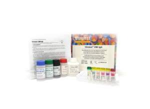
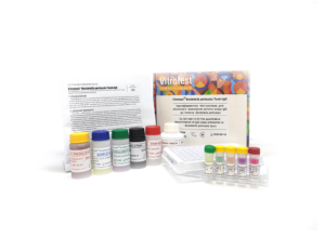
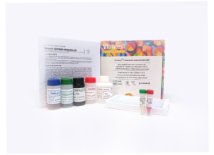
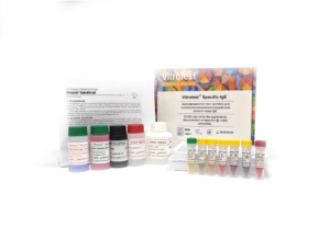
The test kit Vitrotest® CMV-IgG is an enzyme linked immunosorbent assay (ELISA) for the quantitative and semiquantitative determination of IgG class antibodies to Human Cytomegalovirus in human serum or plasma.
Determination of IgG antibodies to human CMV in the test kit Vitrotest® CMV-IgG is based on a solid phase, indirect ELISA in a two-step incubation procedure.
○ ТК005 - 96 tests- Solid phase: breakable microplate ELISA is coated with CMV antigens.
- Conjugate: a monoclonal antibodies to human IgG conjugated to horseradish peroxidase.
- Chromogen: ready to use TMB solution.
- Volume of sample for analysis: 10 μl.
- Assay time: 1h 15 min.
- Linearity range: 0,4 - 8 U/ml.
Human cytomegalovirus (CMV, herpesvirus 5) is a member of the herpesvirus family. This group includes herpes simplex virus types 1 and 2, varicella-zoster virus, and Epstein-Barr virus. CMV is a large enveloped virus; its double-stranded DNA genome codes for over 200 proteins. Viral proteins are regulatory and structural. Among structural proteins, glycoproteins and phosphoproteins are described.
CMV, similarly to other herpes viruses, shares a characteristic ability to remain dormant within the body for life. After initial infection, which may cause few symptoms, CMV becomes latent, residing in cells without causing detectable damage or clinical illness. Severe impairment of the body’s immune system by medication or disease may allow the virus to reactivate from the latent or dormant state and become symptomatic.
CMV infects most humans without harm. However infection with CMV causes serious, life-threatening disease in two circumstances: in immunosuppressed adults and in congenital infections of developing fetuses. In immunocompromised patients (organ transplant recipients, patients with lymphoid cancers, and HIV-infected patients) CMV is a major cause of disease and death. The common manifestations of disease in those patients are pneumonia, retinitis, and gastrointestinal diseases.
Congenital CMV (CCMV) infection is mostly (86% of all cases) asymptomatic. Symptoms, if they develop, include jaundice, pneumonia, a rash, an enlarged liver and spleen, low birth weight, seizures, small head. Mortality of children under symptomatic CCMV is around 20%. Around a half of symptomatic infants and around 12% of asymptomatic ones later develop physical or mental problems. These can include hearing loss, visual impairment or blindness, learning difficulties, epilepsy. -
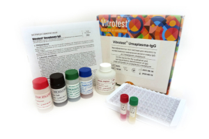
The test kit Vitrotest® Ureaplasma-IgG is an enzyme linked immunosorbent assay (ELISA) for the qualitative and semiquantitative determination of IgG class antibodies to Ureaplasma urealyticum in human serum or plasma.
Determination of IgG antibodies to U.urealyticum in the test kit Vitrotest® Ureaplasma-IgG is based on a solid phase indirect ELISA in a two-step incubation procedure.
○ ТК028 - 96 tests- Solid phase: breakable microplate ELISA is coated with recombinant antigens of U. urealyticum.
- Conjugate: a monoclonal antibodies to human IgG conjugated to horseradish peroxidase.
- Chromogen: ready to use TMB solution.
- Volume of sample for analysis: 20 μl.
- Assay time: 1h 15 min.
The microorganism Ureaplasma urealyticum causes inflammatory diseases of the human genitourinary system and Ureaplasma is often found in women with vaginitis and cystitis.
Penetration of the pathogen into the upper parts of the reproductive system can lead to the disruption of the reproductive functions. In men, U.urealyticum is the cause of nongonococcal urethritis and prostatitis (up to 50% of cases). The role of ureaplasmas in the development of most cases of urolithiasis has been proven. Often U.urealyticum leads to postpartum sepsis in women.
To diagnose ureaplasmosis, both direct methods for identifying ureaplasmas (polymerase chain reaction, immunofluorescence reaction, isolation of pure culture) and serological methods for detecting antibodies specific to Ureaplasma urealyticum are applied. Determination of antibodies in enzyme immunoassay is especially important in chronic ureaplasmosis, as well as in ascending ureaplasma infection. ELISA is a minimally invasive research method that allows for a comprehensive diagnosis of urogenital diseases. -
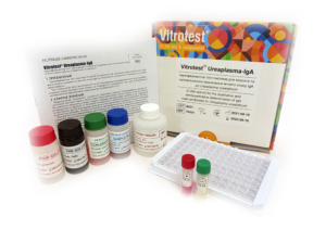
The test kit Vitrotest® Ureaplasma-IgA is an enzyme linked immunosorbent assay (ELISA) for the qualitative and semiquantitative determination of IgA class antibodies to Ureaplasma urealyticum in human serum or plasma.
Determination of IgA antibodies to U.urealyticum in the test kit Vitrotest® Ureaplasma-IgA is based on a solid phase indirect ELISA in a two-step incubation procedure.
○ ТК029 - 96 tests- Solid phase: breakable microplate ELISA is coated with recombinant antigens of U. urealyticum.
- Conjugate: a monoclonal antibodies to human IgA conjugated to horseradish peroxidase.
- Chromogen: ready to use TMB solution.
- Volume of sample for analysis: 20 μl.
- Assay time: 1h 15 min.
The microorganism Ureaplasma urealyticum causes inflammatory diseases of the human genitourinary system and Ureaplasma is often found in women with vaginitis and cystitis.
Penetration of the pathogen into the upper parts of the reproductive system can lead to the disruption of the reproductive functions. In men, U.urealyticum is the cause of nongonococcal urethritis and prostatitis (up to 50% of cases). The role of ureaplasmas in the development of most cases of urolithiasis has been proven. Often U.urealyticum leads to postpartum sepsis in women.
To diagnose ureaplasmosis, both direct methods for identifying ureaplasmas (polymerase chain reaction, immunofluorescence reaction, isolation of pure culture) and serological methods for detecting antibodies specific to Ureaplasma urealyticum are applied. Determination of antibodies in enzyme immunoassay is especially important in chronic ureaplasmosis, as well as in ascending ureaplasma infection. ELISA is a minimally invasive research method that allows for a comprehensive diagnosis of urogenital diseases. -
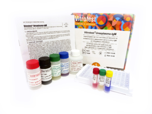
The test kit Vitrotest® Ureaplasma-IgМ is an enzyme linked immunosorbent assay (ELISA) for the qualitative and semiquantitative determination of IgM class antibodies to Ureaplasma urealiticum in human serum or plasma.
Determination of IgM antibodies to Ureaplasma urealiticum in the test kit Vitrotest® Ureaplasma-IgМ is based on a solid phase, «IgM-capture» ELISA in a two-step incubation procedure.
○ ТК096 - 96 tests- Solid phase: breakable microplate ELISA is coated with monoclonal anti-IgM antibodies.
- Conjugate: recombinant U. urealiticum antigen, conjugated to horseradish peroxidase.
- Chromogen: ready to use TMB solution.
- Volume of sample for analysis: 10 μl.
- Assay time: 1h 30 min.
The microorganism Ureaplasma urealyticum causes inflammatory diseases of the human genitourinary system and Ureaplasma is often found in women with vaginitis and cystitis.
Penetration of the pathogen into the upper parts of the reproductive system can lead to the disruption of the reproductive functions. In men, U.urealyticum is the cause of nongonococcal urethritis and prostatitis (up to 50% of cases). The role of ureaplasmas in the development of most cases of urolithiasis has been proven. Often U.urealyticum leads to postpartum sepsis in women.
To diagnose ureaplasmosis, both direct methods for identifying ureaplasmas (polymerase chain reaction, immunofluorescence reaction, isolation of pure culture) and serological methods for detecting antibodies specific to Ureaplasma urealyticum are applied. Determination of antibodies in enzyme immunoassay is especially important in chronic ureaplasmosis, as well as in ascending ureaplasma infection. ELISA is a minimally invasive research method that allows for a comprehensive diagnosis of urogenital diseases. -
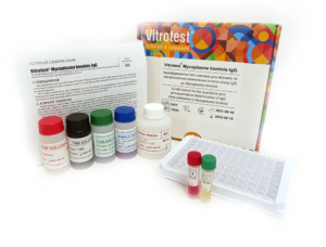
The test kit Vitrotest® Mycoplasma hominis-IgG is an enzyme linked immunosorbent assay (ELISA) for the qualitative and semiquantitative determination of IgG class antibodies to Mycoplasma hominis in human serum or plasma.
Determination of IgG antibodies to M. hominis in the test kit Vitrotest® Mycoplasma hominis-IgG is based on a solid phase indirect ELISA in a two-step incubation procedure.
○ ТК024 - 96 tests- Solid phase: breakable microplate ELISA is coated with native Mycoplasma hominis antigens.
- Conjugate: a monoclonal antibodies to human IgG conjugated to horseradish peroxidase.
- Chromogen: ready to use TMB solution.
- Volume of sample for analysis: 10 μl.
- Assay time: 1h 15 min.
Mycoplasmas are opportunistic pathogens, since they are often detected as part of the normal human microflora. At the same time, these microorganisms may be involved in the inflammatory process during urogenital diseases. Data on the frequency of mycoplasma spread among the population of different countries are contradictory, and infection rates vary from 10 to 80%.
Mycoplasma hominis infects primarily the organs of the genitourinary system and causes various destructive inflammatory processes. In men, M. hominis usually causes urethritis and prostatitis, and in women is provokes urethritis, cervicitis and inflammatory lesions of the pelvic organs. Urogenital mycoplasmosis in pregnant women is especially dangerous, as it can cause miscarriage, premature birth, infection of the fetus and the development of postpartum sepsis.
Clinical manifestations caused by the presence of M. hominis are often similar to symptoms of other diseases of the urogenital tract of bacterial, viral and other etiologies. Therefore, for successful diagnosis of urogenital mycoplasmosis, laboratory tests are required to differentiate them.
Serological methods for diagnosing mycoplasmosis include precipitation and immunofluorescence reactions. To detect serum antibodies to M. hominis, a passive hemagglutination reaction and an enzyme-linked immunosorbent assay are used, which makes it possible to determine the stage and nature of the disease. This is especially important in chronic disease. The presence of IgG antibodies to M. hominis reflects the overall picture of the immune response as a result of prolonged or previous infection. In the latter case, IgG may remain at low levels for many years. -
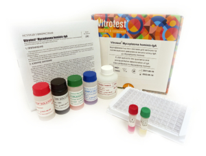
The test kit Vitrotest® Mycoplasma hominis-IgA is an enzyme linked immunosorbent assay (ELISA) for the qualitative and semiquantitative determination of IgA class antibodies to Mycoplasma hominis in human serum or plasma.
Determination of IgA antibodies to M. hominis in the test kit Vitrotest® Mycoplasma hominis-IgA is based on a solid phase indirect ELISA in a two-step incubation procedure.
○ ТК025 - 96 tests- Solid phase: breakable microplate ELISA is coated with native Mycoplasma hominis antigens.
- Conjugate: a monoclonal antibodies to human IgA conjugated to horseradish peroxidase.
- Chromogen: ready to use TMB solution.
- Volume of sample for analysis: 10 μl.
- Assay time: 1h 15 min.
Mycoplasmas are opportunistic pathogens, since they are often detected as part of the normal human microflora. At the same time, these microorganisms may be involved in the inflammatory process during urogenital diseases. Data on the frequency of mycoplasma spread among the population of different countries are contradictory, and infection rates vary from 10 to 80%.
Mycoplasma hominis infects primarily the organs of the genitourinary system and causes various destructive inflammatory processes. In men, M. hominis usually causes urethritis and prostatitis, and in women is provokes urethritis, cervicitis and inflammatory lesions of the pelvic organs. Urogenital mycoplasmosis in pregnant women is especially dangerous, as it can cause miscarriage, premature birth, infection of the fetus and the development of postpartum sepsis.
Clinical manifestations caused by the presence of M. hominis are often similar to symptoms of other diseases of the urogenital tract of bacterial, viral and other etiologies. Therefore, for successful diagnosis of urogenital mycoplasmosis, laboratory tests are required to differentiate them.
Serological methods for diagnosing mycoplasmosis include precipitation and immunofluorescence reactions. To detect serum antibodies to M. hominis, a passive hemagglutination reaction and an enzyme-linked immunosorbent assay are used, which makes it possible to determine the stage and nature of the disease. This is especially important in chronic disease. The presence of IgG antibodies to M. hominis reflects the overall picture of the immune response as a result of prolonged or previous infection. In the latter case, IgG may remain at low levels for many years.
-
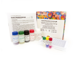
The test kit Vitrotest® Mycoplasma hominis-IgM is an enzyme linked immunosorbent assay (ELISA) for the qualitative and semiquantitative determination of IgM class antibodies to Mycoplasma hominis in human serum or plasma.
Determination of IgM antibodies to Mycoplasma hominis in the test kit Vitrotest® Mycoplasma hominis-IgM is based on a solid phase, «IgM-capture» ELISA in a two-step incubation procedure.
○ ТК097 - 96 tests- Solid phase: breakable microplate ELISA is coated with monoclonal anti-IgM antibodies.
- Conjugate: recombinant M. hominis antigen, conjugated to horseradish peroxidase.
- Chromogen: ready to use TMB solution.
- Volume of sample for analysis: 10 μl.
- Assay time: 1h 30 min.
Mycoplasmas are opportunistic pathogens, since they are often detected as part of the normal human microflora. At the same time, these microorganisms may be involved in the inflammatory process during urogenital diseases. Data on the frequency of mycoplasma spread among the population of different countries are contradictory, and infection rates vary from 10 to 80%.
Mycoplasma hominis infects primarily the organs of the genitourinary system and causes various destructive inflammatory processes. In men, M. hominis usually causes urethritis and prostatitis, and in women is provokes urethritis, cervicitis and inflammatory lesions of the pelvic organs. Urogenital mycoplasmosis in pregnant women is especially dangerous, as it can cause miscarriage, premature birth, infection of the fetus and the development of postpartum sepsis.
Clinical manifestations caused by the presence of M. hominis are often similar to symptoms of other diseases of the urogenital tract of bacterial, viral and other etiologies. Therefore, for successful diagnosis of urogenital mycoplasmosis, laboratory tests are required to differentiate them.
Serological methods for diagnosing mycoplasmosis include precipitation and immunofluorescence reactions. To detect serum antibodies to M. hominis, a passive hemagglutination reaction and an enzyme-linked immunosorbent assay are used, which makes it possible to determine the stage and nature of the disease. This is especially important in chronic disease. The presence of IgG antibodies to M. hominis reflects the overall picture of the immune response as a result of prolonged or previous infection. In the latter case, IgG may remain at low levels for many years. -
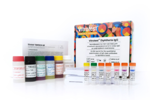
The test kit Vitrotest® Diphtheria-IgG is an enzyme linked immunosorbent assay (ELISA) for the quantitative determination of IgG class antibodies to diphtheria toxoid in human serum or plasma.
Determination of IgG antibodies to diphtheria toxoid in the test kit Vitrotest® Diphtheria-IgG is based on a solid phase, indirect ELISA.
○ ТК027 - 96 tests- Solid phase: breakable microplate ELISA is coated with diphtheria toxoid.
- Conjugate: a monoclonal antibodies to human IgG conjugated to horseradish peroxidase.
- Chromogen: ready to use TMB solution.
- Volume of sample for analysis: 10 μl.
- Assay time: 1h 45 min.
- Analytical sensitivity: 0,0025 IU/ml.
- Linearity range: 0,004 - 1,005 IU/ml.
Diphtheria is an infectious disease that primarily affects children. The causative agent of diphtheria Corynebacterium diphtheriae enters the human body, primarily infecting the throat and upper respiratory tract, and produces a toxin that can affect other organs. The disease is characterized by an acute onset, with the main symptoms being a sore throat, fever, and swollen glands in the neck. In severe cases, the bacteria produce a toxin that causes a thick gray or white film to form on the back of the throat.
The most important virulent factor of C. diphtheriae is the diphtheria toxin (exotoxin), which is secreted by the bacteria, enters the bloodstream, and damages the myocardium, kidneys, and central nervous system. Damage to the heart muscle can lead to arrhythmias, and nerve inflammation can cause paralysis.
Vaccination against diphtheria is a highly effective means of preventing the disease. According to WHO recommendations, all children worldwide should be vaccinated against diphtheria. The primary vaccination with diphtheria toxoid, consisting of three doses of the vaccine, should be administered to infants at the ages of 2, 4, and 6 months (with at least 4-week intervals between vaccinations). This should be followed by three booster immunizations with combined vaccines at the ages of 1-2, 4-7, and 9-15 years. At any age, those who are unvaccinated or incompletely vaccinated against diphtheria should receive the necessary doses to complete the vaccination series.
After the 3-dose primary series of the toxoid-containing vaccine, 94-100% of children show diphtheria antitoxin levels > 0.01 IU/ml, but booster doses of the vaccine are necessary to ensure long-term protection. Quantitative enzyme-linked immunosorbent assay (ELISA) for the determination of IgG antibodies to diphtheria toxoid is widely used to assess the level of protective immunity. -
The test kit Vitrotest® Bordetella pertussis Toxin IgG is an enzyme linked immunosorbent assay (ELISA) for the quantitative determination of IgG class antibodies to Bordetella pertussis toxin in human serum or plasma.
Determination of IgG antibodies to Bordetella pertussis toxin in the test kit Vitrotest® Bordetella pertussis Toxin IgG is based on a solid phase, indirect ELISA in a two-step incubation procedure.
○ ТК125 - 96 tests- Solid phase: breakable microplate ELISA is coated with purified Bordetella pertussis toxin.
- Conjugate: a monoclonal antibodies to human IgG conjugated to horseradish peroxidase.
- Chromogen: ready to use TMB solution.
- Volume of sample for analysis: 10 μl.
- Assay time: 1h 15 min.
- Analytical sensitivity: 3,22 IU/ml.
- Linearity range: 3,5 - 181 IU/ml.
Whooping cough is a dangerous respiratory infection caused by the bacterium Bordetella pertussis. Characteristic symptoms of the disease include paroxysmal spasmodic coughing, a high-pitched "whoop" sound when inhaling, post-cough vomiting, and more. Whooping cough is most severe in infants and young children, often leading to apnea (breathing pauses) and, in some cases, death.
Whooping cough is a globally widespread disease with a cyclic pattern, with peaks in incidence every 3-5 years. Due to the severity of its clinical manifestations, many countries have implemented vaccination programs against whooping cough. As a result of widespread vaccination campaigns conducted in the 1950s and 1960s in developed countries, there was a sharp decrease in both incidence (by more than 90%) and mortality from whooping cough.
However, despite high vaccination coverage, whooping cough remains a public health issue worldwide, with approximately 140,000 cases reported annually.
In Ukraine, according to the National Immunization Schedule, children should be vaccinated against whooping cough at the ages of 2, 4, 6, and 18 months. For vaccinating children against whooping cough during the first year of life, both acellular (aP) and whole-cell (wP) pertussis vaccines can be used. The immunity formed as a result of the full course of vaccination against whooping cough lasts for 5-7 years.
Laboratory methods are used to diagnose whooping cough, with the most common being polymerase chain reaction (PCR) (to detect the pathogen during the first 2-3 weeks of the illness) and enzyme-linked immunosorbent assay (ELISA) (to detect specific antibodies). The presence of IgM antibodies specific to Bordetella pertussis indicates an acute infection or recent vaccination. The detection of IgG antibodies to the pertussis toxin (PT) in the serum of an unvaccinated individual allows for the diagnosis of a past or current infection and provides information about specific immunity after vaccination. -
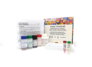
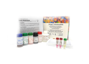
The test kit Vitrotest® Chlamydia-IgG is an enzyme linked immunosorbent assay (ELISA) for the qualitative and semiquantitative determination of IgG class antibodies to Chlamydia trachomatis in human serum or plasma.
Determination of IgG antibodies to Chlamydia trachomatis in the test kit Vitrotest® Chlamydia-IgG is based on a solid phase indirect ELISA in a two-step incubation procedure.
○ ТК014 - 96 tests- Solid phase: breakable microplate ELISA is coated with recombinant Chlamydia trachomatis antigens.
- Conjugate: a monoclonal antibodies to human IgG conjugated to horseradish peroxidase.
- Chromogen: ready to use TMB solution.
- Volume of sample for analysis: 20 μl.
- Assay time: 1h 15 min.
Chlamydiosis is a common sexually transmitted disease (STD) caused by infection with a Gram-negative bacteria Chlamydia trachomatis. The family Chlamydiaceae contains besides C.trachomatis two important pathogenic species, C. psittaci and C.pneumoniae.
The importance of C.trachomatis as a sexually transmitted agent became apparent in the mid-1970-s, with reports of pelvic inflammatory disease (PID), endometritis and salpingitis, resulting in ectopic pregnancy and infertility, as well as conjunctivitis (paratrachoma) and pneumonia of the newborns. In men, untreated chlamydial infections can lead to urethritis and epididymoorchitis, and primarily in men, chlamydiosis is known to cause autoimmune disease, reactive arthritis. Besides these conditions, in poor developing countries C.trachomatis could cause trachoma, a specific ocular disease, which is one of the leading causes of blindness, as well as systemic sexually transmitted disease called lymphogranuloma venereum (LGV).
C.trachomatis develops in biphasic cycle. The bacterium is normally found in two highly specialized morphologic forms — the extracellular, metabolically inactive, infectious elementary body (EB) and the metabolically active, dividing, non-infective intracellular form known as reticulate body (RB). The infectious EB enters the mucosal host cells via endocytosis. Inside endosome EBs reorganize into RBs. RBs divide by binary fission, filling the endosome with hundreds of RBs. Multiplication ceases after 48 – 72 h, and RBs transform into metabolically inactive infectious EBs. The EBs are released from the cell by cell rupture or exocytosis, and target new host cells. In women, C.trachomatis infection may involve not only cervix, but also endometrium and fallopian tubes in around 10% of individuals. The disease sequelae caused by this process include PID and its complications. After PID has occurred, even with treatment, it could cause significant reproductive and gynaecological morbidity, including infertility, ectopic pregnancy, and chronic pelvic pain. In one study, 18% of women developed infertility after clinically diagnosed single episode of PID; the risk of infertility increases considerably after every new episode of PID.
Infertility or subfertility are the result of damage to the cilia lining the fallopian tubes, fallopian tube blockage or closure, or adhesion formation among pelvic organs. The issue of whether chlamydial PID is a consequence of persistent infection, immunopathology, or autoimmune reaction remains unresolved. C.trachomatis can also spread perinatally from an untreated mother to her baby during childbirth, resulting in paratrachoma, urethritis, proctitis, nasopharyngeal infection or pneumonia in many exposed infants. The risk of neonatal chlamydiosis in children born through an infected birth canal is around 15%. Conjunctival infections are detected within the first 22 days of life, eventually followed by nasopharyngeal infections and pneumonia, rectal and vaginal infections during the first several month of life.
Chlamydiosis is known as a ‘silent’ infection because most infected people, especially women, are asymptomatic and lack abnormal physical examination findings. Unfortunately, there might not be a relationship between the severity of symptoms and the severity of the disease. The symptoms of cervicitis may appear 1-3 weeks after infection, and may include mucopurulent endocervical discharge and easily induced endocervical bleeding; if cervicitis develops into PID, abdominal and/or pelvic pain could arise.
C.trachomatis is transmitted if infected secretions from urethra, cervix, rectum, conjunctivae and throat come into contact with mucous membranes of the above-mentioned organs. Chlamydiosis is usually transmitted by sex. Therefore, young people (up to 25 years) are at highest risk of acquiring chlamydiosis. In addition, an infected mother can infect her baby during vaginal delivery. C.trachomatis is grouped into 18 separate serovars, which are distinguished by the antigenic and molecular polymorphism within major outer membrane protein, OmpA. Serovars A-C infect the conjunctive epithelium and lead to ocular infections that can progress to trachoma; serovars L1-L3 are able to infect not only the genital epithelium, but also monocytes and lead to LGV; classical sexually transmitted non-LGV infections are caused by serovars D-K. Protective immunity to chlamydiosis is not absolute, being much stronger to the same serovar; therefore, reinfections are usually by the heterologous serovar. -
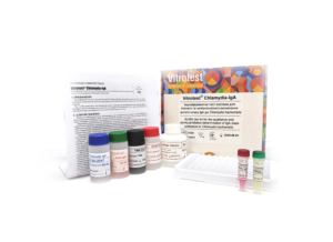
The test kit Vitrotest® Chlamydia-IgА is an enzyme linked immunosorbent assay (ELISA) for the qualitative and semiquantitative determination of IgА class antibodies to Chlamydia trachomatis in human serum or plasma.
Determination of IgА antibodies to Chlamydia trachomatis in the test kit Vitrotest® Chlamydia-IgА is based on a solid phase indirect ELISA in a two-step incubation procedure.
○ ТК015 - 96 tests- Solid phase: breakable microplate ELISA is coated with recombinant Chlamydia trachomatis antigens.
- Conjugate: a monoclonal antibodies to human IgА conjugated to horseradish peroxidase.
- Chromogen: ready to use TMB solution.
- Volume of sample for analysis: 20 μl.
- Assay time: 1h 15 min.
Chlamydiosis is a common sexually transmitted disease (STD) caused by infection with a Gram-negative bacteria Chlamydia trachomatis. The family Chlamydiaceae contains besides C.trachomatis two important pathogenic species, C. psittaci and C.pneumoniae.
The importance of C.trachomatis as a sexually transmitted agent became apparent in the mid-1970-s, with reports of pelvic inflammatory disease (PID), endometritis and salpingitis, resulting in ectopic pregnancy and infertility, as well as conjunctivitis (paratrachoma) and pneumonia of the newborns. In men, untreated chlamydial infections can lead to urethritis and epididymoorchitis, and primarily in men, chlamydiosis is known to cause autoimmune disease, reactive arthritis. Besides these conditions, in poor developing countries C.trachomatis could cause trachoma, a specific ocular disease, which is one of the leading causes of blindness, as well as systemic sexually transmitted disease called lymphogranuloma venereum (LGV).
C.trachomatis develops in biphasic cycle. The bacterium is normally found in two highly specialized morphologic forms — the extracellular, metabolically inactive, infectious elementary body (EB) and the metabolically active, dividing, non-infective intracellular form known as reticulate body (RB). The infectious EB enters the mucosal host cells via endocytosis. Inside endosome EBs reorganize into RBs. RBs divide by binary fission, filling the endosome with hundreds of RBs. Multiplication ceases after 48 – 72 h, and RBs transform into metabolically inactive infectious EBs. The EBs are released from the cell by cell rupture or exocytosis, and target new host cells. In women, C.trachomatis infection may involve not only cervix, but also endometrium and fallopian tubes in around 10% of individuals. The disease sequelae caused by this process include PID and its complications. After PID has occurred, even with treatment, it could cause significant reproductive and gynaecological morbidity, including infertility, ectopic pregnancy, and chronic pelvic pain. In one study, 18% of women developed infertility after clinically diagnosed single episode of PID; the risk of infertility increases considerably after every new episode of PID.
Infertility or subfertility are the result of damage to the cilia lining the fallopian tubes, fallopian tube blockage or closure, or adhesion formation among pelvic organs. The issue of whether chlamydial PID is a consequence of persistent infection, immunopathology, or autoimmune reaction remains unresolved. C.trachomatis can also spread perinatally from an untreated mother to her baby during childbirth, resulting in paratrachoma, urethritis, proctitis, nasopharyngeal infection or pneumonia in many exposed infants. The risk of neonatal chlamydiosis in children born through an infected birth canal is around 15%. Conjunctival infections are detected within the first 22 days of life, eventually followed by nasopharyngeal infections and pneumonia, rectal and vaginal infections during the first several month of life.
Chlamydiosis is known as a ‘silent’ infection because most infected people, especially women, are asymptomatic and lack abnormal physical examination findings. Unfortunately, there might not be a relationship between the severity of symptoms and the severity of the disease. The symptoms of cervicitis may appear 1-3 weeks after infection, and may include mucopurulent endocervical discharge and easily induced endocervical bleeding; if cervicitis develops into PID, abdominal and/or pelvic pain could arise.
C.trachomatis is transmitted if infected secretions from urethra, cervix, rectum, conjunctivae and throat come into contact with mucous membranes of the above-mentioned organs. Chlamydiosis is usually transmitted by sex. Therefore, young people (up to 25 years) are at highest risk of acquiring chlamydiosis. In addition, an infected mother can infect her baby during vaginal delivery. C.trachomatis is grouped into 18 separate serovars, which are distinguished by the antigenic and molecular polymorphism within major outer membrane protein, OmpA. Serovars A-C infect the conjunctive epithelium and lead to ocular infections that can progress to trachoma; serovars L1-L3 are able to infect not only the genital epithelium, but also monocytes and lead to LGV; classical sexually transmitted non-LGV infections are caused by serovars D-K. Protective immunity to chlamydiosis is not absolute, being much stronger to the same serovar; therefore, reinfections are usually by the heterologous serovar. -
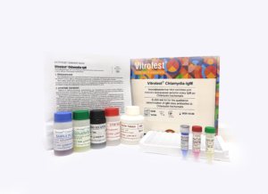
The test kit Vitrotest® Chlamydia-IgМ is an enzyme linked immunosorbent assay (ELISA) for the qualitative determination of IgM class antibodies to Chlamydia trachomatis in human serum or plasma.
Determination of IgM antibodies to Chlamydia trachomatis in the test kit Vitrotest® Chlamydia-IgМ is based on a solid phase «IgM-capture» ELISA in a two-step incubation procedure.
○ ТК036 - 96 tests- Solid phase: breakable microplate ELISA is coated with monoclonal anti-IgM antibodies.
- Conjugate: recombinant antigen MOMP of Chlamydia trachomatis, conjugated to horseradish peroxidase.
- Chromogen: ready to use TMB solution.
- Volume of sample for analysis: 10 μl.
- Assay time: 1h 30 min.
Chlamydiosis is a common sexually transmitted disease (STD) caused by infection with a Gram-negative bacteria Chlamydia trachomatis. The family Chlamydiaceae contains besides C.trachomatis two important pathogenic species, C. psittaci and C.pneumoniae.
The importance of C.trachomatis as a sexually transmitted agent became apparent in the mid-1970-s, with reports of pelvic inflammatory disease (PID), endometritis and salpingitis, resulting in ectopic pregnancy and infertility, as well as conjunctivitis (paratrachoma) and pneumonia of the newborns. In men, untreated chlamydial infections can lead to urethritis and epididymoorchitis, and primarily in men, chlamydiosis is known to cause autoimmune disease, reactive arthritis. Besides these conditions, in poor developing countries C.trachomatis could cause trachoma, a specific ocular disease, which is one of the leading causes of blindness, as well as systemic sexually transmitted disease called lymphogranuloma venereum (LGV).
C.trachomatis develops in biphasic cycle. The bacterium is normally found in two highly specialized morphologic forms — the extracellular, metabolically inactive, infectious elementary body (EB) and the metabolically active, dividing, non-infective intracellular form known as reticulate body (RB). The infectious EB enters the mucosal host cells via endocytosis. Inside endosome EBs reorganize into RBs. RBs divide by binary fission, filling the endosome with hundreds of RBs. Multiplication ceases after 48 – 72 h, and RBs transform into metabolically inactive infectious EBs. The EBs are released from the cell by cell rupture or exocytosis, and target new host cells. In women, C.trachomatis infection may involve not only cervix, but also endometrium and fallopian tubes in around 10% of individuals. The disease sequelae caused by this process include PID and its complications. After PID has occurred, even with treatment, it could cause significant reproductive and gynaecological morbidity, including infertility, ectopic pregnancy, and chronic pelvic pain. In one study, 18% of women developed infertility after clinically diagnosed single episode of PID; the risk of infertility increases considerably after every new episode of PID.
Infertility or subfertility are the result of damage to the cilia lining the fallopian tubes, fallopian tube blockage or closure, or adhesion formation among pelvic organs. The issue of whether chlamydial PID is a consequence of persistent infection, immunopathology, or autoimmune reaction remains unresolved. C.trachomatis can also spread perinatally from an untreated mother to her baby during childbirth, resulting in paratrachoma, urethritis, proctitis, nasopharyngeal infection or pneumonia in many exposed infants. The risk of neonatal chlamydiosis in children born through an infected birth canal is around 15%. Conjunctival infections are detected within the first 22 days of life, eventually followed by nasopharyngeal infections and pneumonia, rectal and vaginal infections during the first several month of life.
Chlamydiosis is known as a ‘silent’ infection because most infected people, especially women, are asymptomatic and lack abnormal physical examination findings. Unfortunately, there might not be a relationship between the severity of symptoms and the severity of the disease. The symptoms of cervicitis may appear 1-3 weeks after infection, and may include mucopurulent endocervical discharge and easily induced endocervical bleeding; if cervicitis develops into PID, abdominal and/or pelvic pain could arise.
C.trachomatis is transmitted if infected secretions from urethra, cervix, rectum, conjunctivae and throat come into contact with mucous membranes of the above-mentioned organs. Chlamydiosis is usually transmitted by sex. Therefore, young people (up to 25 years) are at highest risk of acquiring chlamydiosis. In addition, an infected mother can infect her baby during vaginal delivery. C.trachomatis is grouped into 18 separate serovars, which are distinguished by the antigenic and molecular polymorphism within major outer membrane protein, OmpA. Serovars A-C infect the conjunctive epithelium and lead to ocular infections that can progress to trachoma; serovars L1-L3 are able to infect not only the genital epithelium, but also monocytes and lead to LGV; classical sexually transmitted non-LGV infections are caused by serovars D-K. Protective immunity to chlamydiosis is not absolute, being much stronger to the same serovar; therefore, reinfections are usually by the heterologous serovar. -
The test kit Vitrotest® Chlamydia-IgG/IgA is an enzyme linked immunosorbent assay (ELISA) for the qualitative and semiquantitative determination of IgG and/or IgА class antibodies to Chlamydia trachomatis in human serum or plasma.
Determination of IgG and/or IgА antibodies to Chlamydia trachomatis in the test kit Vitrotest® Chlamydia-IgG/IgA is based on a solid phase indirect ELISA in a two-step incubation procedure.
○ ТК013 - 96 tests- Solid phase: breakable microplate ELISA is coated with recombinant Chlamydia trachomatis antigens.
- Conjugate anti-IgG: a monoclonal antibodies to human IgG conjugated to horseradish peroxidase.
- Conjugate anti-IgA: a monoclonal antibodies to human IgA conjugated to horseradish peroxidase.
- Chromogen: ready to use TMB solution.
- Volume of sample for analysis: 20 μl.
- Assay time: 1h 15 min.
Chlamydiosis is a common sexually transmitted disease (STD) caused by infection with a Gram-negative bacteria Chlamydia trachomatis. The family Chlamydiaceae contains besides C.trachomatis two important pathogenic species, C. psittaci and C.pneumoniae.
The importance of C.trachomatis as a sexually transmitted agent became apparent in the mid-1970-s, with reports of pelvic inflammatory disease (PID), endometritis and salpingitis, resulting in ectopic pregnancy and infertility, as well as conjunctivitis (paratrachoma) and pneumonia of the newborns. In men, untreated chlamydial infections can lead to urethritis and epididymoorchitis, and primarily in men, chlamydiosis is known to cause autoimmune disease, reactive arthritis. Besides these conditions, in poor developing countries C.trachomatis could cause trachoma, a specific ocular disease, which is one of the leading causes of blindness, as well as systemic sexually transmitted disease called lymphogranuloma venereum (LGV).
C.trachomatis develops in biphasic cycle. The bacterium is normally found in two highly specialized morphologic forms — the extracellular, metabolically inactive, infectious elementary body (EB) and the metabolically active, dividing, non-infective intracellular form known as reticulate body (RB). The infectious EB enters the mucosal host cells via endocytosis. Inside endosome EBs reorganize into RBs. RBs divide by binary fission, filling the endosome with hundreds of RBs. Multiplication ceases after 48 – 72 h, and RBs transform into metabolically inactive infectious EBs. The EBs are released from the cell by cell rupture or exocytosis, and target new host cells. In women, C.trachomatis infection may involve not only cervix, but also endometrium and fallopian tubes in around 10% of individuals. The disease sequelae caused by this process include PID and its complications. After PID has occurred, even with treatment, it could cause significant reproductive and gynaecological morbidity, including infertility, ectopic pregnancy, and chronic pelvic pain. In one study, 18% of women developed infertility after clinically diagnosed single episode of PID; the risk of infertility increases considerably after every new episode of PID.
Infertility or subfertility are the result of damage to the cilia lining the fallopian tubes, fallopian tube blockage or closure, or adhesion formation among pelvic organs. The issue of whether chlamydial PID is a consequence of persistent infection, immunopathology, or autoimmune reaction remains unresolved. C.trachomatis can also spread perinatally from an untreated mother to her baby during childbirth, resulting in paratrachoma, urethritis, proctitis, nasopharyngeal infection or pneumonia in many exposed infants. The risk of neonatal chlamydiosis in children born through an infected birth canal is around 15%. Conjunctival infections are detected within the first 22 days of life, eventually followed by nasopharyngeal infections and pneumonia, rectal and vaginal infections during the first several month of life.
Chlamydiosis is known as a ‘silent’ infection because most infected people, especially women, are asymptomatic and lack abnormal physical examination findings. Unfortunately, there might not be a relationship between the severity of symptoms and the severity of the disease. The symptoms of cervicitis may appear 1-3 weeks after infection, and may include mucopurulent endocervical discharge and easily induced endocervical bleeding; if cervicitis develops into PID, abdominal and/or pelvic pain could arise.
C.trachomatis is transmitted if infected secretions from urethra, cervix, rectum, conjunctivae and throat come into contact with mucous membranes of the above-mentioned organs. Chlamydiosis is usually transmitted by sex. Therefore, young people (up to 25 years) are at highest risk of acquiring chlamydiosis. In addition, an infected mother can infect her baby during vaginal delivery. C.trachomatis is grouped into 18 separate serovars, which are distinguished by the antigenic and molecular polymorphism within major outer membrane protein, OmpA. Serovars A-C infect the conjunctive epithelium and lead to ocular infections that can progress to trachoma; serovars L1-L3 are able to infect not only the genital epithelium, but also monocytes and lead to LGV; classical sexually transmitted non-LGV infections are caused by serovars D-K. Protective immunity to chlamydiosis is not absolute, being much stronger to the same serovar; therefore, reinfections are usually by the heterologous serovar. -
The test kit Vitrotest® Chlamydia pneumoniae-IgG is an enzyme linked immunosorbent assay (ELISA) for the qualitative and semiquantitative determination of IgG class antibodies to Chlamydia pneumoniae in human serum or plasma. Determination of IgG antibodies to Chlamydia pneumoniae in the test kit Vitrotest® Chlamydia pneumoniae-IgG is based on a solid phase indirect ELISA in a two-step incubation procedure. ○ ТК081 - 96 tests
- Solid phase: breakable microplate ELISA is coated with Chlamydia pneumoniae antigens.
- Conjugate: a monoclonal antibodies to human IgG conjugated to horseradish peroxidase.
- Chromogen: ready to use TMB solution.
- Volume of sample for analysis: 10 μl.
- Assay time: 1h 15 min.
Chlamydia (Chlamydophila) pneumoniae is one of the most common types of intracellular obligate bacteria transmitted by airborne droplets and capable of causing respiratory tract infections. Most respiratory tract infections caused by C. pneumoniae are asymptomatic or have minor clinical manifestations. Common symptoms that appear after a 3-4 week incubation period are usually runny nose, nasal congestion, fatigue, low-grade fever, hoarseness, sore throat, cough, and headache. However, about 30% of C. pneumoniae infections are characterized by more severe respiratory diseases, such as community-acquired pneumonia with atypical manifestations, bronchitis, etc. In addition, C. pneumoniae is involved in the pathogenesis of multiple inflammatory conditions, including chronic obstructive pulmonary disease (COPD), asthma, lung cancer, atherosclerosis, arthritis, neurological disorders (Alzheimer's disease, multiple sclerosis, schizophrenia). According to statistics, about 30-50% of people under the age of 20 and 80% aged 60 to 70 have antibodies to this pathogen. With primary infection with C. pneumoniae, IgM antibodies usually appear in the human blood after 2-3 weeks, IgG antibodies - somewhat later, after 3-5 weeks from the onset of the disease. The simultaneous detection of IgM and IgG antibodies indicates acute or manifest chronic infection. In case of re-infection or exacerbation of the chronic process, IgM antibodies may not be detected or may be detected in low titers, while the level of IgG class antibodies increases rapidly, which has diagnostic value in the study of paired sera taken at intervals of 10-14 days. For the diagnosis of C. pneumoniae-mediated diseases, the most widespread are the culture method, immunohistochemistry, polymerase chain reaction and serological methods. Among the latter, the most informative and widespread is ELISA. -
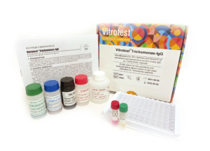
The test kit Vitrotest® Trichomonas-IgG is an enzyme linked immunosorbent assay (ELISA) for the qualitative and semiquantitative determination of IgG class antibodies to Trichomonas vaginalis in human serum or plasma.
Determination of IgG antibodies to Trichomonas vaginalis in the test kit Vitrotest® Trichomonas-IgG is based on a solid phase, indirect ELISA in a two-step incubation procedure.
○ ТК061 - 96 tests- Solid phase: breakable microplate ELISA is coated with Trichomonas vaginalis antigens.
- Conjugate: a monoclonal antibodies to human IgG conjugated to horseradish peroxidase.
- Chromogen: ready to use TMB solution.
- Volume of sample for analysis: 10 μl.
- Assay time: 1h 15 min.
The flagellated protozoan Trichomonas vaginalis is the etiologic agent of one of the most common sexually transmitted diseases in the world. On average, 25% of sexually active individuals are infected with T. vaginalis. According to the latest data, more than 170 million cases of trichomoniasis are reported worldwide each year.
In women, trichomonads cause acute and subacute vaginitis, with symptoms including frothy discharge, itching, and lower abdominal pain. The disease is cyclical – these symptoms subside during menstruation and pregnancy. During pregnancy, trichomoniasis can lead to premature birth and low birth weight in infants. In men, T. vaginalis typically causes urethritis, prostatitis, epididymitis, and dysuria. In most cases, the disease is asymptomatic. The mild symptoms in men are attributed to the flushing out of a significant portion of trichomonads from the urogenital tract during urination. The latent nature of the disease often leads to an underestimation of its spread. Chronic infection almost always results in chronic prostatitis. Trichomoniasis has also been associated with the development of prostate cancer in men.
Currently, the following laboratory methods are used to diagnose trichomoniasis:- microscopy of vaginal or cervical secretions in women and urinary tract secretions in men;
- culture of trichomonads;
- detection of pathogen DNA by polymerase chain reaction (PCR);
- detection of antibodies specific to T. vaginalis by enzyme-linked immunosorbent assay (ELISA).
According to several studies, the sensitivity of direct parasite detection methods ranges from 38% to 82%. It is also known that infected individuals develop a humoral, secretory, and cellular immune response to the parasite, which allows for the use of serological studies for diagnostic purposes. Antibodies to the trichomonad surface antigen p270 and α-actinin are produced in almost all infected individuals, and serum antibody levels correlate with active infection. Serological diagnostic methods are particularly valuable in individuals who do not exhibit clinical manifestations of trichomoniasis and in men who may be asymptomatic carriers of the parasite. -
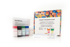
The test-kit Vitrotest® Helicobacter screen is an enzyme linked immunosorbent assay (ELISA) for the detection of IgG, IgA and IgM specific antibodies to Helicobacter pylori in human serum or plasma.
Detection of antibodies to H. pylori in the test-kit Vitrotest® Helicobacter screen is based on a solid phase, indirect ELISA in a two-step incubation procedure.
○ ТК045 - 96 tests- Solid phase: breakable microplate ELISA is coated with recombinant antigens of H. pylori.
- Conjugate: a monoclonal antibodies to human IgA, IgG and IgM conjugated to horseradish peroxidase.
- Chromogen: ready to use TMB solution.
- Volume of sample for analysis: 10 μl.
- Assay time: 1h 15 min.
Helicobacter pylori is a widespread microorganism with which the half of the world’s population has been infected. Its prevalence is very high in developing countries and is quite low in the developed world. According to the World Gastroenterology Organization, the rate of infected adults in Eastern Europe and Asia is about 70-80 %.
Studies during recent decades have shown the key role of bacterium Helicobacter pylori in the pathogenesis of stomach and duodenal lesions. H. pylori is detected almost in 100% of adult patients with duodenal ulcer, approximately in 80 % of patients with peptic ulcer, in 92% of patients with gastric cancer and in 92 % of patients with active chronic gastritis. Research has demonstrated that elimination of helicobacter leads to the disappearance of gastritis and significant reduction in the incidence of duodenal ulcer recurrence.
Helicobacteriosis is a chronic infection with long, often asymptomatic course. Its symptoms do not differ from clinical manifestations of gastro-duodenitis since usual constant pain in the epigastrium occurs. H. pylori is often present in patients with no clinical manifestations of disease.
The infection usually starts from non–acid-secreting antral region of the stomach and stimulates the increased release of gastrin. The increased gastrin levels in turn stimulate excess acid secretion from the more proximal acid-secreting fundic mucosa which is relatively free of inflammation. The increased duodenal acid load damages the duodenal mucosa, causing ulceration. If infection progresses, stomach body is damaged, which could finally cause the development of gastric adenocarcinoma. This tumour is preceded by sequential pathological changes of gastric mucosa, from normal mucosa to superficial gastritis, atrophic gastritis, gastric ulcers and intestinal metaplasia.
Main oncogenic factors are both nitrosating bacteria present in the lumen of hypochloric stomach, capable of generating potentially carcinogenic N-nitrosamines and reactive oxygen species, and H. pylori itself. Duodenal or gastric ulcers are reported to develop in 1 to 10% of infected patients, gastric cancer- in 0.1 to 3%; at the same time, the great majority of patients with H. pylori remain asymptomatic carriers.
H. pylori is usually acquired in childhood. The bacteria are most likely spread through fecal-oral or oral-oral routes from person to person; additional transmission routes, such as water, may be important in developing countries. -
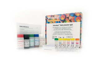
The test kit Vitrotest® Helicobacter-IgG is an enzyme linked immunosorbent assay (ELISA) for the quantitative and semiquantitative determination of IgG class antibodies to CagA protein of Helicobacter pylori in human serum or plasma.
Determination of IgG antibodies to CagA protein of H. pylori in the test kit Vitrotest® Helicobacter-IgG is based on a solid phase, indirect ELISA in a two-step incubation procedure.
○ ТК046 - 96 tests- Solid phase: breakable microplate ELISA is coated with recombinant CagA protein of H. pylori.
- Conjugate: a monoclonal antibodies to human IgG conjugated to horseradish peroxidase.
- Chromogen: ready to use TMB solution.
- Volume of sample for analysis: 10 μl.
- Assay time: 1h 15 min.
Helicobacter pylori is a widespread microorganism with which the half of the world’s population has been infected. Its prevalence is very high in developing countries and is quite low in the developed world. According to the World Gastroenterology Organization, the rate of infected adults in Eastern Europe and Asia is about 70-80 %.
Studies during recent decades have shown the key role of bacterium Helicobacter pylori in the pathogenesis of stomach and duodenal lesions. H. pylori is detected almost in 100% of adult patients with duodenal ulcer, approximately in 80 % of patients with peptic ulcer, in 92% of patients with gastric cancer and in 92 % of patients with active chronic gastritis. Research has demonstrated that elimination of helicobacter leads to the disappearance of gastritis and significant reduction in the incidence of duodenal ulcer recurrence.
Helicobacteriosis is a chronic infection with long, often asymptomatic course. Its symptoms do not differ from clinical manifestations of gastro-duodenitis since usual constant pain in the epigastrium occurs. H. pylori is often present in patients with no clinical manifestations of disease.
The infection usually starts from non–acid-secreting antral region of the stomach and stimulates the increased release of gastrin. The increased gastrin levels in turn stimulate excess acid secretion from the more proximal acid-secreting fundic mucosa which is relatively free of inflammation. The increased duodenal acid load damages the duodenal mucosa, causing ulceration. If infection progresses, stomach body is damaged, which could finally cause the development of gastric adenocarcinoma. This tumour is preceded by sequential pathological changes of gastric mucosa, from normal mucosa to superficial gastritis, atrophic gastritis, gastric ulcers and intestinal metaplasia.
Main oncogenic factors are both nitrosating bacteria present in the lumen of hypochloric stomach, capable of generating potentially carcinogenic N-nitrosamines and reactive oxygen species, and H. pylori itself. Duodenal or gastric ulcers are reported to develop in 1 to 10% of infected patients, gastric cancer- in 0.1 to 3%; at the same time, the great majority of patients with H. pylori remain asymptomatic carriers.
H. pylori is usually acquired in childhood. The bacteria are most likely spread through fecal-oral or oral-oral routes from person to person; additional transmission routes, such as water, may be important in developing countries. -
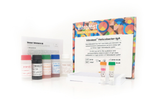
The test kit Vitrotest® Helicobacter-IgА is an enzyme linked immunosorbent assay (ELISA) for the qualitative and semiquantitative determination of IgА class antibodies to CagA protein of Helicobacter pylori in human serum or plasma.
Determination of IgА antibodies to CagA protein of H. pylori in the test kit Vitrotest® Helicobacter-IgА is based on a solid phase, indirect ELISA in a two-step incubation procedure.
○ ТК047 - 96 tests- Solid phase: breakable microplate ELISA is coated with recombinant CagA protein of H. pylori.
- Conjugate: a monoclonal antibodies to human IgА conjugated to horseradish peroxidase.
- Chromogen: ready to use TMB solution.
- Volume of sample for analysis: 10 μl.
- Assay time: 1h 15 min.
Helicobacter pylori is a widespread microorganism with which the half of the world’s population has been infected. Its prevalence is very high in developing countries and is quite low in the developed world. According to the World Gastroenterology Organization, the rate of infected adults in Eastern Europe and Asia is about 70-80 %.
Studies during recent decades have shown the key role of bacterium Helicobacter pylori in the pathogenesis of stomach and duodenal lesions. H. pylori is detected almost in 100% of adult patients with duodenal ulcer, approximately in 80 % of patients with peptic ulcer, in 92% of patients with gastric cancer and in 92 % of patients with active chronic gastritis. Research has demonstrated that elimination of helicobacter leads to the disappearance of gastritis and significant reduction in the incidence of duodenal ulcer recurrence.
Helicobacteriosis is a chronic infection with long, often asymptomatic course. Its symptoms do not differ from clinical manifestations of gastro-duodenitis since usual constant pain in the epigastrium occurs. H. pylori is often present in patients with no clinical manifestations of disease.
The infection usually starts from non–acid-secreting antral region of the stomach and stimulates the increased release of gastrin. The increased gastrin levels in turn stimulate excess acid secretion from the more proximal acid-secreting fundic mucosa which is relatively free of inflammation. The increased duodenal acid load damages the duodenal mucosa, causing ulceration. If infection progresses, stomach body is damaged, which could finally cause the development of gastric adenocarcinoma. This tumour is preceded by sequential pathological changes of gastric mucosa, from normal mucosa to superficial gastritis, atrophic gastritis, gastric ulcers and intestinal metaplasia.
Main oncogenic factors are both nitrosating bacteria present in the lumen of hypochloric stomach, capable of generating potentially carcinogenic N-nitrosamines and reactive oxygen species, and H. pylori itself. Duodenal or gastric ulcers are reported to develop in 1 to 10% of infected patients, gastric cancer- in 0.1 to 3%; at the same time, the great majority of patients with H. pylori remain asymptomatic carriers.
H. pylori is usually acquired in childhood. The bacteria are most likely spread through fecal-oral or oral-oral routes from person to person; additional transmission routes, such as water, may be important in developing countries. -
The test kit Vitrotest® Helicobacter-IgМ is an enzyme linked immunosorbent assay (ELISA) for the qualitative and semiquantitative determination of IgМ class antibodies to CagA protein of Helicobacter pylori in human serum or plasma.
Determination of IgМ antibodies to CagA protein of H. pylori in the test kit Vitrotest® Helicobacter-IgМ is based on a solid phase, indirect ELISA in a two-step incubation procedure.
○ ТК048 - 96 tests- Solid phase: breakable microplate ELISA is coated with recombinant CagA protein of H. pylori.
- Conjugate: a monoclonal antibodies to human IgМ conjugated to horseradish peroxidase.
- Chromogen: ready to use TMB solution.
- Volume of sample for analysis: 10 μl.
- Assay time: 1h 15 min.
Helicobacter pylori is a widespread microorganism with which the half of the world’s population has been infected. Its prevalence is very high in developing countries and is quite low in the developed world. According to the World Gastroenterology Organization, the rate of infected adults in Eastern Europe and Asia is about 70-80 %.
Studies during recent decades have shown the key role of bacterium Helicobacter pylori in the pathogenesis of stomach and duodenal lesions. H. pylori is detected almost in 100% of adult patients with duodenal ulcer, approximately in 80 % of patients with peptic ulcer, in 92% of patients with gastric cancer and in 92 % of patients with active chronic gastritis. Research has demonstrated that elimination of helicobacter leads to the disappearance of gastritis and significant reduction in the incidence of duodenal ulcer recurrence.
Helicobacteriosis is a chronic infection with long, often asymptomatic course. Its symptoms do not differ from clinical manifestations of gastro-duodenitis since usual constant pain in the epigastrium occurs. H. pylori is often present in patients with no clinical manifestations of disease.
The infection usually starts from non–acid-secreting antral region of the stomach and stimulates the increased release of gastrin. The increased gastrin levels in turn stimulate excess acid secretion from the more proximal acid-secreting fundic mucosa which is relatively free of inflammation. The increased duodenal acid load damages the duodenal mucosa, causing ulceration. If infection progresses, stomach body is damaged, which could finally cause the development of gastric adenocarcinoma. This tumour is preceded by sequential pathological changes of gastric mucosa, from normal mucosa to superficial gastritis, atrophic gastritis, gastric ulcers and intestinal metaplasia.
Main oncogenic factors are both nitrosating bacteria present in the lumen of hypochloric stomach, capable of generating potentially carcinogenic N-nitrosamines and reactive oxygen species, and H. pylori itself. Duodenal or gastric ulcers are reported to develop in 1 to 10% of infected patients, gastric cancer- in 0.1 to 3%; at the same time, the great majority of patients with H. pylori remain asymptomatic carriers.
H. pylori is usually acquired in childhood. The bacteria are most likely spread through fecal-oral or oral-oral routes from person to person; additional transmission routes, such as water, may be important in developing countries. -
The test kit Vitrotest® Anti-Lamblia is an enzyme linked immunosorbent assay (ELISA) for the detection of total antibodies to Giardia lamblia (intestinalis) in human serum or plasma.
Determination of total antibodies to Giardia lamblia in the test kit Vitrotest® Anti-Lamblia is based on a solid phase, indirect ELISA in a two-step incubation procedure.
○ ТК030 - 96 tests
○ ТК108 - 192 tests- Solid phase: breakable microplate ELISA is coated Giardia lamblia purified antigens.
- Conjugate: a monoclonal antibodies to human IgG, IgA and IgM conjugated to horseradish peroxidase.
- Chromogen: ready to use TMB solution.
- Volume of sample for analysis 10 μl.
- Assay time: 1h 15 min.
Giardia lamblia (intestinalis) causes giardiasis, a parasitic infestation that can manifest as latent parasitic carriage or in more pronounced forms, such as intestinal dysfunction. Giardiasis has been recorded on all five continents and in most countries worldwide. Infection rates vary from less than 1% to 50%. In many developing countries, where basic sanitary conditions are lacking, infection with Giardia in children by the age of 2 is almost universal. In contrast, in developed countries, the infection rate of G.lamblia is only 3–7%. The disease affects all age groups, but preschool-aged children are the most affected demographic.
The primary route of transmission for G.lamblia is fecal-oral. The parasite has a simple two-stage life cycle. After the host ingests cysts, they release trophozoites in the duodenum, which then attach to the mucosal lining of the small intestine.
Trophozoites exist only on the surface of the mucosa in the upper part of the small intestine. Therefore, Giardia mechanically blocks the mucosa and disrupts membrane digestion and the motor activity of the small intestine. Giardia impairs the absorption of fats, carbohydrates, vitamins C and B12, and leads to secondary bacterial infections. Symptoms of giardiasis can include diarrhea, fatigue, bloating, apathy, weight loss, decreased appetite, pallor, and muscle twitching. From the gastrointestinal tract perspective, giardiasis mainly manifests as enterocolitis with catarrhal symptoms.
Numerous findings indicate the role of the humoral immune response in the elimination of G.lamblia. As shown in an experimental human infection model, IgM antibody levels significantly increased on days 14-21 post-infection and gradually declined following therapy. In contrast, IgG antibody levels remained elevated after successful treatment. The dynamics of IgA levels were similar to those of IgM.
The diagnosis of giardiasis is based on clinical history, symptoms, and the presence of cysts in fecal samples or trophozoites in material obtained from the small intestine during duodenal aspiration or duodenal biopsy. Alternative methods include the detection of G.lamblia antigen in feces and the determination of specific antibodies against Giardia in the patient’s serum. Serological testing is considered a valuable adjunct in the diagnosis of giardiasis. In addition to aiding in diagnosis, it can be useful for assessing the patient’s immune response and for epidemiological studies. -
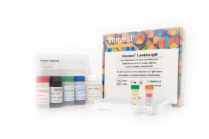
The test kit Vitrotest® Lamblia-IgM is an enzyme linked immunosorbent assay (ELISA) for the detection of IgM class antibodies to Giardia lamblia (intestinalis) in human serum or plasma.
Determination of IgM antibodies to Giardia lamblia in the test kit Vitrotest® Lamblia-IgM is based on a solid phase, indirect ELISA in a two-step incubation procedure.
○ ТК031 - 96 tests- Solid phase: breakable microplate ELISA is coated Giardia lamblia purified antigens.
- Conjugate: a monoclonal antibodies to human IgM conjugated to horseradish peroxidase.
- Chromogen: ready to use TMB solution.
- Volume of sample for analysis 10 μl.
- Assay time: 1h 15 min.
Giardia lamblia (intestinalis) causes giardiasis, a parasitic infestation that can manifest as latent parasitic carriage or in more pronounced forms, such as intestinal dysfunction. Giardiasis has been recorded on all five continents and in most countries worldwide. Infection rates vary from less than 1% to 50%. In many developing countries, where basic sanitary conditions are lacking, infection with Giardia in children by the age of 2 is almost universal. In contrast, in developed countries, the infection rate of G.lamblia is only 3–7%. The disease affects all age groups, but preschool-aged children are the most affected demographic.
The primary route of transmission for G.lamblia is fecal-oral. The parasite has a simple two-stage life cycle. After the host ingests cysts, they release trophozoites in the duodenum, which then attach to the mucosal lining of the small intestine.
Trophozoites exist only on the surface of the mucosa in the upper part of the small intestine. Therefore, Giardia mechanically blocks the mucosa and disrupts membrane digestion and the motor activity of the small intestine. Giardia impairs the absorption of fats, carbohydrates, vitamins C and B12, and leads to secondary bacterial infections. Symptoms of giardiasis can include diarrhea, fatigue, bloating, apathy, weight loss, decreased appetite, pallor, and muscle twitching. From the gastrointestinal tract perspective, giardiasis mainly manifests as enterocolitis with catarrhal symptoms.
Numerous findings indicate the role of the humoral immune response in the elimination of G.lamblia. As shown in an experimental human infection model, IgM antibody levels significantly increased on days 14-21 post-infection and gradually declined following therapy. In contrast, IgG antibody levels remained elevated after successful treatment. The dynamics of IgA levels were similar to those of IgM.
The diagnosis of giardiasis is based on clinical history, symptoms, and the presence of cysts in fecal samples or trophozoites in material obtained from the small intestine during duodenal aspiration or duodenal biopsy. Alternative methods include the detection of G.lamblia antigen in feces and the determination of specific antibodies against Giardia in the patient’s serum. Serological testing is considered a valuable adjunct in the diagnosis of giardiasis. In addition to aiding in diagnosis, it can be useful for assessing the patient’s immune response and for epidemiological studies. -
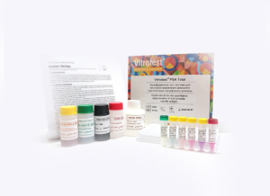
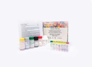
The test kit Vitrotest® PSA Total is an enzyme linked immunosorbent assay (ELISA) for the quantitative determination of total prostate-specific antigen (PSA) in human serum or plasma.
Determination of concentration total PSA in the test kit Vitrotest® PSA Total is based on on a solid-phase "sandwich" ELISA.
○ ТК104 - 96 tests- Solid phase: breakable microplate ELISA is coated with monoclonal antibodies specific to human PSA.
- Conjugate: a monoclonal antibodies to human PSA conjugated to horseradish peroxidase.
- Chromogen: ready to use TMB solution.
- Volume of sample for analysis: 10 μl.
- Assay time: 1h 15 min.
- Analytical sensitivity: 0,134 ng/ml.
- Linearity range: 0,25-30 ng/ml.
In developed countries, prostate cancer ranks second in mortality from malignant diseases among men. According to WHO data, in 2020, prostate cancer mortality in Ukraine reached 4,219 cases.
A valuable biomarker for prostate cancer, which is now widely used for population screening, diagnosis, and monitoring of patients with prostate cancer, is the prostate-specific antigen (PSA). Screening for this tumor marker was introduced in the 1990s in the USA, Canada, Japan, and the UK, where a subsequent trend of decreased prostate cancer mortality has been observed.
Prostate-specific antigen is a glycoprotein produced by the epithelial cells of the prostate gland, the urethral mucosa, and the Cowper’s glands. PSA is secreted into the seminal fluid, ensuring semen liquefaction, but is typically found in very low concentrations in the blood. In blood serum, PSA is present in various forms: complexed with α1-antichymotrypsin (70-90%), free PSA not bound to serum proteins (10-30%), and bound to alpha-2-macroglobulin.
In men’s blood serum, the normal concentration of total PSA does not exceed 4 ng/ml. Elevated levels of this tumor marker may indicate a risk of prostate cancer. According to the American Cancer Society, the likelihood of prostate cancer with serum total PSA levels of 4-10 ng/ml is 25%, while with concentrations above 10 ng/ml, this figure approaches 50%. An increase in PSA levels can also be observed with prostate hypertrophy and prostatitis. Therefore, when elevated serum PSA levels are detected, a digital rectal examination (DRE) of the prostate is recommended to differentiate pathological conditions.
For laboratory identification of prostate cancer, the measurement of total and free prostate-specific antigen in blood serum is used. When PSA concentration is in the range of 4 to 10 ng/ml, determining the percentage of free PSA can be helpful in deciding whether a prostate biopsy is necessary. Typically, a biopsy is recommended when the percentage of free PSA is below 10%, although some doctors consider a threshold level of 25%.
The serum PSA level is an informative marker not only for screening patients for prostate cancer but also for monitoring treatment effectiveness and prognosis after surgical intervention. -
The test kit Vitrotest® PSA Free is an enzyme linked immunosorbent assay (ELISA) for the quantitative determination of free prostate-specific antigen (PSA) in human serum or plasma.
Determination of concentration free PSA in the test kit Vitrotest® PSA Free is based on on a solid-phase "sandwich" ELISA followed by the use of biotin-streptavidin interaction.
○ ТК105 - 96 tests- Solid phase: breakable microplate ELISA is coated with monoclonal antibodies specific to human PSA.
- Biotinylated anti-PSA antibody solution: biotinylated monoclonal antibodies to human PSA.
- Streptavidin-HRP conjugate: streptavidin solution conjugated with horseradish peroxidase.
- Chromogen: ready to use TMB solution.
- Volume of sample for analysis: 25 μl.
- Assay time: 1 hour.
- Analytical sensitivity: 0,096 ng/ml.
- Linearity range: 0,125-8 ng/ml.
In developed countries, prostate cancer ranks second in mortality from malignant diseases among men. According to WHO data, in 2020, prostate cancer mortality in Ukraine reached 4,219 cases.
A valuable biomarker for prostate cancer, which is now widely used for population screening, diagnosis, and monitoring of patients with prostate cancer, is the prostate-specific antigen (PSA). Screening for this tumor marker was introduced in the 1990s in the USA, Canada, Japan, and the UK, where a subsequent trend of decreased prostate cancer mortality has been observed.
Prostate-specific antigen is a glycoprotein produced by the epithelial cells of the prostate gland, the urethral mucosa, and the Cowper’s glands. PSA is secreted into the seminal fluid, ensuring semen liquefaction, but is typically found in very low concentrations in the blood. In blood serum, PSA is present in various forms: complexed with α1-antichymotrypsin (70-90%), free PSA not bound to serum proteins (10-30%), and bound to alpha-2-macroglobulin.
In men’s blood serum, the normal concentration of total PSA does not exceed 4 ng/ml. Elevated levels of this tumor marker may indicate a risk of prostate cancer. According to the American Cancer Society, the likelihood of prostate cancer with serum total PSA levels of 4-10 ng/ml is 25%, while with concentrations above 10 ng/ml, this figure approaches 50%. An increase in PSA levels can also be observed with prostate hypertrophy and prostatitis. Therefore, when elevated serum PSA levels are detected, a digital rectal examination (DRE) of the prostate is recommended to differentiate pathological conditions.
For laboratory identification of prostate cancer, the measurement of total and free prostate-specific antigen in blood serum is used. When PSA concentration is in the range of 4 to 10 ng/ml, determining the percentage of free PSA can be helpful in deciding whether a prostate biopsy is necessary. Typically, a biopsy is recommended when the percentage of free PSA is below 10%, although some doctors consider a threshold level of 25%.
The serum PSA level is an informative marker not only for screening patients for prostate cancer but also for monitoring treatment effectiveness and prognosis after surgical intervention. -
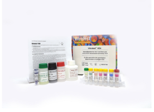
The test kit Vitrotest® HE4 is an enzyme linked immunosorbent assay (ELISA) for the quantitative determination of antigen HE4 in human serum or plasma. Determination of concentration antigen HE4 in the test kit Vitrotest® HE4 is based on a solid-phase “sandwich” ELISA. ○ ТК107 - 96 tests
- Solid phase: breakable microplate ELISA is coated with monoclonal antibodies specific to antigen HE4.
- Conjugate: a monoclonal antibodies to antigen HE4 conjugated to horseradish peroxidase.
- Chromogen: ready to use TMB solution.
- Volume of sample for analysis: 10 μl.
- Assay time: 1h 15 min.
- Analytical sensitivity: 4,85 pmol/l.
- Linearity range: 25-1000 pmol/l.
Ovarian cancer ranks as the second leading cause of cancer-related deaths among women worldwide. Only about 20% of ovarian cancer cases are detected at an early stage. When ovarian cancer is diagnosed at an early stage, approximately 94% of patients survive longer than five years after diagnosis. However, more than 70% of cases are diagnosed at an advanced stage, which significantly worsens the prognosis. For the detection of ovarian cancer in patients without symptoms, two screening tests are most commonly used: transvaginal ultrasound and a blood test for the CA125 tumor marker. However, elevated CA125 levels can be found in 1-2% of healthy women and in patients with other conditions and diseases, including the first trimester of pregnancy, menstruation, endometriosis, uterine fibroids, acute salpingitis, liver disease, and peritonitis. To improve the sensitivity and specificity of laboratory diagnostic tools in patients with benign tumors, the combined determination of serum levels of two tumor markers, CA125 and HE4, is currently used. HE4 (Human Epididymis Protein 4) is a secretory low-molecular-weight glycoprotein predominantly expressed in the epithelial cells of the fallopian tube, as well as normally in the epithelium of the mammary glands, reproductive system organs, intestines, and lungs. Although the physiological functions of this protein have not been fully studied, overexpression of HE4 is observed in serous and endometrioid ovarian carcinomas. Elevated levels of this tumor marker are found both in the early stages of ovarian cancer and in endometrial cancer. Unlike CA125, HE4 levels do not increase in endometriosis, benign gynecological diseases, or ovarian cysts. The serum level of HE4 is an informative marker not only for screening patients for ovarian cancer but also as an important prognostic marker. High concentrations of HE4 in plasma are an independent preoperative marker of poor prognosis for ovarian cancer patients. To improve the assessment of laboratory tests, the Risk of Ovarian Malignancy Algorithm (ROMA) was developed, which is a mathematical model that analyzes HE4 in combination with CA125, taking into account the patient’s menopausal status. The ROMA algorithm allows for a high probability of distinguishing malignant tumors from benign neoplasms in postmenopausal women. -
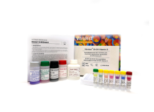
The test kit Vitrotest® 25-OH Vitamin Dis an enzyme linked immunosorbent assay (ELISA) for the quantitative determination of total 25-OH vitamin D (25-OH vitamin D2 and 25-OH vitamin D3) in human serum or plasma.
Determination of total 25-OH vitamin D concentration in the test kit Vitrotest® 25-OH Vitamin D is based on a competitive ELISA with a two-step incubation.
○ TK122 - 96 tests
○ TK124 - 192 tests- Solid phase: a stripped ELISA plate with wells pre-coated with monoclonal antibodies specific to 25-OH vitamin D.
- Biotinylated 25-OH vitamin D solution: biotinylated 25-OH vitamin D.
- Streptavidin-HRP conjugate: streptavidin conjugated with horseradish peroxidase.
- Chromogen: ready to use TMB solution.
- Volume of sample for analysis: 10 µl.
- Assay time: 1 h 30 min.
- Analytical sensitivity: 1,34 ng/ml.
Vitamin D is a fat-soluble steroid prohormone, whose active form is involved in the metabolism of phosphorus and calcium in the human body. Vitamin D, which is produced in the skin (D3, cholecalciferol) or obtained from food (D2, ergocalciferol), is biologically inert and requires two successive hydroxylations. First, 25-OH vitamin D (D2 and D3) is formed in the liver, which, in complex with a binding protein (vitamin D binding protein, VDBP), enters the bloodstream and is transported to the kidneys, where it is converted into the biologically active form 1,25-(OH)2 vitamin D and other metabolites whose roles are still being studied.
25-OH vitamin D is the main storage form of vitamin D in the human body and is present in the blood at much higher concentrations than its active form, 1,25-(OH)2 vitamin D. Its half-life is 2–3 weeks compared to 4-6 hours for 1,25-(OH)2 vitamin D. Additionally, the level of 1,25-(OH)2 vitamin D in serum does not provide information about the vitamin D status and is often normal or even elevated due to secondary hyperparathyroidism associated with vitamin D deficiency. For this reason, 25-OH vitamin D is the analyte of choice for determining the body’s vitamin D status.
According to the literature, the concentration of 25-OH vitamin D in human serum (plasma) within the range of 30-100 ng/ml is sufficient to support all necessary processes in the body that require vitamin D. Most experts consider a level of 25-OH vitamin D < 20 ng/ml to indicate vitamin D deficiency, while 20-30 ng/ml is considered an insufficient level. In Ukraine, only 3.3% of the population have a normal level of vitamin D, 14.9% have an insufficient level, and 81.8% live with its deficiency.
Hypovitaminosis D is widespread in the human population due to minimal exposure to sunlight and low dietary intake of vitamin D. As a result, vitamin D deficiency leads to secondary hyperparathyroidism, followed by loss of bone mass and osteoporosis. There is also evidence that vitamin D deficiency may have other adverse health effects, including an increased risk of malignant neoplasms, cardiovascular diseases, multiple sclerosis, diabetes, and more.
Currently, the reference method for determining the concentration of 25-OH vitamin D is tandem liquid chromatography with mass spectrometry (LC-MS/MS). However, in routine laboratory practice, this method is impractical due to its complexity and the need for specialized personnel. Therefore, immunoassay and immunochemiluminescent methods for determining 25-OH vitamin D have gained widespread use in diagnostic laboratories. -
The test kit Vitrotest® Total-IgE is an enzyme linked immunosorbent assay (ELISA) for the quantitative determination of total immunoglobulin E (IgE) in human serum or plasma.
Determination of total IgE content in the test kit Vitrotest® Total-IgE is based on a solid-phase “sandwich” ELISA.
○ ТК070 - 96 tests- Solid phase: breakable microplate ELISA is coated with monoclonal antibodies specific to human IgE.
- Conjugate: a monoclonal antibodies to human IgЕ conjugated to horseradish peroxidase.
- Chromogen: ready to use TMB solution.
- Volume of sample for analysis: 10 μl.
- Assay time: 1h 15 min.
- Analytical sensitivity: 2,1 IU/ml.
- Linearity range: 0,8-1000 IU/ml.
According to the World Health Organization, 10-20 % of the world’s population suffers from allergic bronchial asthma, allergic rhinitis, atopic dermatitis and other allergic reactions, moreover, the number of such patients increases every year. According to statistics, from 5 to 15% of children have food allergies, which further leads to the development of various allergic diseases.
Allergy or hypersensitivity is a state of altered reactivity of the body in the form of its increased sensitivity to repeated exposure of substances called allergens. As a result of repeated contact with the allergen, an excessively strong immune reaction of the body occurs, accompanied by tissue damage. One of the mechanisms underlying this process is mast cell degranulation mediated by immunoglobulin class E (IgE). According to the literature data, in an adult, the normal concentration of IgE can reach 100 IU/ml and decreases in the elderly. During allergy the IgE level can rise to 2000 IU/ml and above.
Among often diseases that accompanied by an increase in total IgE in the blood, as well as the presence of specific IgE are allergic rhinitis, atopic bronchial asthma, atopic dermatitis, urticaria, aspergillosis and helminthiases, etc. Less common diseases with hyper-IgE syndrome include congenital immunodeficiencies (Job syndrome, DiGeorge syndrome), selective IgA deficiency, WiskottAldridge syndrome and IgEmyeloma. Also, in clinical practice, there are often cases of elevated IgE levels associated with the action of several medications.
Nowadays, the methods of in vitro allergy diagnostics are becoming increasingly popular, one of which is ELISA for the determination of the total IgE content. This method is characterized by several advantages: it has no contraindications to the test, it does not cause additional sensitization and there is no risk of anaphylactic reactions. Most test systems are based on the «sandwich» ELISA, in which the use of monoclonal antibodies to human immunoglobulins E provides high sensitivity and specificity of the analysis. Standardization of the quantitative determination of IgE in human serum or plasma is ensured by using for the manufacture of internal calibrators ELISA kits of the WHO International Standard with an established concentration of human IgE in international units IU/ml.
Since the ELISA method is a highly sensitive and specific analysis and has no contraindications for its application, it can be widely used for the diagnosis of allergic diseases. -
The test kit Vitrotest® Specific-IgE is an enzyme linked immunosorbent assay (ELISA) for the quantitative determination of specific IgE class antibodies in human serum or plasma. The test kit is used together with biotinylated allergens Vitrotest® XX-Biotin, where XX is a code of relevant allergen or mixture.
Quantitative determination of specific IgE antibodies in the Vitrotest® Specific-IgE test kit is based on the principle of capturing IgE antibodies followed by the use of biotin-streptavidin interaction.
○ ТК071 - 192 tests- Solid phase: breakable microplate ELISA is coated with monoclonal antibodies specific to human IgE.
- Biotinylated anti-IgE: biotinylated anti-IgE antibodies.
- Conjugate streptavidin-HRP: streptavidin сonjugated to horseradish peroxidase.
- Chromogen: ready to use TMB solution.
- Volume of sample for analysis: 50 μl.
- Assay time: 1h 30 min.
- Analytical sensitivity: 0,080 IU/ml.
- Linearity range: 0,2-50 IU/ml.
Allergic diseases are among the most common diseases in the world and the severity of these conditions continues to increase. Allergy, or hypersensitivity, is a state of altered reactivity of the body in the form of increased sensitivity to repeated exposure to substances called allergens. As a result of repeated contact with the allergen, an excessively strong immune reaction of the body occurs, accompanied by tissue damage. One mechanism underlying this process is immunoglobulin E (IgE)-mediated mast cell degranulation.
The most common allergic conditions that require proper diagnosis are: anaphylaxis, allergic rhinitis, allergic conjunctivitis, bronchial asthma, etc. Accurate diagnosis coupled with optimal therapy requires the use of appropriate tests to confirm sensitization to a specific allergen and detailed information about the effect of the suspected allergen.
To diagnose in vivo allergies, several types of skin tests are used (skin prick test, intradermal test, patch test). However, the use of these samples has a number of significant limitations, namely, the need for a highly qualified doctor conducting the test and the lack of laboratories conducting these studies. In addition, in vivo diagnosis is rarely performed in children due to possible adverse reactions.
However, in vitro diagnostics of IgE-mediated allergic diseases are useful for identifying the causative allergen and usually include the following laboratory approaches:
1) determination of the level of total IgE;
2) study of the concentration of specific IgE to various allergens - is the most commonly used in vitro
diagnostic approach, performed using a single or multiplex strategy;
3) basophil activation test (BAT), which is quite specific, but difficult to perform, and therefore limited to selected situations.
Today, to detect specific antibodies of the IgE class, the enzyme-linked immunosorbent assay method, which provides high sensitivity and specificity, has become widely used. A separate supply of a wide range of biotinylated allergens, depending on the needs of the diagnostic laboratory, ensures maximum convenience and cost-effectiveness of the study. -
Biotinylated allergens Vitrotest® XX-Biotin for the quantitative determination of specific IgE class antibodies in human serum or plasma. It is used exclusively with the
Vitrotest® Specific-IgE test kit.
○ 1 vial - 30 testsBiotinylated allergens Vitrotest® XX-Biotin, where XX is the code of the relevant allergen or mixture - each microtube contains 1.55 ml of a solution of biotinylated allergens, ready to use. The content of one bottle is enough to conduct 30 studies.
-
The test kit Vitrotest® Free T3 is an enzyme linked immunosorbent assay (ELISA) for the quantitative determination of free triiodothyronine (FT3) in human serum or plasma. Determination of free triiodothyronine concentration in the test kit Vitrotest® Free T3 is based on a competitive ELISA with a two-step incubation.
- TK150 - 96 tests
- Solid phase: strip ELISA plate pre-coated with triiodothyronine.
- Conjugate: buffer solution of monoclonal antibodies specific to triiodothyronine conjugated with horseradish peroxidase.
- Chromogen: ready to use TMB solution.
- Sample volume: 25 μl.
- Assay time: 1 h 20 min.
- Analytical sensitivity: 0,5 pg/ml.
According to data from the World Health Organization (WHO), thyroid diseases are the second most common endocrine disorders after diabetes mellitus. More than 200 million people worldwide suffer from various forms of thyroid dysfunction. In Ukraine, over the past five years, the number of people with thyroid diseases has increased fivefold. The main pathological conditions include hyperthyroidism, hypothyroidism, autoimmune thyroid diseases, benign and malignant neoplasms, etc.
The main hormones produced by the thyroid gland are thyroxine or tetraiodothyronine (T4, containing four iodine atoms) and triiodothyronine (T3, containing three iodine atoms). These hormones are lipophilic and circulate in the blood mainly bound to transport proteins: thyroxine-binding globulin (TBG), transthyretin (also known as thyroxine-binding prealbumin), and albumin. However, only free triiodothyronine and free thyroxine are functionally active, accounting for just 0.3% and 0.03% of total T3 and T4 in blood serum, respectively.
Free triiodothyronine is the biologically active form that directly enters target cells and binds to nuclear receptors to regulate gene expression and cellular function. Free thyroxine has minimal hormonal activity, but its long half-life (8 days) serves as a reservoir or prohormone for free triiodothyronine. All reactions required for the formation and release of T3 and T4 are controlled by thyroid-stimulating hormone (TSH) through a negative feedback mechanism.
Although determination of TSH and FT4 concentrations are considered the primary tests of choice for diagnosing thyroid dysfunction, measurement of FT3 levels is useful for detecting T3-thyrotoxicosis, complex or unusual manifestations of hyperthyroidism, monitoring thyroid disease treatment, and assessing metabolism.
Accumulated evidence indicates that changes in FT3 levels are also closely associated with a number of systemic disorders, such as cardiovascular diseases, dyslipidemia, type 2 diabetes mellitus, and liver dysfunction, highlighting the clinical importance of FT3 monitoring as a functional indicator of thyroid status.
-
The test kit Vitrotest® TSH is an enzyme linked immunosorbent assay (ELISA) for the quantitative determination of thyroid-stimulating hormone (TSH) in human serum or plasma. Determination of thyroid-stimulating hormone concentration in the test kit Vitrotest® TSH is based on a solid-phase sandwich ELISA in a two-step incubation procedure.
- TK148 - 96 tests
- TK149 - 192 tests
- Solid phase: strip ELISA plate pre-coated with the first monoclonal antibodies specific to the β-subunit of human thyroid-stimulating hormone.
- Conjugate: monoclonal antibodies to human TSH conjugated with horseradish peroxidase.
- Chromogen: ready to use TMB solution.
- Sample volume: 50 μl.
- Assay time: 2 h.
- Analytical sensitivity: 0,106 μIU/ml.
- Linearity range: 0,125-16,4 µIU/ml.
According to the World Health Organization (WHO), thyroid diseases are the second most common endocrine disorders after diabetes mellitus. Over 200 million people worldwide suffer from various forms of thyroid dysfunction. In Ukraine, over the past 5 years, the number of people with thyroid diseases has increased fivefold. The main pathological conditions include hyperthyroidism, hypothyroidism, autoimmune thyroid diseases, benign and malignant neoplasms. A connection has also been established between thyroid dysfunction and other diseases such as diabetes, cardiovascular diseases, depression, oral diseases, and cancer.
A valuable biomarker of thyroid functional status widely used for screening, diagnosis, and monitoring of thyroid diseases is thyroid-stimulating hormone (TSH). TSH is a glycoprotein hormone synthesized by the anterior pituitary and is a key regulator of thyroid function. The TSH molecule consists of two different non-covalently bound subunits: the α-subunit, identical in amino acid sequence to the α-subunit of chorionic gonadotropin, luteinizing hormone, and follicle-stimulating hormone, and the hormone-specific β-subunit, which is unique. The main function of TSH is to stimulate the thyroid gland to synthesize and secrete thyroid hormones – thyroxine (T4) and triiodothyronine (T3). TSH binds to the TSH receptor on thyrocytes and activates intracellular signaling cascades that regulate iodine uptake, thyroid metabolism, thyroid growth, and hormone secretion. Through negative feedback, T3 and T4 inhibit TSH secretion.
In healthy adults, TSH serum levels are approximately 0.4 to 4.0 μIU/ml, although narrower ranges may be used to better detect subclinical hypothyroidism. Separate reference intervals for TSH are established for pregnant women, infants, and young children. TSH secretion has pulsatile and circadian patterns, and its concentration depends on factors such as age, sex, ethnicity, iodine intake, reproductive status, and body mass index.
Over the last three decades, laboratory methods used to determine TSH levels have significantly improved. Among immunochemical methods, ELISA has gained wide application due to its convenience, simplicity, high reproducibility, and sensitivity for determining thyroid-stimulating hormone in human serum and plasma.
The standardization of quantitative determination of TSH in human serum or plasma is ensured by the use of the WHO International Standard with assigned TSH concentration in μIU/ml for preparation of internal ELISA calibrators.
-
The test kit Vitrotest® Free T4 is an enzyme linked immunosorbent assay (ELISA) for the quantitative determination of free thyroxine (FT4) in human serum or plasma. Determination of free thyroxine concentration in the test kit Vitrotest® Free T4 is based on a competitive ELISA with a two-step incubation.
- TK151 - 96 tests
- Solid phase: strip ELISA plate pre-coated with monoclonal antibodies specific to thyroxine.
- Conjugate: buffer solution of thyroxine conjugated with horseradish peroxidase.
- Chromogen: ready to use TMB solution.
- Sample volume: 20 μl.
- Assay time: 1 h 20 min.
- Analytical sensitivity: 0,254 ng/dl.
According to WHO, thyroid diseases are the second most common endocrine disorders after diabetes mellitus. Over 200 million people worldwide suffer from various forms of thyroid dysfunction. In Ukraine, the number of people with thyroid diseases has increased fivefold over the past 5 years. The main pathological conditions include hyperthyroidism, hypothyroidism, autoimmune thyroid diseases, benign and malignant neoplasms.
The main hormones produced by the thyroid gland are thyroxine or tetraiodothyronine (T4, containing four iodine atoms) and triiodothyronine (T3, containing three iodine atoms). They are lipophilic and circulate in the blood mainly bound to transport proteins: thyroxine-binding globulin (TBG), transthyretin (thyroxine-binding prealbumin), and albumin. However, the biologically active forms are free triiodothyronine and free thyroxine, which represent only 0.3% and 0.03% of total T3 and T4 in serum, respectively.
Overall, FT4 has minimal hormonal activity, but its long half-life (8 days) serves as a reservoir or prohormone for active free triiodothyronine, which binds to nuclear receptors.
All processes required for the synthesis and release of T3 and T4 are controlled by thyroid-stimulating hormone (TSH), secreted by pituitary thyrotropic cells. TSH secretion is regulated through pituitary negative feedback: elevated free T4 and T3 levels suppress TSH synthesis and secretion, while decreased levels increase TSH secretion.
Determination of TSH and free thyroxine concentrations are the primary tests for diagnosing thyroid dysfunction. FT4 is an important marker for differentiating subclinical hyperthyroidism from overt hyperthyroidism or hypothyroidism, for investigating suspected abnormal TSH secretion, TSH-secreting pituitary adenomas, and for monitoring treatment of thyroid diseases.
-
The test kit Vitrotest Anti-TPO is an enzyme linked immunosorbent assay (ELISA) for the quantitative determination of autoantibodies to thyroid peroxidase (TPO) in human serum or plasma. Determination of autoantibodies to thyroid peroxidase in the test kit Vitrotest Anti-TPO is based on a solid phase, indirect ELISA in a two-step incubation procedure.
- TK152 - 96 tests
- Solid phase: strip ELISA plate pre-coated with recombinant thyroid peroxidase.
- Conjugate: buffer solution of monoclonal antibodies to human IgG conjugated with horseradish peroxidase.
- Chromogen: ready to use TMB solution.
- Sample volume: 10 μl.
- Assay time: 1 h 20 min.
- Analytical sensitivity: 2,17 IU/ml.
- Linearity range: 3,125 - 410 IU/ml.
According to WHO, thyroid diseases are the second most widespread endocrine disorders after diabetes mellitus. Over 200 million people worldwide suffer from various types of thyroid dysfunction. In Ukraine, over the past 5 years, the number of people with thyroid diseases has increased fivefold.
Autoimmune thyroid diseases represent a diverse group of organ-specific autoimmune disorders, the most common of which are Hashimoto’s thyroiditis and Graves’ disease. The pathological process is associated with the formation of autoantibodies to thyroid peroxidase and thyroglobulin.
Thyroid peroxidase is a membrane-bound enzyme responsible for iodine oxidation and iodination of tyrosyl residues in the thyroglobulin molecule during the synthesis of thyroid hormones T3 and T4. Most anti-TPO antibodies belong to the IgG1 subclass, which activates complement. Additionally, anti-TPO antibodies can bind through their Fc fragment to natural killer cells, which in turn cause cytotoxic damage to thyrocytes.
Damage to thyroid cells, as well as direct enzyme inhibition, can lead to insufficient hormone production (hypothyroidism), sometimes preceded by transient hyperthyroidism. Besides thyroid disorders, elevated anti-TPO titers may also occur in a wide range of diseases: pernicious anemia, systemic lupus erythematosus, rheumatoid arthritis, insulin-dependent diabetes mellitus, breast cancer, and others. A correlation has also been established between anti-TPO antibodies and obstetric complications, such as postpartum thyroiditis or postpartum depression.
Anti-TPO antibodies may be present in individuals without clinical or laboratory signs of thyroid dysfunction. The presence of anti-TPO without overt disease is associated with a higher risk of developing autoimmune thyroiditis in the future and, together with TSH levels, is used to predict the development of hypo- and hyperthyroidism.
In modern laboratory diagnostics, ELISA is widely used for determining the concentration of autoantibodies to thyroid peroxidase due to its simplicity, convenience, high sensitivity, and specificity. Standardization of quantitative determination of anti-TPO in human serum or plasma is ensured by the use of the WHO International Standard with assigned concentration in IU/ml for preparation of internal ELISA calibrators.
-
The Vitrotest® Total-IgG immunodiffusion kit is designed for the quantitative determination of total immunoglobulin G (IgG) in human serum or plasma by radial immunodiffusion in a Mancini technique.
The determination of IgG antibodies in the Vitrotest® Total-IgG immunodiffusion kit is based on an immunoprecipitation reaction. Immunoglobulins of the test sample diffuse in a semi-solid agarose gel containing immune polyclonal serum and form a precipitation ring when equivalent concentrations are reached. The diameter of the ring depends on the concentration of IgG. Comparison with the control allows to determine the concentration of immunoglobulins in the tested serum.
○ TK100 - 70 tests- Immunodiffusion medium - ready-to-use agar gel with wells containing highly specific to human IgG immune serum.
- Control - is a mixture of purified human immunoglobulins of classes G, A, and M with known concentrations.
- The volume of sample for analysis - 3 μL.
- Assay time - 24 hours.
Immunoglobulins (antibodies) are a group of glycoproteins found in mammalian plasma and tissue fluid. Five classes of immunoglobulins are found in humans - IgG, IgA, IgM, IgD, and IgE - which differ in function, molecular size, charge, and carbohydrate content.
IgG is the main isotype of immunoglobulins in normal human serum, comprising for 70-75% of the total amount of serum immunoglobulins. Its concentration normally ranges from 6.0 to 16.0 g/L. Class G immunoglobulins are major class of antibodies of the secondary immune response, they are synthesized for a longer time after an antigenic stimulus and have the unique property of initiating anti- and pro-inflammatory reactions. Antibodies of this class bind also soluble antigens, so they are the main part of antitoxins. In humans, IgG is transported from mother to fetus across the placenta, creating passive immunity for the neonatal period. Excessive production of immunoglobulins G may be associated with an active infectious process, certain types of immunoproliferative diseases, or other conditions that lead to hyperstimulation of B-cell clones that produce IgG. IgG deficiency can be primary (congenital), which is rare, or secondary (acquired), caused by factors that suppress humoral immunity.
To determine the content of total human IgG in diagnostic laboratories, the radial immunodiffusion (RID) method is widely used, which is considered the standard for measuring of different classes immunoglobulins in human serum and plasma. -
The Vitrotest® Total-IgA immunodiffusion kit is designed for the quantitative determination of total immunoglobulin A (IgA) in human serum or plasma by radial immunodiffusion in a Mancini technique.
The determination of IgA antibodies in the Vitrotest® Total-IgA immunodiffusion kit is based on the immunoprecipitation reaction. Immunoglobulins of the test serum diffuse in a semi-solid agar gel containing immune polyclonal serum and form a precipitation ring when equivalent concentrations are reached. The diameter of the ring depends on the concentration of IgA. Comparison with the control allows to determine the concentration of immunoglobulins in the tested serum.
○ TK101 - 70 tests- Immunodiffusion medium - ready-to-use agar gel with wells containing highly specific immune serum to human IgA.
- Control - a mixture of purified polyclonal antibodies of classes G, A, and M of human with known concentrations.
- Sample volume for analysis - 3 µL.
- Analysis time - 24 hours.
Immunoglobulins (antibodies) are a group of glycoproteins found in the blood plasma and tissue fluid of mammals. Five classes of immunoglobulins are found in humans - IgG, IgA, IgM, IgD, and IgE - which differ in molecular size, charge, and carbohydrate content.
IgA presents in two forms - serum and secretory, in serum comprise up 15% of all immunoglobulins, and can be both in monomeric form and as a dimer. Its concentration in normal ranges from 0,8 to 3,0 g/L. Serum immunoglobulin A can activate complement in an alternative pathway and neutralize microbes and toxins circulating in the blood, but its action is weaker than secretory IgA.
Secretory IgA neutralizes bacterial toxins, localizes viruses, and stimulates phagocytosis, providing local resistance to infection. An increased IgA concentration is observed in many inflammatory diseases: acute and chronic bacterial, fungal, parasite infection, chronic liver disease, and cirrhosis. A reduced IgA level is observed at physiologic hypogammaglobulinemia in children (at the age of 3-5 months), congenital hypogammaglobulinemia or agammaglobulinemia, neoplasms of the immune system, treatment with cytostatics and immunosuppressants.
To determine the content of total human IgA in diagnostic laboratories, the radial immunodiffusion (RID) method is widely used, which is considered the standard for measuring of different classes immunoglobulins in human serum and plasma.
-
The Vitrotest® Total-IgM immunodiffusion kit is designed for the quantitative determination of total immunoglobulin M (IgM) in human serum or plasma by radial immunodiffusion in a Mancini technique. The determination of IgA antibodies in the Vitrotest® Total-IgA immunodiffusion kit is based on an immunoprecipitation reaction. Immunoglobulins of the test serum diffuse in a semi-solid agarose gel containing immune polyclonal serum and form a precipitation ring when equivalent concentrations are reached. The diameter of the ring depends on the concentration of IgM. Comparison with the control allows the concentration of immunoglobulins in the test serum to be determined. ○ TK102 - 70 tests
- Immunodiffusion medium - ready-to-use agar gel with wells containing highly specific human IgM immune serum.
- Control - a mixture of purified polyclonal antibodies of classes G, A, and M with known concentrations.
- Sample volume for analysis - 3 μL.
- Analysis time - 48 hours.
Immunoglobulins (antibodies) are a group of glycoproteins found in mammalian plasma and tissue fluid. In humans, as well as in most higher mammals, five classes of immunoglobulins are found - IgG, IgA, IgM, IgD, and IgE - which differ in molecular size, charge, and carbohydrate content. IgM is the first immunoglobulin to be synthesized in response to primary antigenic stimulation and is a marker of the primary immune response. The high avidity of IgM antibodies makes them particularly effective at binding antigens present at low levels and non-protein antigens, such as carbohydrates or lipids, present on microbial surfaces. A very important property of IgM is the activation of phagocytosis. Polyreactive “natural” IgM, which is detected in human serum starting from the 20th week of pregnancy, plays an important role in the innate defense against infectious pathogens. The content of IgM is 5-10% of all serum immunoglobulins. Normally, the concentration of M-class immunoglobulins in the blood ranges from 0.4 to 2.5 g/L. Elevated IgM level is observed in acute and chronic infections, rheumatoid arthritis, acute and chronic lymphocytosis, myeloma, Waldenström's macroglobulinemia, endothelioma, osteosarcoma, candidiasis, and cystic fibrosis. Also, a significant increase in the amount of IgM occurs in hyperimmunoglobulinemia M. Decreased IgM concentration is observed at physiological hypogammaglobulinemia in children (aged 3-5 months), congenital hypogammaglobulinemia or agammaglobulinemia, neoplasms of the immune system, treatment with cytostatics and immunosuppressants, and ionizing radiation exposure. To determine the content of total human IgM in diagnostic laboratories, the radial immunodiffusion (RID) method is widely used, which is considered the standard for measuring of different classes immunoglobulins in human serum and plasma. -
The test kit Vitrotest® Brucella-IgG is an enzyme linked immunosorbent assay (ELISA) for the qualitative and semiquantitative determination of igG class antibodies to Brucella in human serum or plasma.
Determination of IgG antibodies to Brucella in the test kit Vitrotest® Brucella-IgG is based on a solid phase, indirect ELISA in a two-step incubation procedure.
- TK153 - 96 tests
- Solid phase: strip ELISA plate pre-coated with antigens of Brucella.
- Conjugate: monoclonal antibodies to human IgG conjugated with horseradish peroxidase.
- Chromogen: ready to use TMB solution.
- Sample volume: 100 μl.
- Assay time: 1 h 15 min.
Brucellosis is a zoonotic infectious disease caused by bacteria of the genus Brucella. Globally, 1.6–2.1 million human cases of brucellosis are reported annually. Among the countries with the highest reported incidence of brucellosis are Iran, Kyrgyzstan, Tajikistan, Kazakhstan, Azerbaijan, Turkmenistan, Armenia, and Uzbekistan.
The species most pathogenic to humans include Brucella melitensis, Brucella abortus, Brucella suis, and Brucella canis. Different species of Brucella vary in their reservoir hosts and degree of virulence: B. melitensis is considered the most pathogenic for humans, whereas B. abortus more commonly causes relatively milder forms of the disease.
Human infection occurs through contact with infected animals and their biological fluids, as well as through the consumption of animal-derived products (raw milk and insufficiently heat-treated meat), and by the airborne route during occupational exposure. The pathogen enters the human body through mucous membranes or damaged skin. Subsequently, the bacteria are transported by macrophages to lymphoid tissues, spread through the lymphatic system, and may potentially proliferate in multiple organs, causing localized and systemic infections. By persisting within host cells such as macrophages, Brucella spp. employ strategies to evade the host immune response, resulting in prolonged infection.
The incubation period of brucellosis usually ranges from 1 to 4 weeks but may extend to several months. Clinical manifestations are characterized by undulating fever, sweating (especially at night), weakness, and enlargement of the lymph nodes, liver, and spleen. In chronic cases, the musculoskeletal, nervous, and cardiovascular systems may also be affected.
Due to the nonspecific nature of its symptoms, the diagnosis of brucellosis is challenging and requires a comprehensive approach, including the evaluation of clinical findings, epidemiological history, and laboratory test results (bacterial culture, serological assays, and PCR). Serological methods used for the diagnosis of brucellosis include the agglutination test, complement fixation test, and enzyme-linked immunosorbent assay (ELISA).
Using the ELISA method, IgM antibodies are detected during the early stages of infection, whereas IgG antibodies are detected later and may persist for a long period after previous infection or successful treatment. A rapid decline in IgG antibody titers usually indicates a favorable response to antibiotic therapy, while persistently high or increasing titers may indicate treatment failure, residual disease, or an impending clinical relapse. Therefore, the detection of IgG antibodies specific to Brucella is an important marker of current or past infection, particularly in subacute and chronic forms of brucellosis.
Productsvitroadmin2025-09-19T11:29:17+03:00